biomechanics – NIH Director's Blog (original) (raw)
Why Flies and Humans Freeze When Startled
Posted on February 6th, 2020 by Dr. Francis Collins
When faced with something unexpected and potentially ominous, like a sudden, loud noise or a threat of danger, humans often freeze before we act. This is colloquially referred to as the “deer in the headlights” phenomenon. The movie of fruit flies that you see above may help explain the ancient origins of the “startle response” and other biomechanical aspects of motion.
In this video, which shows a footrace between two flies (Drosophila melanogaster), there are no winners or losers. Their dash across the screen provides a world-class view of the biomechanics of walking in these tiny, 3 millimeter-long insects that just won’t sit still.
The fly at the top zips along at about 25 millimeters per second, the normal walking speed for Drosophila. As a six-legged hexapod, the fly walks with a “tripod gait,” alternating between its stance phase—right fore (RF), left middle (LM), and right hind (RH) —and its swing phase sequence of left fore (LF), right middle (RM), and left hind (LH).
The slowpoke at the bottom of the video clocks in at a mere 15 millimeters per second. This fly’s more-tentative gait isn’t due to an injury or a natural lack of speed. What is causing the delay is the rapid release of the chemical messenger serotonin into its nervous system, which models a startle response.
You may have already heard about serotonin because of its role in regulating mood and appetite in humans. Now, a team led by Richard S. Mann and Clare Howard, Columbia University’s Zuckerman Institute, New York, has discovered that fruit flies naturally release serotonin to turn on neural circuits that downshift and steady the speed of their gait.
As detailed recently in Current Biology [1], serotonin is active under myriad conditions to tell flies to slow things down. For example, serotonin helps flies weather the stress of extreme temperatures, conserve energy during bouts of hunger, and even walk upside down on the ceiling.
But the research team, which was supported by the NIH-led Brain Research through Advancing Innovative Neurotechnologies® (BRAIN) Initiative, found that serotonin’s most-powerful effect came during an actual startle response, prompted by a sudden, jolting vibration. Scientists suspect the release of serotonin activates motor neurons much like an emergency brake, stiffening and locking up the fly’s leg joints. When the researchers blocked the fly’s release of serotonin, it interrupted their normal startle response.
In years past, such a detailed, high-resolution “action video” of Drosophila, one of the most-popular model organisms in biology, would have been impossible to produce. Fruit flies are tiny and possess extremely high energy.
But a few years ago, the Mann lab developed the approach used in this video to bring the hurried gait of fruit flies into tight focus [2]. Their system combines an optical touch sensor and high-speed video imaging that records the footfalls of all six of a fly’s feet.
Then, using the lab’s unique software program called FlyWalker , the researchers can extract various biomechanical parameters of walking in time and space. These include step length, footprint alignment, and, as the letters in the video show, the natural sequence of a tripod gait.
Drosophila may be a very distant relative of humans. But these ubiquitous insects that sometimes buzz around our fruit bowls contain many fundamental clues into human biology, whether the area of research is genetics, nutrition, biomechanics, or even the underlying biology of the startle response.
Reference:
[1] Serotonergic Modulation of Walking in Drosophila. Howard CE, Chen CL, Tabachnik T, Hormigo R, Ramdya P, Mann RS. Curr Biol. 2019 Nov 22.
[2] Quantification of gait parameters in freely walking wild type and sensory deprived Drosophila melanogaster. Mendes CS, Bartos I, Akay T, Márka S, Mann RS. Elife. 2013 Jan 8;2:e00231.
Links:
Brain Research through Advancing Innovative Neurotechnologies® (BRAIN) Initiative (NIH)
Mann Lab (Columbia University’s Zuckerman Institute, New York)
MouseWalker Colored Feet (YouTube)
NIH Support: National Institute for Neurological Disorders and Stroke; National Institute of General Medical Sciences
Posted In: Cool Videos
Tags: biomechanics, BRAIN Initiative, Brain Research through Advancing Innovative Neurotechnologies Initiative, Drosophila melanogaster, fruit fly, gait, model organism, neuroscience, neurotransmitters, serotonin, startle response, tripod gait, walking
From Juggling to Biomechanics
Posted on August 10th, 2018 by Dr. Francis Collins
For any aspiring juggler, the path to greatness requires mastering the dreaded “five-club backcross.” It’s a move that begins by juggling five clubs in front of your body and transitions to doing the same thing behind your back! Dr. Noah Cowan has nailed it once, and vows to do it again one day.
But this NIH-funded neuroscientist and bioengineer, who directs the Locomotion in Mechanical and Biological Systems (LIMBS) Laboratory at Johns Hopkins University’s Whiting School of Engineering in Baltimore, doesn’t have much time to practice his juggling these days. Instead, he is focusing on ways to use virtual juggling, such as the ball-and-paddle system shown in the video above, to explore the biomechanics of motion. His ultimate goal? To apply what he’s learned to advance the fields of robotics, prosthetics development, and physical therapy.
Watching Cancer Cells Play Ball
Posted on July 12th, 2018 by Dr. Francis Collins
Credit: Ning Wang, University of Illinois at Urbana-Champaign
As tumor cells divide and grow, they push, pull, and squeeze one another. While scientists have suspected those mechanical stresses may play important roles in cancer, it’s been tough to figure out how. That’s in large part because there hadn’t been a good way to measure those forces within a tissue. Now, there is.
As described in Nature Communications, an NIH-funded research team has developed a technique for measuring those subtle mechanical forces in cancer and also during development [1]. Their ingenious approach is called the elastic round microgel (ERMG) method. It relies on round elastic microspheres—similar to miniature basketballs, only filled with fluorescent nanoparticles in place of air. In the time-lapse video above, you see growing and dividing melanoma cancer cells as they squeeze and spin one of those cell-sized “balls” over the course of 24 hours.
Posted In: Cool Videos
Tags: 3D imaging, biomechanical force, biomechanics, biophysics, cancer, cell biology, compression, development, developmental biology, elastic microspheres, elastic round microgel method, ERMG method, fluorescent light microscopy, imaging, melanoma, nanoparticles, video, zebrafish
Robotic Exoskeleton Could Be Right Step Forward for Kids with Cerebral Palsy
Posted on September 5th, 2017 by Dr. Francis Collins
More than 17 million people around the world are living with cerebral palsy, a movement disorder that occurs when motor areas of a child’s brain do not develop correctly or are damaged early in life. Many of those affected were born extremely prematurely and suffered brain hemorrhages shortly after birth. One of the condition’s most common symptoms is crouch gait, which is an excessive bending of the knees that can make it difficult or even impossible to walk. Now, a new robotic device developed by an NIH research team has the potential to help kids with cerebral palsy walk better.
What’s really cool about the robotic brace, or exoskeleton, which you see demonstrated above, is that it’s equipped with computerized sensors and motors that can detect exactly where a child is in the walking cycle—delivering bursts of support to the knees at just the right time. In fact, in a small study of seven young people with crouch gait, the device enabled six to stand and walk taller in their very first practice session!
Posted In: Health, Science, technology, Video
Tags: bioengineering, biomechanics, birth injury, brain injury, cerebral palsy, childhood disorder, CP, crouch gait, gait, knees, mobility, motor skills, movement disorders, muscular dystrophy, neurological disorders, orthotic ankle braces, physical disability, premature birth, rehabilitation medicine, robotic exoskeleton, technology
A Look Inside a Beating Heart Cell
Posted on May 3rd, 2016 by Dr. Francis Collins
Caption: Microtubules (blue) in a beating heart muscle cell, or cardiomyocyte. Credit: Lab of Ben Prosser, Ph.D., Perelman School of Medicine, University of Pennsylvania
You might expect that scientists already know everything there is to know about how a healthy heart beats. But researchers have only recently had the tools to observe some of the dynamic inner workings of heart cells as they beat. Now an NIH-funded team has captured video to show that a component of a heart muscle cell called microtubules—long thought to be very rigid—serve an unexpected role as molecular shock absorbers.
As described for the first time recently in the journal Science, the microtubules buckle under the force of each contraction of the muscle cell before springing back to their original length and form. The team also details a biochemical process that allows a cell to fine-tune the level of resistance that the microtubules provide. The findings have important implications for understanding not only the mechanics of a healthy beating heart, but how the abnormal stiffening of heart cells might play a role in various forms of cardiac disease.
Posted In: Health, Science, Video
Tags: biomechanics, cardiac disease, cardiology, cardiomyocyte, heart, heart contraction, heart disease, heart muscle cells, heartbeat, hypertrophic cardiomyopathy, microscopy, microtubule contractility, microtubules, tyrosination, tyrosine
Snapshots of Life: Green Eggs and Heart Valves
Posted on February 25th, 2016 by Dr. Francis Collins

Credit: Jessica Ryvlin, Stephanie Lindsey, and Jonathan Butcher, Cornell University, Ithaca, NY
What might appear in this picture to be an exotic, green glow worm served up on a collard leaf actually comes from something we all know well: an egg. It’s a 3-day-old chicken embryo that’s been carefully removed from its shell, placed in a special nutrient-rich bath to keep it alive, and then photographed through a customized stereo microscope. In the middle of the image, just above the blood vessels branching upward, you can see the outline of a transparent, developing eye. Directly to the left is the embryonic heart, which at this early stage is just a looped tube not yet with valves or pumping chambers.
Developing chicks are one of the most user-friendly models for studying normal and abnormal heart development. Human and chick hearts have a lot in common structurally, with four chambers and four valves pumping two circulations of blood in parallel. Unlike mammalian embryos tucked away in the womb, researchers have free range to study the chick heart in or out of the egg as it develops from a simple looped tube to a four-chambered organ.
Jonathan Butcher and his NIH-supported research group at Cornell University, Ithaca, NY, snapped this photo, a winner in the Federation of American Societies for Experimental Biology’s 2015 BioArt competition, to monitor differences in blood flow through the developing chick heart. You can get a sense of these differences by the varying intensities of green fluorescence in the blood vessels. The Butcher lab is interested in understanding how the force of the blood flow triggers the switching on and off of genes responsible for making functional heart valves. Although the four valves aren’t yet visible in this image, they will soon elongate into flap-like structures that open and close to begin regulating the normal flow of blood through the heart.
Tags: biomechanics, blood vessels, cardiology, chicken, chicken embryo, congenital heart defects, congenital heart disease, egg, FASEB Bioart 2015, fertilized egg, heart, heart development, heart valve
Cool Videos: Better Computation, Better Hope for Movement Disorders
Posted on July 30th, 2015 by Dr. Francis Collins
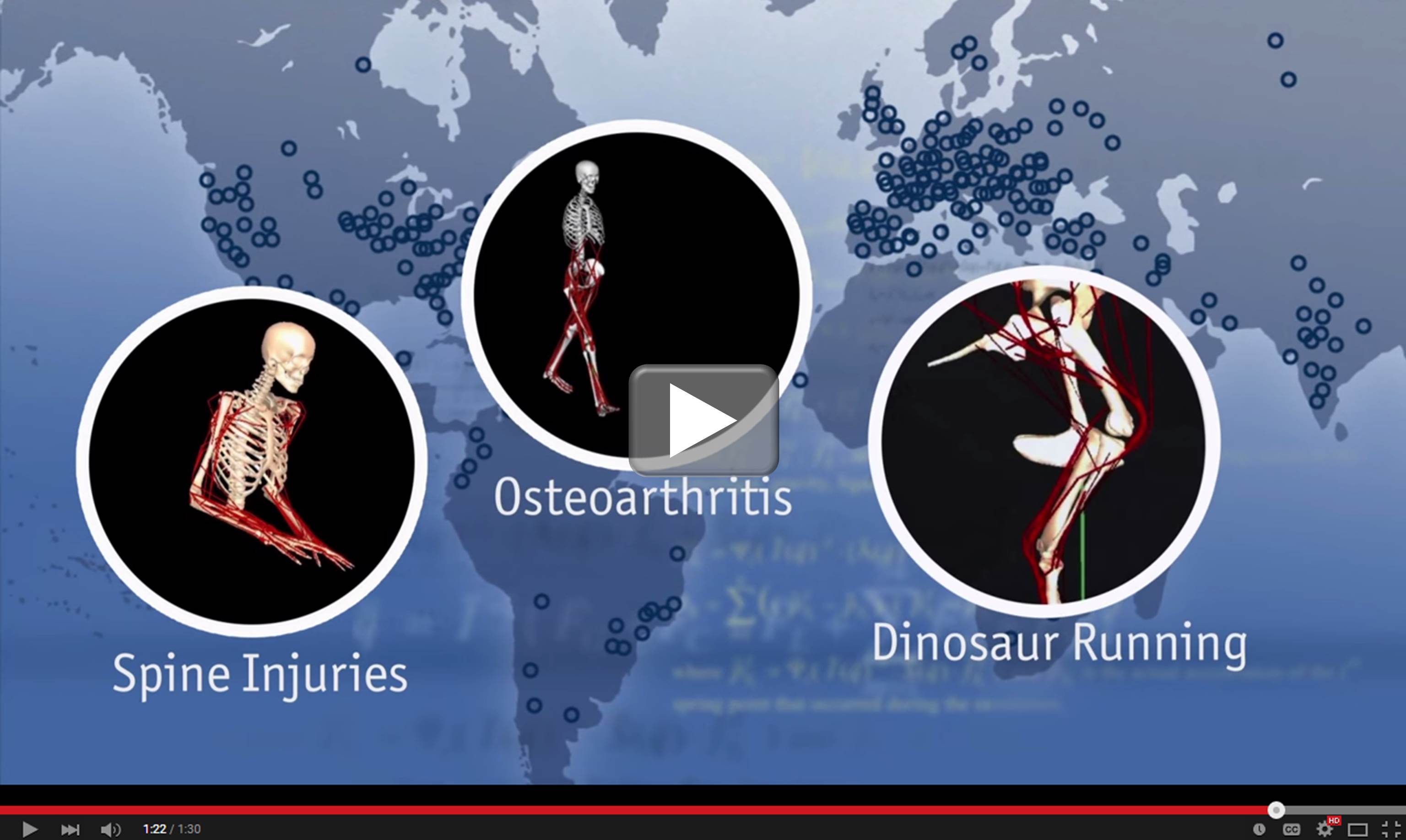 Avatar. Pick your Sim. The entertainment world has done an amazing job developing software that generates animated characters with strikingly realistic movement. But scientists have taken this one step further to create models that can help kids with cerebral palsy walk better, delay the onset of osteoarthritis, and even answer a question in the minds of children of all ages: How exactly did T. rex run?
That’s what the researchers behind this video—an entrant in the NIH Common Fund’s recent video competition—have done. They’ve developed OpenSim: a free software tool that combines state-of-the-art musculoskeletal modeling and dynamic computer simulations to produce highly accurate representations of the underlying biomechanics of motion. OpenSim was designed at the NIH-supported center for physics-based Simulation of Biological Structures (Simbios) at Stanford University, Palo Alto, CA. And now, researchers around the world are using OpenSim to find more effective interventions for a variety of movement disorders.
Links:
NIH Common Fund Video Competition
OpenSim (Stanford University, Palo Alto, CA)
NIH Support: Common Fund; Eunice Kennedy Shriver National Institute of Child Health and Human Development; National Institute for General Medical Sciences
Posted In: Health, Science, Video
Tags: biomechanics, cerebral palsy, computer simulations, dinosaurs, motion, movement disorders, musculoskeletal modeling, OpenSim, osteoarthritis, simulation, software, T. rex