cryo-EM – Page 2 – NIH Director's Blog (original) (raw)
Electricity-Conducting Bacteria May Inspire Next-Gen Medical Devices
Posted on July 18th, 2019 by Dr. Francis Collins

Credit: Edward H. Egelman
Technological advances with potential for improving human health sometimes come from the most unexpected places. An intriguing example is an electricity-conducting biological nanowire that holds promise for powering miniaturized pacemakers and other implantable electronic devices.
The nanowires come from a bacterium called Geobacter sulfurreducens, shown in the electron micrograph above. This rod-shaped microbe (white) was discovered two decades ago in soil collected from an unlikely place: a ditch outside of Norman, Oklahoma. The bug can conduct electricity along its arm-like appendages, and, in the hydrocarbon-contaminated, oxygen-depleted soil in which it lives, such electrical inputs and outputs are essentially the equivalent of breathing.
Scientists fascinated with G. sulfurreducens thought that its electricity had to be flowing through well-studied microbial appendages called pili. But, as the atomic structure of these nanowires (multi-colors, foreground) now reveals, these nanowires aren’t pili at all! Instead, the bacteria have manufactured unique submicroscopic arm-like structures. These arms consist of long, repetitive chains of a unique protein, each surrounding a core of iron-containing molecules.
The surprising discovery, published in the journal Cell, was made by an NIH-funded team involving Edward Egelman, University of Virginia Health System, Charlottesville. Egelman’s lab has had a long interest in what’s called a type 4 pili. These strong, adhering appendages help certain infectious bacteria enter tissues and make people sick. In fact, they enable bugs like Neisseria meningitidis to cross the blood-brain barrier and cause potentially deadly bacterial meningitis. While other researchers had proposed that those same type 4 pili allowed G. sulfurreducens to conduct electricity, Egelman wasn’t so sure.
So, he took advantage of recent advances in cryo-electron microscopy, which involves flash-freezing molecules at extremely low temperatures before bombarding them with electrons to capture their images with a special camera. The cryo-EM images allowed his team to nail down the atomic structure of the nanowires, now called OmcS filaments.
Using those images and sophisticated bioinformatics, Egelman and team determined that OmcS proteins uniquely fit into the nanowires’ long repetitive chains, spacing their iron-bearing cores at regular intervals to transfer electrons and convey electricity. In fact, bacteria unable to produce OmcS proteins make filaments that conduct electricity 100 times less efficiently.
With these cryo-EM structures in hand, Egelman says his team will continue to explore their conductive properties. Such knowledge might someday be used to build biologically-inspired nanowires, measuring 1/100,000th the width of a human hair, to connect miniature electronic devices directly to living tissues. This is one more example of how nature’s ability to invent is pretty breathtaking—surely one wouldn’t have predicted the discovery of nanowires in a bacterium that lives in contaminated ditches.
Reference:
[1] Structure of Microbial Nanowires Reveals Stacked Hemes that Transport Electrons over Micrometers. Wang F, Gu Y, O’Brien JP, Yi SM, Yalcin SE, Srikanth V, Shen C, Vu D, Ing NL, Hochbaum AI, Egelman EH, Malvankar NS. Cell. 2019 Apr 4;177(2):361-369.
Links:
Electroactive microorganisms in bioelectrochemical systems. Logan BE, Rossi R, Ragab A, Saikaly PE. Nat Rev Microbiol. 2019 May;17(5):307-319.
High Resolution Electron Microscopy (National Cancer Institute/NIH)
Egelman Lab (University of Virginia, Charlottesville)
NIH Support: National Institute of General Medical Sciences; National Institute of Allergy and Infectious Diseases; Common Fund
Posted In: Snapshots of Life
Tags: bacteria, bacterial meningitis, basic science, bioengineering, bioinformatics, cryo-electron microscopy, cryo-EM, electricity, G. sulfurreducens, Geobacter sulfurreducens, medical device, meningitis, nanotechnology, nanowire, Neisseria meningitidis, Oklahoma, OmcS filaments, pacemaker, soil
Looking to Llamas for New Ways to Fight the Flu
Posted on November 13th, 2018 by Dr. Francis Collins
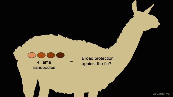 Researchers are making tremendous strides toward developing better ways to reduce our risk of getting the flu. And one of the latest ideas for foiling the flu—a “gene mist” that could be sprayed into the nose—comes from a most surprising source: llamas.
Like humans and many other creatures, these fuzzy South American relatives of the camel produce immune molecules, called antibodies, in their blood when exposed to viruses and other foreign substances. Researchers speculated that because the llama’s antibodies are so much smaller than human antibodies, they might be easier to use therapeutically in fending off a wide range of flu viruses. This idea is now being leveraged to design a new type of gene therapy that may someday provide humans with broader protection against the flu [1].
Posted In: News
Tags: adeno-associated virus, antibody, antibody treatment, bioengineering, bird flu, cryo-EM, flu, flu pandemic, flu shot, gene mist, gene therapy, hemagglutinin, imaging, immunity, influenza, influenza type A, influenza type B, llama, nanobody, virus, x-ray crystallography
MicroED: From Powder to Structure in a Half-Hour
Posted on October 30th, 2018 by Dr. Francis Collins
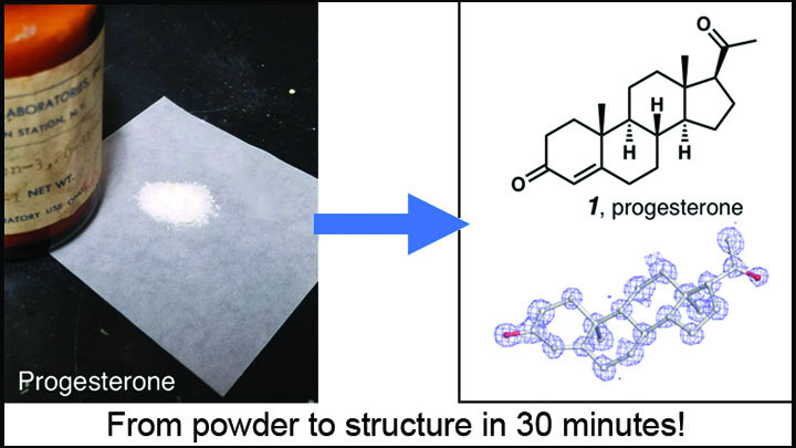
Credit: Adapted from Jones et al. ChemRxiv.org
Over the past few years, there’s been a great deal of excitement about the power of cryo-electron microscopy (cryo-EM) for mapping the structures of large biological molecules like proteins and nucleic acids. Now comes word of another absolutely incredible use of cryo-EM: determining with great ease and exquisite precision the structure of the smaller organic chemical compounds, or “small molecules,” that play such key roles in biological exploration and drug development.
The new advance involves a cryo-EM technique called microcrystal-electron diffraction (MicroED). As detailed in a preprint on ChemRxiv.org [1] and the journal Angewandte Chemie [2], MicroED has enabled researchers to take the powdered form of commercially available small molecules and generate high-resolution data on their chemical structures in less than a half-hour—dramatically faster than with traditional methods!
Posted In: News
Tags: acetaminophen, ACS Central Science, carbamazepine, cryo-electron microscopy, cryo-EM, cryo-EM service centers, crystals, drug development, ibuprofen, imaging, micro-electron diffraction, MicroED, microscopy, NMR, nuclear magnetic resonance, organic chemistry, preprints, progesterone, protein structure, small molecules, structural biology, x-ray crystallography
A Ray of Molecular Beauty from Cryo-EM
Posted on September 20th, 2018 by Dr. Francis Collins

Credit: Subramaniam Lab, National Cancer Institute, NIH
Walk into a dark room, and it takes a minute to make out the objects, from the wallet on the table to the sleeping dog on the floor. But after a few seconds, our eyes are able to adjust and see in the near-dark, thanks to a protein called rhodopsin found at the surface of certain specialized cells in the retina, the thin, vision-initiating tissue that lines the back of the eye.
This illustration shows light-activating rhodopsin (orange). The light photons cause the activated form of rhodopsin to bind to its protein partner, transducin, made up of three subunits (green, yellow, and purple). The binding amplifies the visual signal, which then streams onward through the optic nerve for further processing in the brain—and the ability to avoid tripping over the dog.
Posted In: Snapshots of Life
Tags: cell biology, cell signaling, cryo-EM, drug development, eyes, G proteins, G-protein coupled receptors, GPCR, imaging, retina, retinitis pigmentosa, rhodopsin, structural biology, transducin, vision
A Lean, Mean DNA Packaging Machine
Posted on June 7th, 2018 by Dr. Francis Collins
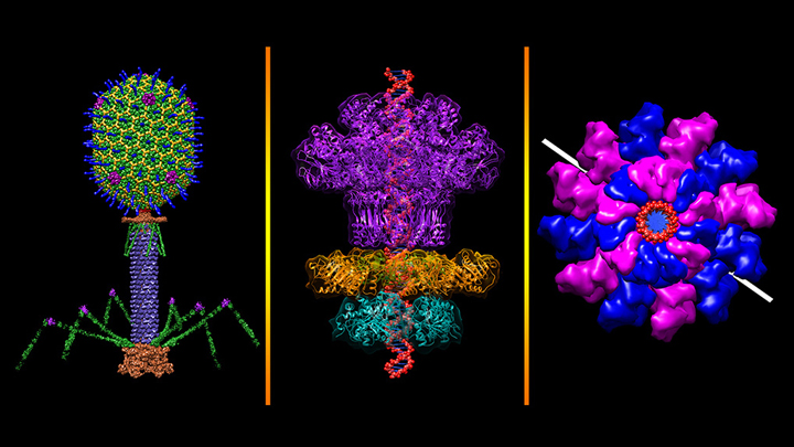
Credit: Victor Padilla-Sanchez, The Catholic University of America, Washington, D.C.
All plants and animals are susceptible to viral infections. But did you know that’s also true for bacteria? They get nailed by viruses called bacteriophages, and there are thousands of them in nature including this one that resembles a lunar lander: bacteriophage T4 (left panel). It’s a popular model organism that researchers have studied for nearly a century, helping them over the years to learn more about biochemistry, genetics, and molecular biology [1].
The bacteriophage T4 infects the bacterium Escherichia coli, which normally inhabits the gastrointestinal tract of humans. T4’s invasion starts by touching down on the bacterial cell wall and injecting viral DNA through its tube-like tail (purple) into the cell. A DNA “packaging machine” (middle and right panels) between the bacteriophage’s “head” and “tail” (green, yellow, blue spikes) keeps the double-stranded DNA (middle panel, red) at the ready. All the vivid colors you see in the images help to distinguish between the various proteins or protein subunits that make up the intricate structure of the bacteriophage and its DNA packaging machine.
Posted In: Snapshots of Life
Tags: bacteria, bacteriophage, cancer, Chimera Visualization Software, cryo-electron microscopy, cryo-EM, E. coli, Escherichia coli, FASEB Bioart 2017, HIV, structural biology, T4 bacteriophage, virus, x-ray crystallography
Cryo-EM Images Capture Key Enzyme Tied to Cancer, Aging
Posted on May 1st, 2018 by Dr. Francis Collins
Each time your cells divide, telomeres—complexes of specialized DNA sequences, RNA, and protein that protect the tips of your chromosomes—shorten just a bit. And, as the video shows, that shortening renders the genomic information on your chromosomes more vulnerable to changes that can drive cancer and other diseases of aging.
Consequently, over the last few decades, much research has focused on efforts to understand telomerase, a naturally occurring enzyme that helps to replace the bits of telomere lost during cell division. But there’s been a major hitch: until recently, scientists hadn’t been able to determine telomerase’s molecular structure in detail—a key step in figuring out exactly how the enzyme works. Now, thanks to better purification methods and an exciting technology called cryo-electron microscopy (cryo-EM), NIH-funded researchers and their colleagues have risen to the challenge to produce the most detailed view yet of human telomerase in its active form [1].
This structural biology advance is a critical step toward learning more about the role of telomerase in cancers, as well as genetic conditions linked to telomerase deficiencies. It’s also an important milestone in the quest for drugs targeting telomerase in different ways, perhaps to slow the growth of cancerous cells or to boost the proliferative capacity of life-giving adult stem cells.
One reason telomerase has been so difficult to study in humans is that the enzyme isn’t produced at detectable levels in the vast majority of our cells. To get around this problem, the team led by Eva Nogales and Kathleen Collins at the University of California, Berkeley, first coaxed human cells in the lab to produce larger quantities of active telomerase. They then used fluorescent microscopy, along with extensive knowledge of the enzyme’s biochemistry, to develop a multi-step purification process that yielded relatively homogenous samples of active telomerase.
The new study is also yet another remarkable example of how cryo-EM microscopy has opened up new realms of scientific possibility. That’s because, in comparison to other methods, cryo-EM enables researchers to solve complex macromolecular structures even when only tiny amounts of material are available. It can also produce detailed images of molecules, like telomerase, that are extremely flexible and hard to keep still while taking a picture of their structure.
As described in Nature, the researchers used cryo-EM to capture the structure of human telomerase in unprecedented detail. Their images reveal two lobes, held together by a flexible RNA tether. One of those lobes contains the highly specialized core enzyme. It uses an internal RNA template as a guide to make the repetitive, telomeric DNA that’s added at the tips of chromosomes. The second lobe, consisting of a complex of RNA and RNA-binding proteins, plays important roles in keeping the complex stable and properly in place.
This new, more-detailed view helps to explain how mutations in particular genes may lead to telomerase-related health conditions, including bone marrow failure, as well as certain forms of anemia and pulmonary fibrosis. For example, it reveals that a genetic defect known to cause bone marrow failure affects an essential protein in a spot that’s especially critical for telomerase’s proper conformation and function.
This advance will also be a big help for designing therapies that encourage telomerase activity. For example, it could help to boost the success of bone marrow transplants by rejuvenating adult stem cells. It might also be possible to reinforce the immune systems of people with HIV infections. While telomerase-targeted treatments surely won’t stop people from growing old, new insights into this important enzyme will help to understand aging better, including why some people appear to age faster than others.
As remarkable as these new images are, the researchers aren’t yet satisfied. They’ll continue to refine them down to the minutest structural details. They say they’d also like to use cryo-EM to understand better how the complex attaches to chromosomes to extend telomeres. Each new advance in the level of atomic detail will not only make for amazing new videos, it will help to advance understanding of human biology in health, aging, and disease.
References:
[1] Cryo-EM structure of substrate-bound human telomerase holoenzyme. Nguyen THD, Tam J, Wu RA, Greber BJ, Toso D, Nogales E, Collins K. Nature. 2018 April 25. [Epub ahead of publication]
Links:
High Resolution Electron Microscopy (National Cancer Institute/NIH)
Nogales Lab (University of California, Berkeley)
Collins Lab (University of California, Berkeley)
NIH Support: National Institute of General Medical Sciences
Posted In: Cool Videos, News
Tags: aging, anemia, biochemistry, bone marrow transplant, cancer, cell division, chromosome, Cool Videos, cryo-EM, DNA, drug design, holoenzyme, imaging, immunity, microscopy, structural biology, telomerase, telomeres
What a Year It Was! A Look Back at Research Progress in 2017
Posted on January 2nd, 2018 by Dr. Francis Collins
I want to wish everyone a Happy New Year! Hope your 2018 is off to a great start.
Over the holidays, the journal Science published its annual, end-of-the-year list of research breakthroughs, from anthropology to zoology. I always look forward to seeing the list and reflecting on some of the stunning advances reported in the past 12 months. Last year was no exception. Science’s 2017 Breakthrough of the Year, as chosen by its editors, was in the field of astrophysics. Scientists were able to witness the effects of the collision of two neutron stars—large stars with collapsed inner cores—smacking into each other 130 million light years away. How cool is that!
Numbered prominently among the nine other breakthroughs were five from biomedicine: gene therapy, gene editing, cancer immunotherapy, cryo-EM, and biology preprints. All involved varying degrees of NIH support, and all drew great interest from readers. In fact, three of the top four vote-getters in the “People’s Choice” category came from biomedicine. That includes the People’s 2017 Breakthrough of the Year: gene therapy success. And so, in what has become a Director’s Blog tradition, I’ll kick off our new year of posts by taking a closer look at these biomedical breakthroughs—starting with the little girl in the collage above, and moving clockwise around the images:
Tags: 2017 Nobel Prize in Chemistry, ALL, axicabtagene ciloleucel, B-cell acute lymphoblastic leukemia, cancer, cancer immunotherapy, car t-cell therapy, CRISPR/Cas9, cryo-electron microscopy, cryo-EM, gene editing, gene therapy, Huntington's disease, immunotherapy, inherited retinal degenerations, Kymriah, mismatch repair, nusineren, pembrolizumab, preprints, RNA editing, Science’s 2017 Breakthrough of the Year, sickle cell disease, spinraza, tisagenlecleucel, wearable devices, Yescarta
Twinkle, Twinkle Little Cryo-EM Star
Posted on December 21st, 2017 by Dr. Francis Collins
The stars are out and shining this holiday season. But there are some star-shaped structures now under study in the lab that also give us plenty of reason for hope. One of them is a tiny virus called bacteriophage phi-6, which researchers are studying in an effort to combat a similar, but more-complex, group of viruses that can cause life-threatening dehydration in young children.
Thanks to a breakthrough technology called cryo-electron microscopy (cryo-EM), NIH researchers recently captured, at near atomic-level of detail, the 3D structure of this immature bacteriophage phi-6 particle in the process of replication. At the points of its “star,” key proteins (red) are positioned to transport clipped, single-stranded segments of the virus’ own genetic information into its newly made shell, or procapsid (blue). Once inside the procapsid, an enzyme (purple) will copy the segments to make the genetic information double-stranded, while another protein (yellow) will help package them. As the procapsid matures, it undergoes dramatic structural changes.
Posted In: Health, Science, Video
Tags: bacteriophage, bacteriophage phi-6, cryo-electron microscopy, cryo-EM, double-stranded RNA viruses, gastrointestinal disease, global health, imaging, procapsid, rotavirus, rotavirus vaccine, virus
Creative Minds: Preparing for Future Pandemics
Posted on April 20th, 2017 by Dr. Francis Collins
Jonathan Abraham / Credit: ChieYu Lin
Growing up in Queens, NY, Jonathan Abraham developed a love for books and an interest in infectious diseases. One day Abraham got his hands on a copy of Laurie Garrett’s The Coming Plague, a 1990s bestseller warning of future global pandemics, and he sensed his life’s calling. He would help people around the world survive deadly viral outbreaks, particularly from Ebola, Marburg, and other really bad bugs that cause deadly hemorrhagic fevers.
Abraham, now a physician-scientist at Brigham and Women’s Hospital, Boston, continues to chase that dream. With support from an NIH Director’s 2016 Early Independence Award, Abraham has set out to help design the next generation of treatments to enable more people to survive future outbreaks of viral hemorrhagic fever. His research strategy: find antibodies in the blood of known survivors that helped them overcome their infections. With further study, he hopes to develop purified forms of the antibodies as potentially life-saving treatments for people whose own immune systems may not make them in time. This therapeutic strategy is called passive immunity.
Tags: antibodies, cryo-electron microscopy, cryo-EM, Ebola, hemorrhagic fever, infectious diseases, junin virus, Marburg virus, neutralizing antibodies, New World hemorrhagic fever, NIH Director’s 2016 Early Independence Award, pandemic, passive immunity, viral hemorrhagic fever, viral pandemics, virology, virus, x-ray crystallography
Creative Minds: Breaking Size Barriers in Cryo-Electron Microscopy
Posted on May 26th, 2016 by Dr. Francis Collins

Dmitry Lyumkis
When Dmitry Lyumkis headed off to graduate school at The Scripps Research Institute, La Jolla, CA, he had thoughts of becoming a synthetic chemist. But he soon found his calling in a nearby lab that imaged proteins using a technique known as single-particle cryo-electron microscopy (EM). Lyumkis was amazed that the team could take a purified protein, flash-freeze it in liquid nitrogen, and then fire electrons at the protein, capturing the resulting image with a special camera. Also amazing was the sophisticated computer software that analyzed the raw 2D camera images, merging the data and reconstructing it into 3D representations of the protein.
The work was profoundly complex, but Lyumkis thrives on solving extremely difficult puzzles. He joined the Scripps lab to become a structural biologist and a few years later used single-particle cryo-EM to help determine the atomic structure of a key protein on the surface of the human immunodeficiency virus (HIV), the cause of AIDS. The protein had been considered one of the greatest challenges in structural biology and a critical target in developing an AIDS vaccine [1].
Now, Lyumkis has plans to take single-particle cryo-EM to a whole new level—literally. He wants to develop new methods that allow it to model the atomic structures of much smaller proteins. Right now, single-particle cryo-EM has worked with proteins as small as roughly 150 kilodaltons, a measure of a protein’s molecular weight (the approximate average mass of a protein is 53 kDa). Lyumkis plans to drop that number well below 100 kDa, noting that if his new methods work as he hopes, there should be very little, if any, lower size limit to get the technique to work. He envisions generating within a matter of days or weeks the precise structure of an average-sized protein involved in a disease, and then potentially handing it off as an atomic model for drug developers to target for more effective treatment.
Tags: 2015 NIH Director’s Early Independence Award, 3D computational analysis, atomic structure, computation, cryo-electron microscopy, cryo-EM, drug design, drug discovery, drugs, electron microscopy, HIV, human immunodeficiency virus, IKK complex, protein structure, proteins, single-particle cryo-EM, small proteins, structural biology