Human Connectome Project – NIH Director's Blog (original) (raw)
Aided by AI, Study Uncovers Hidden Sex Differences in Dynamic Brain Function
Posted on February 29th, 2024 by Dr. Monica M. Bertagnolli

Credit: Adobe/Feodora
We’re living in an especially promising time for biomedical discovery and advances in the delivery of data-driven health care for everyone. A key part of this is the tremendous progress made in applying artificial intelligence to study human health and ultimately improve clinical care in many important and sometimes surprising ways.1 One new example of this comes from a fascinating study, supported in part by NIH, that uses AI approaches to reveal meaningful sex differences in the way the brain works.
As reported in the Proceedings of the National Academy of Sciences, researchers led by Vinod Menon at Stanford Medicine, Stanford, CA, have built an AI model that can—nine times out of ten—tell whether the brain in question belongs to a female or male based on scans of brain activity alone.2 These findings not only help resolve long-term debates about whether reliable differences between sexes exist in the human brain, but they’re also a step toward improving our understanding of why some psychiatric and neurological disorders affect women and men differently.
The prevalence of certain psychiatric and neurological disorders in men and women can vary significantly, leading researchers to suspect that sex differences in brain function likely exist. For example, studies have found that females are more likely to experience depression, anxiety, and eating disorders, while autism, Attention-Deficit/Hyperactivity Disorder, and schizophrenia are seen more often in males. But earlier research to understand sex differences in the brain have focused mainly on anatomical and structural studies of brain regions and their connections. Much less is known about how those structural differences translate into differences in brain activity and function.
To help fill those gaps in the new study, Menon’s team took advantage of vast quantities of brain activity data from MRI scans from the NIH-supported Human Connectome Project. The data was captured from hundreds of healthy young adults with the goal of studying the brain and how it changes with growth, aging, and disease. To use this data to explore sex differences in brain function, the researchers developed what’s known as a deep neural network model in which a computer “learned” how to recognize patterns in brain activity data that could distinguish a male from a female brain.
This approach doesn’t rely on any preconceived notions about what features might be important. A computer is simply shown many examples of brain activity belonging to males and females and, over time, can begin to pick up on otherwise hidden differences that are useful for making such classifications accurately. One of the things that made this work different from earlier attempts was it relied on dynamic scans of brain activity, which capture the interplay among brain regions.
After analyzing about 1,500 brain scans, a computer could usually (although not always) tell whether a scan came from a male or female brain. The findings also showed the model worked reliably well in different datasets and in brain scans for people in different places in the U.S. and Europe. Overall, the findings confirm that reliable sex differences in brain activity do exist.
Where did the model find those differences? To get an idea, the researchers turned to an approach called explainable AI, which allowed them to dig deeper into the specific features and brain areas their model was using to pick up on sex differences. It turned out that one set of areas the model was relying on to distinguish between male and female brains is what’s known as the default mode network. This area is responsible for processing self-referential information and constructing a coherent sense of the self and activates especially when people let their minds wander.3 Other important areas included the striatum and limbic network, which are involved in learning and how we respond to rewards, respectively.
Many questions remain, including whether such differences arise primarily due to inherent biological differences between the sexes or what role societal circumstances play. But the researchers say that the discovery already shows that sex differences in brain organization and function may play important and overlooked roles in mental health and neuropsychiatric disorders. Their AI model can now also be applied to begin to explain other kinds of brain differences, including those that may affect learning or social behavior. It’s an exciting example of AI-driven progress and good news for understanding variations in human brain functions and their implications for our health.
References:
[1] Bertagnolli, MM. Advancing health through artificial intelligence/machine learning: The critical importance of multidisciplinary collaboration. PNAS Nexus. DOI: 10.1093/pnasnexus/pgad356 (2023).
[2] Ryali S, et al. Deep learning models reveal replicable, generalizable, and behaviorally relevant sex differences in human functional brain organization. Proc Natl Acad Sci. DOI: 10.1073/pnas.2310012121 (2024).
[3] Menon, V. 20 years of the default mode network: A review and synthesis. Neuron.DOI: 10.1016/j.neuron.2023.04.023 (2023).
NIH Support: National Institute of Mental Health, National Institute of Biomedical Imaging and Bioengineering, Eunice Kennedy Shriver National Institute of Child Health and Human Development, National Institute on Aging
Changes in Normal Brain Connections Linked to Eating Disorders
Posted on April 11th, 2023 by Lawrence Tabak, D.D.S., Ph.D.
Anyone who has ever had a bad habit knows how vexingly difficult breaking it can be. The reason is the repeated action, initially linked to some type of real or perceived reward, over time changes the way our very brains are wired to work. The bad habit becomes automatic, even when the action does us harm or we no longer wish to do it.
Now an intriguing new study shows that the same bundled nerve fibers, or brain circuits, involved in habit formation also can go awry in people with eating disorders. The findings may help to explain why eating disorders are so often resistant to will power alone. They also may help to point the way to improved approaches to treating eating disorders, suggesting strategies that adjust the actual brain circuitry in helpful ways.
These latest findings, published in the journal Science Translational Medicine, come from the NIH-supported Casey Halpern, University of Pennsylvania’s Perelman School of Medicine, Philadelphia, and Cara Bohon, Stanford University School of Medicine, Stanford, CA [1].
Halpern, Bohon, and colleagues were interested in a growing body of evidence linking habitual behaviors to mental health conditions, most notably substance use disorders and addictions. But what especially intrigued them was recent evidence also suggesting a possible role for habitual behaviors in the emergence of eating disorders.
To look deeper into the complex circuitry underlying habit formation and any changes there that might be associated with eating disorders, they took advantage of a vast collection of data from the NIH-funded Human Connectome Project (HCP). It was completed several years ago and now serves as a valuable online resource for researchers.
The HCP offers a detailed wiring map of a normal human brain. It describes all the structural and functional neural connections based on careful analyses of hundreds of high-resolution brain scans. These connections are then layered with genetic, behavioral, and other types of data. This incredible map now allows researchers to explore and sometimes uncover the roots of neurological and mental health conditions within the brain’s many trillions of connections.
In the new study, Halpern, Bohon, and colleagues did just that. First, they used sophisticated mapping methods in 178 brain scans from the HCP data to locate key portions of a brain region called the striatum, which is thought to be involved in habit formation. What they really wanted to know was whether circuits operating within the striatum were altered in some way in people with binge eating disorder or bulimia nervosa.
To find out, the researchers recruited 34 women who have an eating disorder and, with their consent, imaged their brains using a variety of techniques. Twenty-one participants were diagnosed with binge eating disorder, and 13 had bulimia nervosa. For comparison purposes, the researchers looked at the same brain circuits in 19 healthy volunteers.
The two groups were otherwise similar in terms of their ages, weights, and other features. But the researchers suspected they might find differences between the healthy group and those with an eating disorder in brain circuits known to have links to habitual behaviors. And, indeed, they did.
In comparison to a “typical” brain, those from people with an eating disorder showed striking changes in the connectivity of a portion of the striatum known as the putamen. That’s especially notable because the putamen is known for its role in learning and movement control, including reward, thinking, and addiction. What’s more, those observed changes in the brain’s connections and circuitry in this key brain area were more evident in people whose eating disorder symptoms and emotional eating were more frequent and severe.
Using other brain imaging methods in 10 of the volunteers (eight with binge eating disorder and two healthy controls), the researchers also connected those changes in the habit-forming brain circuits to high levels of a protein receptor that responds to dopamine. Dopamine is an important chemical messenger in the brain involved in pleasure, motivation, and learning. They also observed in those with eating disorders structural changes in the architecture of the densely folded, outer layer of the brain known as grey matter.
While there’s much more to learn, the researchers note the findings may lead to future treatments aimed to modify the brain circuitry in beneficial ways. Indeed, Halpern already has encouraging early results from a small NIH-funded clinical trial testing the ability of deep brain stimulation (DBS) in people with binge eating disorder to disrupt signals that drive food cravings in another portion of the brain associated with reward and motivation, known as the nucleus accumbens, [2]. In DBS, doctors implant a pacemaker-like device capable of delivering harmless therapeutic electrical impulses deep into the brain, aiming for the spot where they can reset the abnormal circuitry that’s driving eating disorders or other troubling symptoms or behaviors.
But the latest findings published in Science Translational Medicine now suggest other mapped brain circuits as potentially beneficial DBS targets for tackling binge eating, bulimia nervosa, or other life-altering, hard-to-treat eating disorders. They also may ultimately have implications for treating other conditions involving various other forms of compulsive behavior.
These findings should come as a source of hope for the family and friends of the millions of Americans—many of them young people—who struggle with eating disorders. The findings also serve as an important reminder for the rest of us that, despite common misconceptions that disordered eating is a lifestyle choice, these conditions are in fact complex and serious mental health problems driven by fundamental changes in the brain’s underlying circuitry.
Finding new and more effective ways to treat serious eating disorders and other compulsive behaviors is a must. It will require equally serious ongoing efforts to unravel their underlying causes and find ways to alter their course—and this new study is an encouraging step in that direction.
References:
[1] Human habit neural circuitry may be perturbed in eating disorders. Wang AR, Kuijper FM, Barbosa DAN, Hagan KE, Lee E, Tong E, Choi EY, McNab JA, Bohon C, Halpern CH. Sci Transl Med. 2023 Mar 29;15(689):eabo4919.
[2] Pilot study of responsive nucleus accumbens deep brain stimulation for loss-of-control eating. Shivacharan RS, Rolle CE, Barbosa DAN, Cunningham TN, Feng A, Johnson ND, Safer DL, Bohon C, Keller C, Buch VP, Parker JJ, Azagury DE, Tass PA, Bhati MT, Malenka RC, Lock JD, Halpern CH. Nat Med. 2022 Sep;28(9):1791-1796.
Links:
Eating Disorders (National Institute of Mental Health/NIH)
Casey Halpern (Penn Medicine, Philadelphia)
Cara Bohon (Stanford University, Stanford, CA)
NIH Support: National Institute of Mental Health; National Institute of Neurological Disorders and Stroke
Posted In: News
Tags: binge eating disorder, brain, brain circuits, brain imaging, bulimia nervosa, clinical trial, connectomics, DBS, deep brain stimulation, eating, eating disorders, habitual behaviors, habitual eating, HCP, Human Connectome Project, imaging, mental health, neuroanatomy, nucleus accumbens, putamen, striatum
The Amazing Brain: A Sharper Image of the Pyramidal Tract
Posted on August 17th, 2021 by Dr. Francis Collins
Flip the image above upside down, and the shape may remind you of something. If you think it resembles a pyramid, then you and a lot of great neuroscientists are thinking alike. What you are viewing is a colorized, 3D reconstruction of a pyramidal tract, which are bundles of nerve fibers that originate from the brain’s cerebral cortex and relay signals to the brainstem or the spinal cord. These signals control many important activities, including the voluntary movement of our arms, legs, head, and face.
For a while now, it’s been possible to combine a specialized form of magnetic resonance imaging (MRI) with computer modeling tools to produce 3D reconstructions of complicated networks of nerve fibers, such as the pyramidal tract. Still, for technical reasons, the quality of these reconstructions has remained poor in parts of the brain where nerve fibers cross at angles of 40 degrees or less.
The video above demonstrates how adding a sophisticated algorithm, called Orientation Distribution Function (ODF)-Fingerprinting, to such modeling can help overcome this problem when reconstructing a pyramidal tract. It has potential to enhance the reliability of these 3D reconstructions as neurosurgeons begin to use them to plan out their surgeries to help ensure they are carried out with the utmost safety and precision.
In the first second of the video, you see gray, fuzzy images from a diffusion MRI of the pyramidal tract. But, very quickly, a more colorful, detailed 3D reconstruction begins to appear, swiftly filling in from the top down. Colors are used to indicate the primary orientations of the nerve fibers: left to right (red), back to front (green), and top to bottom (blue). The orange, magenta, and other colors represent combinations of these primary directional orientations.
About three seconds into the video, a rough draft of the 3D reconstruction is complete. The top of the pyramidal tract looks pretty good. However, looking lower down, you can see distortions in color and relatively poor resolution of the nerve fibers in the middle of the tract—exactly where the fibers cross each other at angles of less than 40 degrees. So, researchers tapped into the power of their new ODF-Fingerprinting software to improve the image—and, starting about nine seconds into the video, you can see an impressive final result.
The researchers who produced this amazing video are Patryk Filipiak and colleagues in the NIH-supported lab of Steven Baete, Center for Advanced Imaging Innovation and Research, New York University Grossman School of Medicine, New York. The work paired diffusion MRI data from the NIH Human Connectome Project with the ODF-Fingerprinting algorithm, which was created by Baete to incorporate additional MRI imaging data on the shape of nerve fibers to infer their directionality [1].
This innovative approach to imaging recently earned Baete’s team second place in the 2021 “Show Us Your BRAINs” Photo and Video contest, sponsored by the NIH-led Brain Research through Advancing Innovative Neurotechnologies® (BRAIN) Initiative. But researchers aren’t stopping there! They are continuing to refine ODF-Fingerprinting, with the aim of modeling the pyramidal tract in even higher resolution for use in devising new and better ways of helping people undergoing neurosurgery.
Reference:
[1] Fingerprinting Orientation Distribution Functions in diffusion MRI detects smaller crossing angles. Baete SH, Cloos MA, Lin YC, Placantonakis DG, Shepherd T, Boada FE. Neuroimage. 2019 Sep;198:231-241.
Links:
Brain Research through Advancing Innovative Neurotechnologies® (BRAIN) Initiative (NIH)
Human Connectome Project (University of Southern California, Los Angeles)
Steven Baete (Center for Advanced Imaging Innovation and Research, New York University Grossman School of Medicine, New York)
Show Us Your BRAINs! Photo and Video Contest (BRAIN Initiative/NIH)
NIH Support: National Institute of Biomedical Imaging and Bioengineering; National Institute of Neurological Disorders and Stroke; National Cancer Institute
Posted In: Generic
Tags: 3D imaging, brain, brain imaging, BRAIN Initiative, Brain Research through Advancing Innovative Neurotechnologies Initiative, brainstem, cerebral cortex, diffusion MRI, Human Connectome Project, MRI, neurons, neuroscience, neurosurgery, ODF-Fingerprinting, pyramidal tract, Show Us Your BRAINs!, spinal cord, tractography
The Amazing Brain: Mapping Brain Circuits in Vivid Color
Posted on August 20th, 2019 by Dr. Francis Collins
Hop aboard as we fly up, down, left, and right through the information highways of the human brain! This captivating and eye-catching video was one of the winners of the 2019 “Show us Your Brain!” contest sponsored by the NIH-led Brain Research through Advancing Innovative Neurotechnologies® (BRAIN) Initiative.
The video travels through several portions of the brain’s white matter—bundles of fiber that carry nerve signals between the brain and the body, as well as within the brain itself. Fiber colors indicate directionality: left-right fibers (red), front-back fibers (green), and top-bottom fibers (blue).
Looking from the back, we start our journey deep within the brain in the limbic system, the area that helps control emotion, learning, and memory. About three seconds in, visual fibers pop into view extending from the eyes to various brain areas into the occipital lobe (one of four major brain lobes) in the back of the brain.
About two seconds later, flying over top as the brain starts rotating, we see various fiber bundles spray upward throughout the cerebral cortex, communicating information related to language processing, short-term memory, and other functions. About halfway through the video, several green bundles emerge arching across the brain’s midline. These bundles, called the corpus callosum, house the fibers enabling communication between left and right sides of the brain. Finally, the video closes as we see many different fiber bundles lighting up all over, enabling communication between different cortical and subcortical portions of the brain through association and projection pathways.
Dynamic maps like these are created using a 3D imaging technique called diffusion MRI tractography [1]. The technique tracks subtle pathways of water movement in the brain, and allows researchers to model the physical properties (connectional anatomy) that underlie the brain’s electrical properties (neuronal signaling). Postdoctoral researcher Ryan Cabeen and Arthur Toga, director of the University of Southern California Mark and Mary Stevens Neuroimaging and Informatics Institute, Los Angeles, used the method to study how white matter changes in developing and aging brains, as well as in brains affected by neurodegenerative or neurological disorders.
Scientific animator Jim Stanis produced the video with Cabeen and Toga. The team first created a population-averaged brain using high-quality diffusion MRI datasets from the Human Connectome Project ,and then used sophisticated computational tools to delineate each bundle manually .
The tractography technique lets scientists visualize and quantitatively analyze the brain’s wiring patterns, complementing our understanding of how the brain functions. Such methods are especially useful to learn about the organization of deep-brain areas that remain out of reach for scientists using current tools and imaging techniques.
Reference:
[1] Kernel regression estimation of fiber orientation mixtures in diffusion MRI. Cabeen RP, Bastin ME, Laidlaw DH. Neuroimage. 2016 Feb 15;127:158-172.
Links:
Arthur Toga (USC Mark and Mary Stevens Neuroimaging and Informatics Institute, Los Angeles)
Ryan Cabeen (USC Mark and Mary Stevens Neuroimaging and Informatics Institute)
qitwiki—Information about the Quantitative Imaging Toolkit (USC)
Human Connectome Project (USC)
Show Us Your Brain Contest! (BRAIN Initiative/NIH)
Brain Research through Advancing Innovative Neurotechnologies® (BRAIN) Initiative (NIH)
NIH Support: National Institute of Neurological Disorders and Stroke; National Institute of Mental Health
Posted In: Cool Videos
Tags: brain, BRAIN Initiative, brain lobes, cerebral cortex, corpus callosum, Human Connectome Project, limbic system, MRI, neuroanatomy, neurofibers, neuroscience, occipital lobe, quantitative imaging, Show Us Your Brain, tractography
Cool Videos: Starring the Wiring Diagram of the Human Brain
Posted on February 7th, 2017 by Dr. Francis Collins

The human brain contains distinct geographic regions that communicate throughout the day to process information, such as remembering a neighbor’s name or deciding which road to take to work. Key to such processing is a vast network of densely bundled nerve fibers called tracts. It’s estimated that there are thousands of these tracts, and, because the human brain is so tightly packed with cells, they often travel winding, contorted paths to form their critical connections. That situation has previously been difficult for researchers to image three-dimensional tracts in the brain of a living person.
That’s now changing with a new approach called tractography, which is shown with the 3D data visualization technique featured in this video. Here, researchers zoom in and visualize some of the neural connections detected with tractography that originate or terminate near the hippocampus, which is a region of the brain essential to learning and memory. If you’re wondering about what the various colors represent, they indicate a tract’s orientation within the brain: side to side is red, front to back is green, and top to bottom is blue.
Posted In: Health, Science, Video
Tags: brain, brain imaging, BRAIN Initiative, Brain-Art Competition 2016, hippocampus, Human Connectome Project, imaging, magnetic resonance imaging, MRI, nerve fibers, neuroimaging, neurology, supercomputing, tractography, tracts
Big Data and Imaging Analysis Yields High-Res Brain Map
Posted on July 26th, 2016 by Dr. Francis Collins

Caption: Map of 180 areas in the left and right hemispheres of the cerebral cortex.
Credit: Matthew F. Glasser, David C. Van Essen, Washington University Medical School, Saint Louis, Missouri
Neuroscientists have been working for a long time to figure out how the human brain works, and that has led many through the years to attempt to map its various regions and create a detailed atlas of their complex geography and functions. While great progress has been made in recent years, existing brain maps have remained relatively blurry and incomplete, reflecting only limited aspects of brain structure or function and typically in just a few people.
In a study reported recently in the journal Nature, an NIH-funded team of researchers has begun to bring this map of the human brain into much sharper focus [1]. By combining multiple types of cutting-edge brain imaging data from more than 200 healthy young men and women, the researchers were able to subdivide the cerebral cortex, the brain’s outer layer, into 180 specific areas in each hemisphere. Remarkably, almost 100 of those areas had never before been described. This new high-resolution brain map will advance fundamental understanding of the human brain and will help to bring greater precision to the diagnosis and treatment of many brain disorders.
Tags: Autism Spectrum Disorder, big data, brain, brain imaging, brain mapping, brain scan, cerebral cortex, connectome, connectomics, fMRI, Functional magnetic resonance imaging, human brain, Human Connectome Project, imaging, neuroimaging, neuroscience, NIH Blueprint for Neuroscience Research, schizophrenia
Making the Connections: Study Links Brain’s Wiring to Human Traits
Posted on October 6th, 2015 by Dr. Francis Collins

Caption: The wiring diagram of a human brain, measured in a healthy individual, where the movement of water molecules is measured by diffuse tensor magnetic resonance imaging, revealing the connections. This is an example of the type of work being done by the Human Connectome Project.
Source: Courtesy of the Laboratory of Neuro Imaging and Martinos Center for Biomedical Imaging, Consortium of the Human Connectome Project
For questions about why people often think, act, and perceive the world so differently, the brain is clearly an obvious place to look for answers. However, because the human brain is packed with tens of billions of neurons, which together make trillions of connections, knowing exactly where and how to look remains profoundly challenging.
Undaunted by these complexities, researchers involved in the NIH-funded Human Connectome Project (HCP) have been making progress, as shown by some intriguing recent discoveries. In a study published in Nature Neuroscience [1], an HCP team found that the brains of individuals with “positive” traits—such as strong cognitive skills and a healthy sense of well-being—show stronger connectivity in certain areas of the brain than do those with more “negative” traits—such as tendencies toward anger, rule-breaking, and substance use. While these findings are preliminary, they suggest it may be possible one day to understand, and perhaps even modify, the connections within the brain that are associated with human behavior in all its diversity.
Tags: behavior, brain, brain connectivity, BRAIN Initiative, cerebral cortex, cognition, default mode network, human behavior, human brain, Human Connectome Project, lifestyle, memory, MRI, neurons, stem cells
Cool Videos: Reconstructing the Cerebral Cortex
Posted on September 17th, 2015 by Dr. Francis Collins
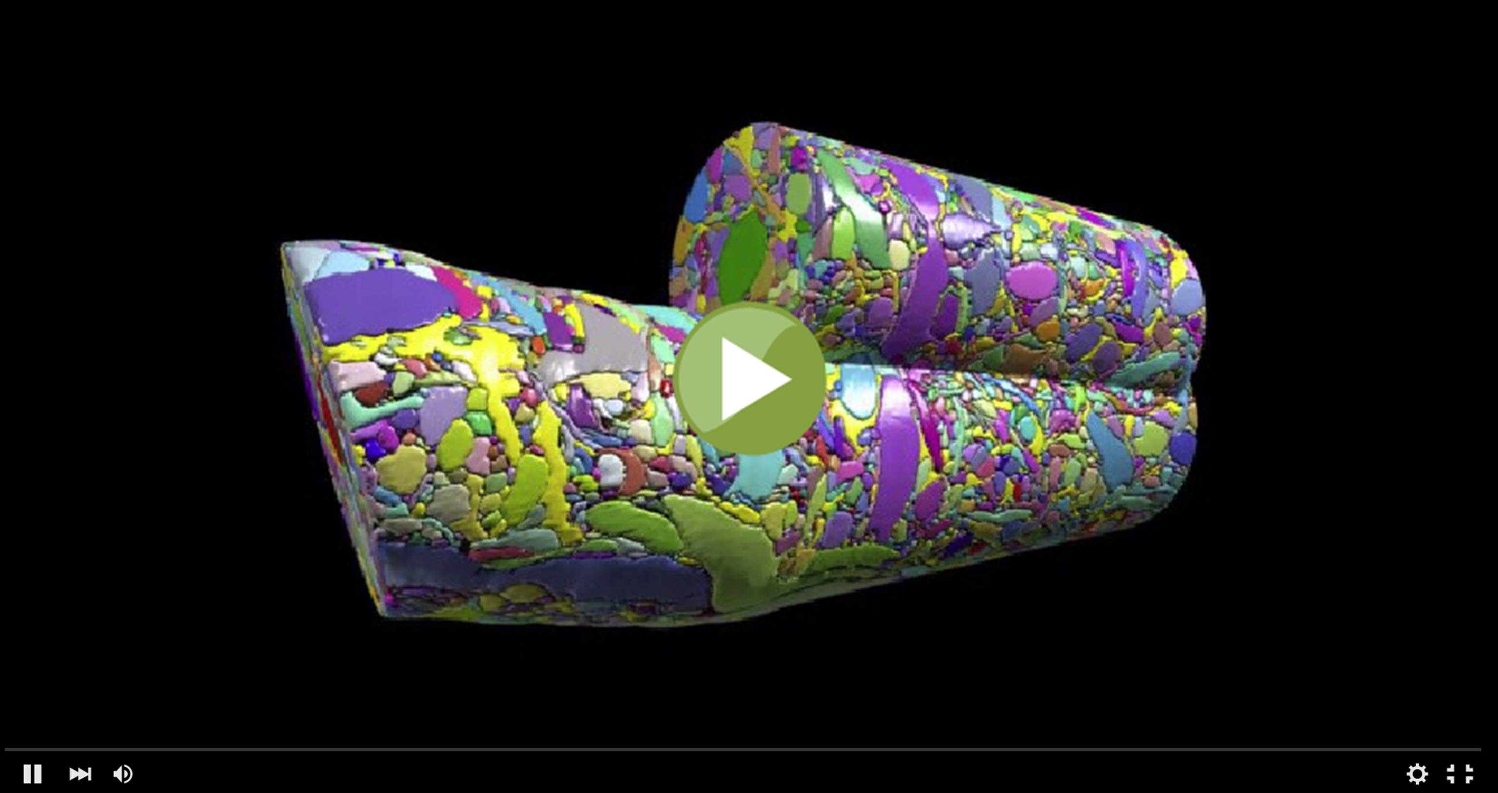
This colorful cylinder could pass for some sort of modern art sculpture, but it actually represents a sneak peak at some of the remarkable science that we can look forward to seeing from the Brain Research through Advancing Innovative Neurotechnologies (BRAIN) Initiative. In a recent study in the journal Cell [1], NIH grantee Jeff Lichtman of Harvard University, Cambridge, MA and his colleagues unveiled the first digitized reconstruction of tissue from the mammalian cerebral cortex—the outermost part of the brain, responsible for complex behaviors.
Specifically, Lichtman’s group mapped in exquisite detail a very small cube of a mouse’s cerebral cortex. In fact, the cube is so tiny (smaller than a grain of sand!) that it contained no whole cells, just a profoundly complex tangle of finger-like nerve cell extensions called axons and dendrites. And what you see in this video is just one cylindrical portion of that tissue sample, in which Licthtman and colleagues went full force to identify and label every single cellular and intracellular element. The message-sending axons are delineated in an array of pastel colors, while more vivid hues of red, green, and purple mark the message-receiving dendrites and bright yellow indicates the nerve-insulating glia. In total, the cylinder contains parts of about 600 axons, 40 different dendrites, and 500 synapses, where nerve impulses are transmitted between cells.
Posted In: Health, Science, Video
Tags: axons, brain, BRAIN Initiative, brain mapping, brain reconstruction, brain research, cerebral cortex, connectomics, dendrites, Human Connectome Project, nerve cells, neural tissue, neurology, neurons, neurotransmitter, synapse