miscarriage – NIH Director's Blog (original) (raw)
Zooming In on Meiosis
Posted on December 6th, 2018 by Dr. Francis Collins
Credit: Simone Köhler, Michal Wojcik, Ke Xu, and Abby Dernburg, University of California, Berkeley
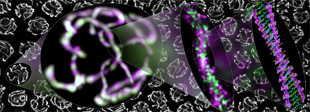
Meiosis—the formation of egg and sperm cells—is a highly choreographed process that creates genetic diversity in all plants and animals, including humans, to make each of us unique. This kaleidoscopic image shows cells from a worm exchanging DNA during meiosis.
You can see a protein-based polymer tether (green) from what’s called the synaptonemal complex. The complex holds together partner chromosomes (magenta) to facilitate DNA exchange in nuclei (white). Moving from left to right are views of the molecular assembly that progressively zoom in on the DNA, revealing in exquisite detail (far right) the two paired partner chromosomes perfectly aligned. This is not just the familiar DNA double helix. This is a double helix made up of two double helices!
Posted In: Snapshots of Life
Tags: art, Biophysical Society’s 2018 Art of Science Image Contest, C. elegans, chromosome, developmental biology, developmental disabilities, DNA, DNA exchange, double helix, egg cells, imaging, meiosis, miscarriage, PALM, photo-activated localization microscopy, sperm, stochastic optical reconstruction microscopy, STORM, superresolution imaging, synaptonemal complex, worm