model organism – NIH Director's Blog (original) (raw)
The Perfect Cytoskeletal Storm
Posted on February 13th, 2020 by Dr. Francis Collins
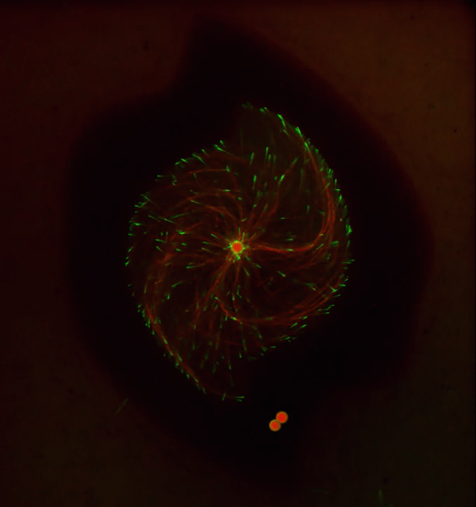
Ever thought about giving cell biology a whirl? If so, I suggest you sit down and take a look at this full-blown cytoskeletal “storm,” which provides a spectacular dynamic view of the choreography of life.
Before a cell divides, it undergoes a process called mitosis that copies its chromosomes and produces two identical nuclei. As part of this process, microtubules, which are structural proteins that help make up the cell’s cytoskeleton, reorganize the newly copied chromosomes into a dense, football-shaped spindle. The position of this mitotic spindle tells the cell where to divide, allowing each daughter cell to contain its own identical set of DNA.
To gain a more detailed view of microtubules in action, researchers designed an experimental system that utilizes an extract of cells from the African clawed frog (Xenopus laevis). As the video begins, a star-like array of microtubules (red) radiate outward in an apparent effort to prepare for cell division. In this configuration, the microtubules continually adjust their lengths with the help of the protein EB-1 (green) at their tips. As the microtubules grow and bump into the walls of a lab-generated, jelly-textured enclosure (dark outline), they buckle—and the whole array then whirls around the center.
Abdullah Bashar Sami, a Ph.D. student in the NIH-supported lab of Jesse “Jay” Gatlin, University of Wyoming, Laramie, shot this movie as a part his basic research to explore the still poorly understood physical forces generated by microtubules. The movie won first place in the 2019 Green Fluorescent Protein Image and Video Contest sponsored by the American Society for Cell Biology. The contest honors the 25th anniversary of the discovery of green fluorescent protein (GFP), which transformed cell biology and earned the 2008 Nobel Prize in Chemistry for three scientists who had been supported by NIH.
Like many movies, the setting was key to this video’s success. The video was shot inside a microfluidic chamber, designed in the Gatlin lab, to study the physics of microtubule assembly just before cells divide. The tiny chamber holds a liquid droplet filled with the cell extract.
When the liquid is exposed to an ultra-thin beam of light, it forms a jelly-textured wall, which traps the molecular contents inside [1]. Then, using time-lapse microscopy, the researchers watch the mechanical behavior of GFP-labeled microtubules [2] to see how they work to position the mitotic spindle. To do this, microtubules act like shapeshifters—scaling to adjust to differences in cell size and geometry.
The Gatlin lab is continuing to use their X. laevis system to ask fundamental questions about microtubule assembly. For many decades, both GFP and this amphibian model have provided cell biologists with important insights into the choreography of life, and, as this work shows, we can expect much more to come!
References:
[1] Microtubule growth rates are sensitive to global and local changes in microtubule plus-end density. Geisterfer ZM, Zhu D, Mitchison T, Oakey J, Gatlin JC. November 20, 2019.
[2] Tau-based fluorescent protein fusions to visualize microtubules. Mooney P, Sulerud T, Pelletier JF, Dilsaver MR, et al. Cytoskeleton (Hoboken). 2017 Jun;74(6):221-232.
Links:
Mitosis (National Human Genome Research Institute/NIH)
Gatlin Lab (University of Wyoming, Laramie)
Green Fluorescent Protein Image and Video Contest (American Society for Cell Biology, Bethesda, MD)
2008 Nobel Prize in Chemistry (Nobel Foundation, Stockholm, Sweden)
NIH Support: National Institute of General Medical Sciences
Posted In: Cool Videos
Tags: 2019 Green Fluorescent Protein Image and Video Contest, American Society for Cell Biology, biophysics, cell biology, cell division, chromosome, cytoskeleton, DNA, EB-1, frog, GFP, green fluorescent protein, microfluidics, microtubule growth rate, microtubules, mitosis, mitotic spindle, model organism, Nobel Prize, Xenopus laevis
Why Flies and Humans Freeze When Startled
Posted on February 6th, 2020 by Dr. Francis Collins
When faced with something unexpected and potentially ominous, like a sudden, loud noise or a threat of danger, humans often freeze before we act. This is colloquially referred to as the “deer in the headlights” phenomenon. The movie of fruit flies that you see above may help explain the ancient origins of the “startle response” and other biomechanical aspects of motion.
In this video, which shows a footrace between two flies (Drosophila melanogaster), there are no winners or losers. Their dash across the screen provides a world-class view of the biomechanics of walking in these tiny, 3 millimeter-long insects that just won’t sit still.
The fly at the top zips along at about 25 millimeters per second, the normal walking speed for Drosophila. As a six-legged hexapod, the fly walks with a “tripod gait,” alternating between its stance phase—right fore (RF), left middle (LM), and right hind (RH) —and its swing phase sequence of left fore (LF), right middle (RM), and left hind (LH).
The slowpoke at the bottom of the video clocks in at a mere 15 millimeters per second. This fly’s more-tentative gait isn’t due to an injury or a natural lack of speed. What is causing the delay is the rapid release of the chemical messenger serotonin into its nervous system, which models a startle response.
You may have already heard about serotonin because of its role in regulating mood and appetite in humans. Now, a team led by Richard S. Mann and Clare Howard, Columbia University’s Zuckerman Institute, New York, has discovered that fruit flies naturally release serotonin to turn on neural circuits that downshift and steady the speed of their gait.
As detailed recently in Current Biology [1], serotonin is active under myriad conditions to tell flies to slow things down. For example, serotonin helps flies weather the stress of extreme temperatures, conserve energy during bouts of hunger, and even walk upside down on the ceiling.
But the research team, which was supported by the NIH-led Brain Research through Advancing Innovative Neurotechnologies® (BRAIN) Initiative, found that serotonin’s most-powerful effect came during an actual startle response, prompted by a sudden, jolting vibration. Scientists suspect the release of serotonin activates motor neurons much like an emergency brake, stiffening and locking up the fly’s leg joints. When the researchers blocked the fly’s release of serotonin, it interrupted their normal startle response.
In years past, such a detailed, high-resolution “action video” of Drosophila, one of the most-popular model organisms in biology, would have been impossible to produce. Fruit flies are tiny and possess extremely high energy.
But a few years ago, the Mann lab developed the approach used in this video to bring the hurried gait of fruit flies into tight focus [2]. Their system combines an optical touch sensor and high-speed video imaging that records the footfalls of all six of a fly’s feet.
Then, using the lab’s unique software program called FlyWalker , the researchers can extract various biomechanical parameters of walking in time and space. These include step length, footprint alignment, and, as the letters in the video show, the natural sequence of a tripod gait.
Drosophila may be a very distant relative of humans. But these ubiquitous insects that sometimes buzz around our fruit bowls contain many fundamental clues into human biology, whether the area of research is genetics, nutrition, biomechanics, or even the underlying biology of the startle response.
Reference:
[1] Serotonergic Modulation of Walking in Drosophila. Howard CE, Chen CL, Tabachnik T, Hormigo R, Ramdya P, Mann RS. Curr Biol. 2019 Nov 22.
[2] Quantification of gait parameters in freely walking wild type and sensory deprived Drosophila melanogaster. Mendes CS, Bartos I, Akay T, Márka S, Mann RS. Elife. 2013 Jan 8;2:e00231.
Links:
Brain Research through Advancing Innovative Neurotechnologies® (BRAIN) Initiative (NIH)
Mann Lab (Columbia University’s Zuckerman Institute, New York)
MouseWalker Colored Feet (YouTube)
NIH Support: National Institute for Neurological Disorders and Stroke; National Institute of General Medical Sciences
Posted In: Cool Videos
Tags: biomechanics, BRAIN Initiative, Brain Research through Advancing Innovative Neurotechnologies Initiative, Drosophila melanogaster, fruit fly, gait, model organism, neuroscience, neurotransmitters, serotonin, startle response, tripod gait, walking
Study in Africa Yields New Diabetes Gene
Posted on July 30th, 2019 by Dr. Francis Collins
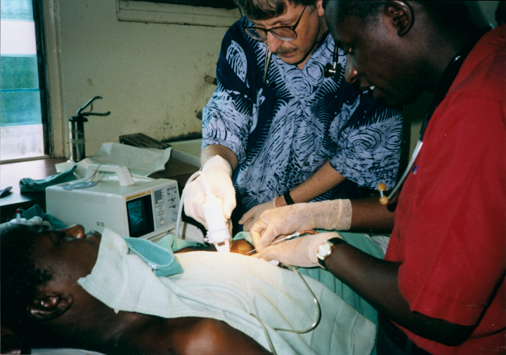
Caption: Volunteering my medical services in Nigeria three decades ago inspired me to learn more about type 2 diabetes in Africa and beyond. Credit: Margaret Collins
When I volunteered to serve as a physician at a hospital in rural Nigeria more than 25 years ago, I expected to treat a lot of folks with infectious diseases, such as malaria and tuberculosis. And that certainly happened. What I didn’t expect was how many people needed care for type 2 diabetes (T2D) and the health problems it causes. Surprisingly, these individuals were generally not overweight, and the course of their illness seemed different than in the West.
The experience inspired me to join with other colleagues at Howard University, Washington, DC, to help found the Africa America Diabetes Mellitus (AADM) study. It aims to uncover genomic risk factors for T2D in Africa and, using that information, improve understanding of the condition around the world.
So, I’m pleased to report that, using genomic data from more than 5,000 volunteers, our AADM team recently discovered a new gene, called ZRANB3, that harbors a variant associated with T2D in sub-Saharan Africa [1]. Using sophisticated laboratory models, the team showed that a malfunctioning ZRANB3 gene impairs insulin production to control glucose levels in the bloodstream.
Since my first trip to Nigeria, the number of people with T2D has continued to rise. It’s now estimated that about 8 to 10 percent of Nigerians have some form of diabetes [2]. In Africa, diabetes affects more than 7 percent of the population, more than twice the incidence in 1980 [3].
The causes of T2D involve a complex interplay of genetic, environmental, and lifestyle factors. I was particularly interested in finding out whether the genetic factors for T2D might be different in sub-Saharan Africa than in the West. But at the time, there was a dearth of genomic information about T2D in Africa, the cradle of humanity. To understand complex diseases like T2D fully, we need all peoples and continents represented in the research.
To begin to fill this research gap, the AADM team got underway and hasn’t looked back. In the latest study, led by Charles Rotimi at NIH’s National Human Genome Research Institute, in partnership with multiple African diabetes experts, the AADM team enlisted 5,231 volunteers from Nigeria, Ghana, and Kenya. About half of the study’s participants had T2D and half did not.
As reported in Nature Communications, their genome-wide search for T2D gene variants turned up three interesting finds. Two were in genes previously linked to T2D risk in other human populations. The third involved a gene that codes for ZRANB3, an enzyme associated with DNA replication and repair that had never been reported in association with T2D.
To understand how ZRANB3 might influence a person’s risk for developing T2D, the researchers turned to zebrafish (Danio rerio), an excellent vertebrate model for its rapid development. The researchers found that the ZRANB3 gene is active in insulin-producing beta cells of the pancreas. That was important to know because people with T2D frequently have reduced numbers of beta cells, which compromises their ability to produce enough insulin.
The team next used CRISPR/Cas9 gene-editing tools either to “knock out” or reduce the expression of ZRANB3 in young zebrafish. In both cases, it led to increased loss of beta cells.
Additional study in the beta cells of mice provided more details. While normal beta cells released insulin in response to high levels of glucose, those with suppressed ZRANB3 activity couldn’t. Together, the findings show that ZRANB3 is important for beta cells to survive and function normally. It stands to reason, then, that people with a lower functioning variant of ZRANB3 would be more susceptible to T2D.
In many cases, T2D can be managed with some combination of diet, exercise, and oral medications. But some people require insulin to manage the disease. The new findings suggest, particularly for people of African ancestry, that the variant of the ZRANB3 gene that one inherits might help to explain those differences. People carrying particular variants of this gene also may benefit from beginning insulin treatment earlier, before their beta cells have been depleted.
So why wasn’t ZRANB3 discovered in the many studies on T2D carried out in the United States, Europe, and Asia? It turns out that the variant that predisposes Africans to this disease is extremely rare in these other populations. Only by studying Africans could this insight be uncovered.
More than 20 years ago, I helped to start the AADM project to learn more about the genetic factors driving T2D in sub-Saharan Africa. Other dedicated AADM leaders have continued to build the research project, taking advantage of new technologies as they came along. It’s profoundly gratifying that this project has uncovered such an impressive new lead, revealing important aspects of human biology that otherwise would have been missed. The AADM team continues to enroll volunteers, and the coming years should bring even more discoveries about the genetic factors that contribute to T2D.
References:
[1] ZRANB3 is an African-specific type 2 diabetes locus associated with beta-cell mass and insulin response. Adeyemo AA, Zaghloul NA, Chen G, Doumatey AP, Leitch CC, Hostelley TL, Nesmith JE, Zhou J, Bentley AR, Shriner D, Fasanmade O, Okafor G, Eghan B Jr, Agyenim-Boateng K, Chandrasekharappa S, Adeleye J, Balogun W, Owusu S, Amoah A, Acheampong J, Johnson T, Oli J, Adebamowo C; South Africa Zulu Type 2 Diabetes Case-Control Study, Collins F, Dunston G, Rotimi CN. Nat Commun. 2019 Jul 19;10(1):3195.
[2] Diabetes mellitus in Nigeria: The past, present and future. Ogbera AO, Ekpebegh C. World J Diabetes. 2014 Dec 15;5(6):905-911.
[3] Global report on diabetes. Geneva: World Health Organization, 2016. World Health Organization.
Links:
Diabetes (National Institute of Diabetes ad Digestive and Kidney Diseases/NIH)
Diabetes and African Americans (Department of Health and Human Services)
Why Use Zebrafish to Study Human Diseases (Intramural Research Program/NIH)
Charles Rotimi (National Human Genome Research Institute/NIH)
NIH Support: National Human Genome Research Institute; National Institute of Diabetes and Digestive and Kidney Diseases; National Institute on Minority Health and Health Disparities
Posted In: News
Tags: AADM, Africa, Africa America Diabetes Mellitus, beta cells, CRISPR/Cas9, diabetes, gene editing, genomics, Ghana, global health, Kenya, minority health, model organism, Nigeria, pancreas, sub-Saharan Africa, T2D, type 2 diabetes, zebrafish, ZRANB3
Watch Flowers Spring to Life
Posted on April 25th, 2019 by Dr. Francis Collins
Spring has sprung! The famous Washington cherry blossoms have come and gone, and the tulips and azaleas are in full bloom. In this mesmerizing video, you’ll get a glimpse of the early steps in how some spring flowers bloom.
Floating into view are baby flowers, their cells outlined (red), at the tip of the stem of the mustard plant Arabidopsis thaliana. Stem cells that contain the gene STM (green) huddle in the center of this fast-growing region of the plant stem—these stem cells will later make all of the flower parts.
As the video pans out, slightly older flowers come into view. These contain organs called sepals (red, bumpy outer regions) that will grow into leafy support structures for the flower’s petals.
Movie credits go to Nathanaёl Prunet, an assistant professor at the University of California, Los Angeles, who shot this video while working in the NIH-supported lab of Elliot Meyerowitz at the California Institute of Technology, Pasadena. Prunet used confocal microscopy to display the different ages and stages of the developing flowers, generating a 3D data set of images. He then used software to produce a bird’s-eye view of those images and turned it into a cool movie. The video was one of the winners in the Federation of American Societies for Experimental Biology’s 2018 BioArt competition.
Beyond being cool, this video shows how a single gene, STM, plays a starring role in plant development. This gene acts like a molecular fountain of youth, keeping cells ever-young until it’s time to grow up and commit to making flowers and other plant parts.
Like humans, most plants begin life as a fertilized cell that divides over and over—first into a multi-cell embryo and then into mature parts, or organs. Because of its ease of use and low cost, Arabidopsis is a favorite model for scientists to learn the basic principles driving tissue growth and regrowth for humans as well as the beautiful plants outside your window. Happy Spring!
Links:
Meyerowitz Lab (California Institute of Technology, Pasadena)
Prunet Lab (University of California, Los Angeles)
The Arabidosis Information Resource (Phoenix Bioinformatics, Fremont, CA)
BioArt Scientific Image and Video Competition (Federation of American Societies for Experimental Biology, Bethesda, MD)
NIH Support: National Institute of General Medical Sciences
Posted In: Cool Videos
Tags: 2018 BioArt Scientific Image & Video Competition, Arabidopsis, Arabidopsis thaliana, BioArt, development, developmental biology, flowers, model organism, mustard plant, plants, stem cells, STM, video
Students Contribute to Research Through Ovarian Art
Posted on February 21st, 2019 by Dr. Francis Collins

Credit: Crystal D. Rogers and Mariano Loza-Coll, California State University, Northridge
Seeing the development of an organ under a microscope for the first time can be a truly unforgettable experience. But for a class taught by Crystal Rogers at California State University, Northridge, it can also be an award-winning moment.
This image, prepared during a biology lab course, was one of the winners in the 2018 BioArt Scientific Image & Video Competition, sponsored by the Federation of American Societies for Experimental Biology (FASEB). This colorful image shows the tip of an ovary from a fruit fly (Drosophila melanogaster), provided by Mariano Loza-Coll. You can see that the ovary is packed with oocytes (DNA stained blue). The orderly connective structure (pink) and signal-transmitting molecules like STAT (yellow) are common to egg maturation and reproductive processes in humans.
What makes this image unique among this year’s BioArt winners is that the prep work was done by undergraduate students just learning how to work in a lab. They did the tissue dissections, molecular labeling, and beautiful stainings in preparation for Rogers to “snap” the photo on her research lab’s optical-sectioning microscope.
What’s also fantastic is that many of Rogers’s students are from groups traditionally underrepresented in biomedicine. Many are considering careers in research and, from the looks of things, they are off to a beautiful start.
After teaching classes, Rogers also has an NIH-supported lab to run. She and her team study salamanders and chickens to determine how biological “glue” proteins, called cadherins, help to create neural crest cells, a critical cell type that arises very early in development [1].
For developmental biologists, it’s essential to understand what prompts these neural crest cells to migrate to locations throughout the body, from the heart to the skin to the cranium, or head. For example, cranial neural crest cells at first produce what appears to be the same generic, undifferentiated facial template in vertebrate species. And yet, neural crest cells and the surrounding ectodermal cells go on to generate craniofacial structures as distinct as the beak of a toucan, the tusk of a boar, or the horn of a rhinoceros.
But if the organ of interest is an ovary, the fruit fly has long been a go-to organism to learn more. Not only does the fruit fly open a window into ovarian development and health issues like infertility, it showcases the extraordinary beauty of biology.
Reference:
[1] A catenin-dependent balance between N-cadherin and E-cadherin controls neuroectodermal cell fate choices. Rogers CD, Sorrells LK, Bronner ME. Mech Dev. 2018 Aug;152:44-56.
Links:
Rogers Lab (California State University, Northridge)
BioArt Scientific Image & Video Competition (Federation of American Societies for Experimental Biology, Bethesda, MD)
NIH Support: Eunice Kennedy Shriver National Institute of Child Health and Human Development
Posted In: Snapshots of Life
Tags: BioArt, cadherin, development, developmental biology, Drosophila melanogaster, embryology, embryos, fruit fly, microscopy, model organism, neural crest cells, oocyte, ovary, STAT
Finding Brain Circuits Tied to Alertness
Posted on November 14th, 2017 by Dr. Francis Collins
Everybody knows that it’s important to stay alert behind the wheel or while out walking on the bike path. But our ability to react appropriately to sudden dangers is influenced by whether we feel momentarily tired, distracted, or anxious. How is it that the brain can transition through such different states of consciousness while performing the same routine task, even as its basic structure and internal wiring remain unchanged?
A team of NIH-funded researchers may have found an important clue in zebrafish, a popular organism for studying how the brain works. Using a powerful new method that allowed them to find and track brain circuits tied to alertness, the researchers discovered that this mental state doesn’t work like an on/off switch. Rather, alertness involves several distinct brain circuits working together to bring the brain to attention. As shown in the video above that was taken at cellular resolution, different types of neurons (green) secrete different kinds of chemical messengers across the zebrafish brain to affect the transition to alertness. The messengers shown are: serotonin (red), acetylcholine (blue-green), and dopamine and norepinephrine (yellow).
What’s also fascinating is the researchers found that many of the same neuronal cell types and brain circuits are essential to alertness in zebrafish and mice, despite the two organisms being only distantly related. That suggests these circuits are conserved through evolution as an early fight-or-flight survival behavior essential to life, and they are therefore likely to be important for controlling alertness in people too. If correct, it would tell us where to look in the brain to learn about alertness not only while doing routine stuff but possibly for understanding dysfunctional brain states, ranging from depression to post-traumatic stress disorder (PTSD).
Posted In: Health, Science, technology
Tags: acetylcholine, alertness, brain, brain circuits, brain imaging, brain states, Danio rerio, depression, dopamine, evolution, evolutionary biology, locus coeruleus, mice, model organism, Multi-MAP, neurology, neuromodulation, neurotransmitter, norepinephrine, optogenetics, PTSD, serotonin, zebrafish
Creative Minds: Building a CRISPR Gene Drive Against Malaria
Posted on November 9th, 2017 by Dr. Francis Collins
Valentino Gantz/Credit: Erik Jepsen
Researchers have used Drosophila melanogaster, the common fruit fly that sometimes hovers around kitchens, to make seminal discoveries involving genetics, the nervous system, and behavior, just to name a few. Could a new life-saving approach to prevent malaria be next? Valentino Gantz, a researcher at the University of California, San Diego, is on a path to answer that question.
Gantz has received a 2016 NIH Director’s Early Independence Award to use Drosophila to hone a new bioengineered tool that acts as a so-called “gene drive,” which spreads a new genetically encoded trait through a population much faster than would otherwise be possible. The lessons learned while working with flies will ultimately be applied to developing a more foolproof system for use in mosquitoes with the hope of stopping the transmission of malaria and potentially other serious mosquito-borne diseases.
Posted In: Health, Science, technology
Tags: 2016 NIH Director’s Early Independence Award, Anopheles stephensi, CRISPR/Cas9, Drosophila melanogaster, ecology, fruit fly, gene drive, gene editing, genetic engineering, genome editing, insects, malaria, model organism, mosquitoes, mutagenic chain reaction, neglected tropical diseases
Snapshots of Life: Coming Face to Face with Development
Posted on January 26th, 2017 by Dr. Francis Collins
Credit: Oscar Ruiz and George Eisenhoffer, University of Texas MD Anderson Cancer Center, Houston
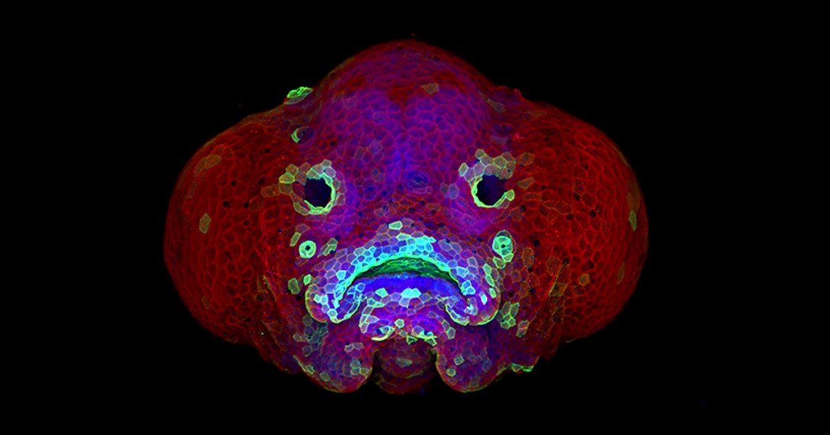
Zebrafish (Danio rerio) is a favorite model for studying development, in part because its transparent embryos make it possible to produce an ever-growing array of amazingly informative images. For one recent example, check out this Federation of American Societies for Experimental Biology’s 2016 BioArt winner, which shows the developing face of a 6-day-old zebrafish larva.
Yes, those downturned “lips” are indeed cells that will go on to become the fish’s mouth. But all is not quite what it appears: the two dark circles that look like eyes are actually developing nostrils. Both the nostrils and mouth express high levels of F-actin (green), a structural protein that helps orchestrate cell movement. Meanwhile, the two bulging areas on either side of the fish’s head, which are destined to become eyes and skin, express keratin (red).
Oscar Ruiz, who works in the lab of George Eisenhoffer at The University of Texas MD Anderson Cancer Center, Houston, used a confocal microscope to create this image. What was most innovative about his work was not the microscope itself, but how he prepared the sample for imaging. With traditional methods, researchers can only image the faces of zebrafish larvae from the side or the bottom. However, the Eisenhoffer lab has devised a new method of preparing fish larvae that makes it possible to image their faces head-on. This has enabled the team to visualize facial development at much higher resolution than was previously possible.
Tags: art, birth defects, cancer, cleft lip, cleft lip and palate, cleft palate, confocal microscope, craniofacial development, Danio rerio, development, epithelial cancers, epithelium, F-actin, FASEB Bioart 2016, model organism, Science, zebrafish
Creative Minds: The Worm Tissue-ome Teaches Developmental Biology for Us All
Posted on November 17th, 2016 by Dr. Francis Collins
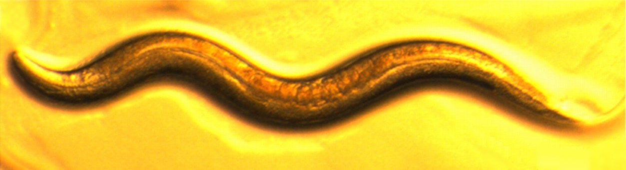
Caption: An adult Caenorhabditis elegans, 5 days
Credit: Coleen Murphy, Princeton University, Princeton, NJ
In the nearly 40 years since Nobel Prize-winning scientist Sydney Brenner proposed using a tiny, transparent soil worm called Caenorhabditis elegans as a model organism for biomedical research_,_ C. elegans has become one of the most-studied organisms on the planet. Researchers have determined that C. elegans has exactly 959 cells, 302 of which are neurons. They have sequenced and annotated its genome, developed an impressive array of tools to study its DNA, and characterized the development of many of its tissues.
But what researchers still don’t know is exactly how all of these parts work together to coordinate this little worm’s response to changes in nutrition, environment, health status, and even the aging process. To learn more, 2015 NIH Director’s Pioneer Award winner Coleen Murphy of Princeton University, Princeton, NJ, has set out to analyze which genes are active, or transcribed, in each of the major tissues of adult C. elegans, building the framework for what’s been dubbed the C. elegans “tissue-ome.”
Tags: 2015 NIH Director’s Pioneer Award, adult worms, aging, aging research, C. elegans, Caenorhabditis elegans, daf-2, developmental biology, genetics, genomics, gerontology, learning, lifespan, metabolism, model organism, nutrition, tissue-ome, transcriptome, worm
Snapshots of Life: Fish Awash in Color
Posted on March 31st, 2016 by Dr. Francis Collins

Credit: Chen-Hui Chen, Duke University
If this image makes you think of a modern art, you’re not alone. But what you’re actually seeing are hundreds of live cells from a tiny bit (0.0003348 square inches) of skin on the tail fin of a genetically engineered adult zebrafish. Zebrafish are normally found in tropical freshwater and are a favorite research model to study vertebrate development and tissue regeneration. The cells have been labeled with a cool, new fluorescent imaging tool called Skinbow. It uniquely color codes cells by getting them to express genes encoding red, green, and blue fluorescent proteins at levels that are randomly determined. The different ratios of these colorful proteins mix to give each cell a distinctive hue when imaged under a microscope. Here, you can see more than 70 detectable Skinbow colors that make individual cells as visually distinct from one another as jellybeans in a jar.
Skinbow is the creation of NIH-supported scientists Chen-Hui Chen and Kenneth Poss at Duke University, Durham, NC, with imaging computational help from collaborators Stefano Di Talia and Alberto Puliafito. As reported recently in the journal Developmental Cell [1], Skinbow’s distinctive spectrum of color occurs primarily in the outermost part of the skin in a layer of non-dividing epithelial cells. Using Skinbow, Poss and colleagues tracked these epithelial cells, individually and as a group, over their entire 2 to 3 week lifespans in the zebrafish. This gave them an unprecedented opportunity to track the cellular dynamics of wound healing or the regeneration of lost tissue over time. While Skinbow only works in zebrafish for now, in theory, it could be adapted to mice and maybe even humans to study skin and possibly other organs.
Posted In: Science
Tags: Brainbow, cell biology, cells, dermatology, fish, gene expression, genes, model organism, skin, Skinbow, tissue regeneration, wound healing, zebra fish, zebrafish