UMS – NIH Director's Blog (original) (raw)
Snapshots of Life: Muscling in on Development
Posted on July 27th, 2017 by Dr. Francis Collins
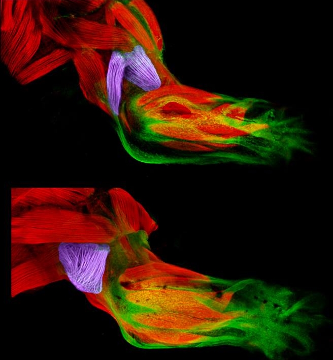
Credit: Mary P. Colasanto, University of Utah, Salt Lake City
Twice a week, I do an hour of weight training to maintain muscle strength and tone. Millions of Americans do the same, and there’s always a lot of attention paid to those upper arm muscles—the biceps and triceps. Less appreciated is another arm muscle that pumps right along during workouts: the brachialis. This muscle—located under the biceps—helps your elbow flex when you are doing all kinds of things, whether curling a 50-pound barbell or just grabbing a bag of groceries or your luggage out of the car.
Now, scientific studies of the triceps and brachialis are providing important clues about how the body’s 40 different types of limb muscles assume their distinct identities during development [1]. In these images from the NIH-supported lab of Gabrielle Kardon at the University of Utah, Salt Lake City, you see the developing forelimb of a healthy mouse strain (top) compared to that of a mutant mouse strain with a stiff, abnormal gait (bottom).
Tags: brachialis, connective tissue, development, FluoRender, forelimb muscles, genetics, genomics, lateral triceps, limb muscles, limbs, mouse, mouse genetics, muscle, musculoskeletal disorder, rare disease, Tbx3, transcription factor, triceps, ulnar-mammary syndrome, UMS, University of Utah’s 2016 Research as Art
