Tooth wear: the view of the anthropologist (original) (raw)
Introduction
Over the past century, anthropological research of many contemporary and pre-contemporary populations including hunter-gatherer, agricultural, medieval and current has concluded that tooth wear is a normal physiological phenomenon where teeth, although worn, remain functional throughout life [7]. Variations to the degree and pattern of tooth wear between populations is attributed to the abrasiveness of the diet and the use of teeth as tools [16]. Such direct associations between diet and wear have also been substantiated within other species [22]. Furthermore, it has been documented that the stomatognathic system changes or adapts in response to progressive wear, indicating a dynamic craniofacial complex [13, 19].
In contrast to the anthropological approach, it can be argued that dentistry evolved into a science over the past century at a time when populations were overwhelmed by dental caries, periodontal disease and broken down dentitions. As a result, dentistry progressively developed the expertise to restore and rehabilitate dentitions to their original morphology based on an underlying premise that the newly erupted tooth was the ideal functional form. Although dental opinion is partly moving away from past rigid geometric concepts, there nevertheless was an early period where any type of tooth wear was considered pathological.
This manuscript is not a literature review but an attempt to cross disciplines and provide a different model to the overall understanding of dental wear. This paradigm encompasses an anthropological perspective with the discipline of comparative anatomy while still taking into account current clinical observations made by dentists.
Terminology and the mechanisms of wear
For many years, anthropologists often used the terms abrasion, attrition and even erosion interchangeably to denote the same thing: tooth wear caused mainly by the diet and tool use. In actual fact, they were inadvertently referring to abrasion. Conversely, in recent years, dentists seem to agree that the terms attrition, abrasion and erosion define different mechanisms; however, there seems to be subtle variations on how these mechanisms are understood. The definitions outlined will follow those of Every [8].
Attrition
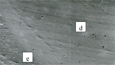
Attrition occurs from tooth-to-tooth contact without the presence of food (i.e. tooth grinding) and typically is characterised by the facet that is matched by a corresponding facet on a tooth in the opposing arch. When dentine is exposed, it remains flat with no ‘cupping’ or ‘scooping,’ and the microwear detail observed under magnification is that of parallel striations typically occurring within the facet border (Fig. 1). In general, well-defined, shiny facets is a good measure for active attrition. Furthermore, the prevalence of faceting is high among pre-contemporary Australian Aboriginal populations [11].
Fig. 1
Microwear detail of a facet showing parallel striations. The dentine (d) is not scooped out and is at the same level as the enamel (e)
Abrasion
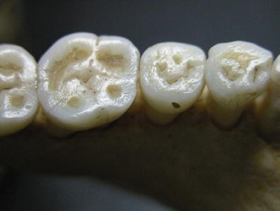
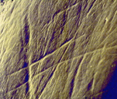
Abrasion occurs by the friction of exogenous material forced over tooth surfaces. Although a multitude of foreign bodies (including the toothbrush) can cause abrasion, the most common yet most overlooked is food. The action of food on a tooth surface is ‘non-anatomically specific’; that is, the action generally occurs over the whole occlusal surface producing a wear area as opposed to a facet. In contrast to attrition, exposed dentine will scoop. The action of abrasive food on enamel or dentine will cause pitting, gouge marks and other characteristics of mechanical breakdown (Fig. 2). Under magnification, the typical microwear detail is identified by haphazard scratch marks (Fig. 3) that reflect the type of diet consumed or foreign body forced over the tooth surface [8]. In general, the buccal cusps of the lower molars and the palatal cusps of the upper molars wear faster (Fig. 2).
Fig. 2
An example showing the effect of an abrasive diet on the teeth of a pre-contemporary Australian Aboriginal. Note the gouged and pitted enamel and the scooping of the dentine. Note how the buccal cusps of the lower molars are wearing faster
Fig. 3
Microwear detail of an abrasion area showing haphazard scratch marks. The depth, length and breadth of these scratch marks reflect the type of diet consumed
Abrasion is the predominant wear mechanism caused by the mastication of hard fibrous material, typical of hunter-gatherer populations, especially those living in desert environments. In such populations, individuals of all ages show evidence of abrasion on both enamel and in dentine when exposed [4]. It is also well established that abrasion has a direct linear association with age [17]. Although this relationship tends to becomes exponential with molar wear during old age, it nevertheless highlights that the general diet and hence its abrasive potential remains relatively constant within a set environment.
It is interesting to note that scooped dentine is not sensitive, and microscopically, there is a mechanical smear layer over the surface occluding dentinal tubules. Although mechanical smear layers have been reported to result from dental burs, mechanical action during mastication produces similar results. It seems that dentine can be ‘burnished.’ Secondly, in contrast to erosion, dentinal scooping from abrasion is relatively shallow following a relatively fixed depth-to-breadth ratio that seems to remain constant as the wear progresses, provided the diet remains the same [6]. As the masticatory stroke widens with progressive wear, the site of maximum depth tends to localise on the buccal aspect of the scoop of the lowers and the palatal aspect of the upper teeth.
Erosion
Erosion can be defined as the chemical dissolution of tooth substance without the presence of plaque. The clinical appearance has been well documented [15, 23], and although dentinal scooping is a common feature, in contrast to abrasion, there are essential differences. If the erosion is active, the dentinal tubules remain open resulting in sensitivity, and the depth of scooping perpetually increases.
It must be emphasised that although researchers have documented different clinical erosive patterns depending on source (i.e. intrinsic and extrinsic) and that the patterns are moderated by salivary flow and the presence of pellicle, the continual action of acid over time will affect more than just an occlusal surface. Hence, multiple surfaces often become affected to various degrees, removing all traces of biofilm and leaving the dentition in a state as if the oral hygiene is excellent.
Interplay between the various mechanisms of wear
The mechanisms of attrition, abrasion and erosion act together, each with different intensity and duration to produce a multitude of different wear patterns. The interplay between attrition and abrasion has been demonstrated with casts from longitudinal growth studies of the same individuals of pre-contemporary aboriginal populations [10]. Homogeneous groups in unchanging harsh environments show constant wear rates (abrasion). However, such casts also show evidence of attrition, where the definition of facets come and go over time, showing an intermittent, although a common, occurrence of grinding. This is why the definition of attrition is ‘tooth-to-tooth contact without the presence of food.’ If facets occurred from tooth-to-tooth contact during mastication as some allude to, then facets by inference should always be present and well defined among hunter-gatherer societies that vigorously masticate their food.
Physiological adaptation
The anthropological approach to tooth wear can be summarised as interplay between genes and environment. The model highlights how genetic factors independently influence initial tooth morphology and occlusion, while the occlusion and food consistency influence the masticatory pattern. Over time, mastication causes tooth wear, which in turn affects tooth form and hence the occlusion and again in time the masticatory pattern. The cycle then continues until occasionally among older individuals, teeth may become ‘worn out’ and lost [3]. Modern dentistry would describe this progression as a continuum from a ‘canine-rise’ occlusion, to group function, to a flat occlusal plane with an associated edge-to-edge anterior bite.
There are numerous examples that associate gradual tooth wear with perpetual physiological adaptation. For example, as cusps reduced in height, the ‘teardrop’ nature of the masticatory pattern becomes wider [4] with associated ‘re-modelling’ and flattening of the glenoid fossa [18]. In addition, the physiological process of continual eruption occurs as a compensatory mechanism to wear. The interplay between wear and continual eruption determines the occlusal vertical dimension.
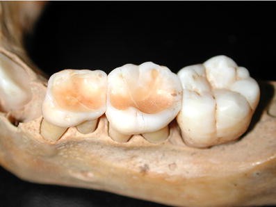
There is also a direct relationship between occlusal load, interproximal wear (Fig. 4) and the mesial migration of teeth [5, 24]. Heavy occlusal load from vigorous mastication of fibrous and often hard food not only causes abrasion but is also responsible for the relative movement of adjacent teeth to one another producing inter-proximal wear. It is interesting to note that this relative movement between teeth at times causes the mesial of teeth to wear faster than the distal of their adjacent neighbour, producing a mesial concavity rather than a flat surface [9].
Fig. 4
Heavy occlusal wear during the mixed dentition stage of a desert dwelling Australian Aboriginal. The interproximal wear and the interproximal concavity developing on the mesial of the lower left first permanent molar. The deepest point of wear is on the buccal aspect of the scooped area
Begg’s orthodontic principles were underpinned by his theory of ‘attritional occlusion’ that was based on arch length reduction caused by mesial tooth migration during inter-proximal wear (here attrition refers to dietary wear). Begg maintained that arch length reduction provided posterior space for the emergence of the third molars without impaction, a common feature among pre-contemporary aboriginal populations. Begg further believed that the higher prevalence for crowding among modern societies closely related to the relative lack of inter-proximal wear. The concept of removing premolars for orthodontic reasons first originated from Begg’s premise that the reduction in arch length over a lifetime was equivalent to the mesio-distal diameter of a premolar tooth. Although this has been challenged since, the theory nevertheless influenced orthodontic clinical practice for many years.
Form and function
Comparative studies have also documented the above physiological processes in many different mammalian species lending support to the idea of perpetual change.
Wear has existed ever since the first dental structures evolved, millions of years ago. It can therefore be argued from an evolutionary perspective that wear was one of the prime selective forces that shaped not only the anatomy of teeth but the properties of the dental tissues themselves. The anatomical relationship and different wear characteristics of dentine and enamel, together with the wear process, is essential for masticatory efficiency.
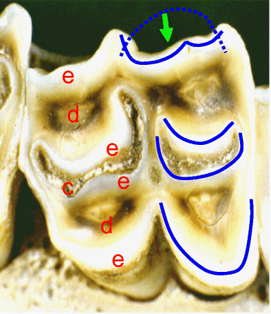
The immediate scooping of the dentine and even cementum soon after exposure has been documented in many different species, in particular herbivores. For example, Fig. 5 shows the worn occlusal surface of a sheep’s tooth with scooped-out dentine and cementum. This ‘differential wear’ promotes the development of a sickle-shaped enamel pattern that is diametrically opposite between teeth in opposing arches. That is, the concavity of each sickle ‘blade’ faces towards the lingual on the lower teeth and towards the buccal on the uppers. The tooth form functions when the mandible moves with a wide, ‘lateral’ masticatory cycle to produce a shearing action made possible by a shallow glenoid fossa. That is, the opposing enamel blades move past one another to produce what is called ‘scissorial point cutting,’ which is a very efficient masticatory action [8, 10, 21]. This functional pattern was first demonstrated by Every [8] more than 50 years ago in many different species including herbivores, carnivores, primates and humans. Hence, the wider masticatory action seen among humans with progressing wear fits this paradigm.
Fig. 5
Shows a sheep’s tooth with scooped out dentine (d) and cementum (c) leaving ‘sickle’-shaped enamel blades (e) (shown in blue). The green arrow shows the movement direction of the blade system (dotted green line) from teeth in the opposing arch that causes scissorial point cutting where the blades contact
Another common feature observed in many species is that of continual eruption, where roots remain incomplete throughout most of life, producing enamel, dentine and even cementum perpetually. Nature seems to have improved and sustained masticatory efficiency through differential wear and has even compensated for wear through continual eruption.
Physiological vs pathological tooth wear
It seems that attrition and particularly abrasion has been evident among human populations since early Homo, at about 2 million years. Only in recent times has the prevalence of abrasion reduced dramatically in our modern industrialised societies because of the consumption of processed softer foods.
Alternatively, erosion seems to be a modern-day disease. Although it can be argued that our early hunter-gatherer ancestors were exposed to acids through their diet (e.g. acidic fruits, berries etc.), these exposures were seasonal and therefore transient with no apparent effect. Saliva would have played a significant protective role as did the presence of biofilms as physical barriers. In such harsh environments, water would have been the main liquid consumed. In theory, perhaps acid would have occasionally affected the occlusal surfaces of teeth of hunter-gatherer populations; however, with abrasion being such a predominant mechanism, the erosion would not be clinically evident. It can be argued that the prevalence of erosion is too low to be of significance. If erosion was in any way excessive, non-occlusal surfaces would have been affected. Furthermore, non-carious cervical lesions, whether they are dished shaped, wedge shaped or other, to date have not been observed in hunter-gatherer societies, especially Australian Aboriginal. This includes ancient American skulls [1] as well as European prehistoric and historic skeletal remains [2].
With the advent of agriculture and through the middle ages, although the fermentation of food products became a more common occurrence, the prevalence of erosion was relatively low, until we reach our modern, affluent existence where erosion is reaching very high proportions. It is interesting to note that in one study of pre-European contact, Maori did show evidence of occlusal erosion; however, there was also strong evidence in this population that a significant shift in diet had occurred [14] including the fermentation of some foods before consumption (personal communication).
In contrast to mechanical processes (e.g. abrasion) that have always been present, erosion at the scale observed today is a modern-day condition. This argument identifies this extreme consumption of acids as an imbalance to the oral environment and perhaps identifies the mechanism of erosion as pathological.
Wear rates and the clinical perspective
Although the clinical assessment of wear is often subjective, such information is essential before operative treatment is undertaken. In general, the extent of tooth wear has an association with age. Wear in a young individual may be considered pathological if the teeth will not last throughout life, while the same amount of wear in an older person my be quite physiological. Superimposed over this is the real problem of aesthetics in today’s society, which can result in ‘pre-mature’ operative treatment. The presence of erosion, however, must be approached with a slightly different understanding. For example, very mild erosion that is successfully stopped using preventive measures may be correctly left without operative intervention by dental practitioners. However, this does not make the wear mechanism physiological.
One aspect of tooth wear that is overlooked among dental circles is the degree of activity of the mechanism(s) in question during the clinical examination of patients. For example, if hypothetically a patient at the age of 50 years has lost say 10% of the clinical crown in the last year because of some change in lifestyle, then if this rate continues, the patient will loose the whole crown by the age of 60 years. Although the wear may look insignificant, this can be considered as pathological. This is why quantifying ‘current’ tooth wear rates are also important during patient assessment before treatment is undertaken.
An affective method that provides a relatively rapid measure of activity for the general practitioner and can be used for all wear mechanisms is a scratch test where a No12 scalpel blade can be used to very lightly scratch an affected surface [12]. Observation of the scratch at various time intervals (e.g. a few days) will easily show activity. If the scratch is not sharp or well defined or non-existent, then the mechanism(s) is active.
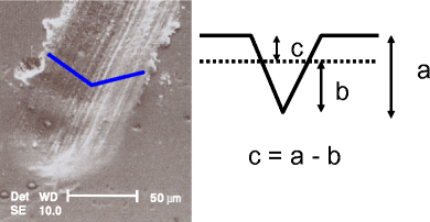
For more precise quantification, a micro-impression of an initial scratch using either resin composite [20], where the negative is observed under a scanning electron microscope (SEM), or simply a positive replica from a rubber impression, then observed under a SEM, will both produce very accurate results. During scanning, the depth of the scratch is determined at various points along the scratch, and a mean depth is obtained. Repeating the procedure of the same scratch on a subsequent appointment (a few days) will give the patient’s wear rate in microns (Fig. 6). This allows a prognostic assessment, which although still somewhat subjective, is still valuable in treatment planning decisions.
Fig. 6
Showing an electron micrograph of a scratch in a tooth surface. If the depth of the initial scratch is measured (a), then re-measured on a subsequent appointment (b), the amount of tooth loss (c) over that time period and hence the current wear rate is determined
Conclusion
There is no doubt that the disciplines of dental anthropology and dentistry is converging. Anthropologists seem to have generally accepted attrition, abrasion and erosion as distinctly separate mechanisms, while dentists are generally moving away from static concepts, acknowledging dynamic change as an inevitable progression throughout life.
References
- Aaron GM (2004) The prevalence of non-carious cervical lesions in modern and ancient American skulls: lack of evidence for an occlusal aetiology. M.S. thesis, The University of Florida, Florida
- Aubry M, Maffart B, Donat B, Brau JJ (2003) Brief communication: Study of noncarious cervical tooth lesions in samples of prehistoric, historic and modern populations from the South of France. Am J Phys Anthropol 121:10–14
Article PubMed Google Scholar - Barrett MJ (1969) Functioning occlusion. Ann Aust Col Dent Sur 2:68–80
Google Scholar - Barrett MJ (1977) Masticatory and non-masticatory uses of teeth. In: Wright RVS (ed) Stone tools as cultural markers: change, evolution and complexity. Australian Institute of Aboriginal Studies, Canberra, pp 17–23
Google Scholar - Begg PR (1954) Stone age man’s dentition. Am J Orthod 40:298–312, 373–383, 462–475, 517–531
Article Google Scholar - Bell EJ, Kaidonis JA, Townsend GC, Richards LC (1998) Comparison of exposed dentinal surfaces resulting from abrasion and erosion. Aust Dent J 43:362–366
Article PubMed Google Scholar - Brace CL (1977) Occlusion to the anthropological eye. In: McNamara JA Jr (ed) The biology of occlusal development. Monograph no. 7, Craniofacial Growth Series. The University of Michigan, Ann Arbor, pp 179–209
Google Scholar - Every RG (1972) A new terminology for mammalian teeth: founded on the phenomenon of thegosis. Parts 1 & 2. Pegasus, Christchurch, New Zealand
Google Scholar - Kaidonis JA, Townsend GC, Richards LC (1992) Brief communication: interproximal tooth wear: a new observation. Am J Phys Anthropol 88:105–107
Article PubMed Google Scholar - Kaidonis JA, Townsend GC, Richards LC (1992) Abrasion: an evolutionary and clinical view. Aust Prosthodont J 6:9–16
PubMed Google Scholar - Kaidonis JA, Townsend GC, Richards LC (1993) Nature and frequency of dental wear facets in an Australian Aboriginal population. J Oral Rehabil 20:333–340
Article PubMed Google Scholar - Kaidonis JA, Townsend GC, Richards LC (2004) Non-carious changes to tooth crowns. In: Mount GJ, Hume WR (eds) Preservation and restoration of tooth structure, 2nd edn. Knowledge, UK, pp 47–60
Google Scholar - Kaifu Y, Kasai K, Townsend GC, Richards LC (2003) Tooth wear and the “design” of the human dentition: a perspective from evolutionary medicine. Yearbook of Phys Anthropol 46:47–61
Article Google Scholar - Kieser JA, Dennison KJ, Kaidonis JA, Huang D, Herbison PGB, Tayles NG (2001) Patterns of dental wear in the early Maori dentition. Int J Osteoarchaeol 11:206–217
Article Google Scholar - Lussi A (2006) Dental erosion. From diagnosis to therapy. In: Whitford GM (ed) Monographs in oral science. Karger, Basel
Google Scholar - Molnar S (1972) Tooth wear and culture: a survey of tooth functions among some prehistoric populations. Curr Anthropol 13:511–526
Article Google Scholar - Richards LC, Brown T (1981) Dental attrition and age relationships in Australian Aboriginals. Archaeol Phys Anthropol Ocean 16:94–98
Google Scholar - Richards LC (1984) Form and function of the masticatory system. In: Ward GK (ed) 54th Congress of the Australian and New Zealand Association for the Advancement of Science, Canberra, pp 96–109
- Richards LC (1985) Dental attrition and craniofacial morphology in two Australian Aboriginal populations. J Dent Res 64:1311–1315
PubMed Google Scholar - Scally KB (1980) Replicating wet fragile dental tissues using Bis-GMA resin. Micron 11:473–474
Google Scholar - Smith JM, Savage RJG (1959) The mechanics of mammalian jaws. Sch Sci Rev 40:289–301
Google Scholar - Teaford MF, Walker A (1984) Quantitative differences in dental microwear between primate species with different diets and a comment on the presumed diet of Sivapithecus. Am J Phys Anthropol 64:191–200
Article PubMed Google Scholar - Yip KHK, Smales RJ, Kaidonis JA (eds) (2006) Tooth erosion: prevention and treatment. Jaypee Brothers, New Delhi
- Wolpoff MH (1971) Interstitial wear. Am J Phys Anthropol 34:205–228
Article PubMed Google Scholar
