LTRPC7 is a Mg·ATP-regulated divalent cation channel required for cell viability (original) (raw)
Accession codes
Accessions
GenBank/EMBL/DDBJ
Data deposits
The GenBank accession numbers for murine and human LTRPC7 cDNA and protein sequences are AY032951 and AY032950, respectively.
References
- Harteneck, C., Plant, T. D. & Schultz, G. From worm to man: three subfamilies of TRP channels. Trends Neurosci. 23, 159– 166 (2000).
Article CAS Google Scholar - Ryazanov, A. G. et al. Identification of a new class of protein kinases represented by eukaryotic elongation factor-2 kinase. Proc. Natl Acad. Sci. USA 94, 4884– 4889 (1997).
Article ADS CAS Google Scholar - Romani, A. M. & Scarpa, A. Regulation of cellular magnesium. Front. Biosci. 5, D720– 734 (2000).
Article CAS Google Scholar - Luthi, D., Gunzel, D. & McGuigan, J. A. Mg-ATP binding: its modification by spermine, the relevance to cytosolic Mg2+ buffering, changes in the intracellular ionized Mg2+ concentration and the estimation of Mg2+ by 31P-NMR. Exp. Physiol. 84, 231– 252 (1999).
CAS PubMed Google Scholar - Runnels, L. W., Yue, L. & Clapham, D. E. TRP-PLIK, a bifunctional protein with kinase and ion channel activities. Science 291, 1043– 1047 (2001).
Article ADS CAS Google Scholar
Acknowledgements
We thank D. Tani and M. Monteilh-Zoller for technical assistance. This work was funded in part by a Beth Israel Pathology Foundation grant and a BIDMC Fireman Fellowship award to A.M.S., and a NIH grant. A.-L.P. is supported by the German Academic Exchange Service (DAAD).
Author information
Author notes
- Andrew M. Scharenberg
Present address: Department of Pediatrics and Immunology, University of Washington and Children's Hospital and Medical Center, Seattle, Washington, 98195-6320, USA - Monica J. S. Nadler, Meredith C. Hermosura, Kazunori Inabe, Anne-Laure Perraud and Tomohiro Kurosaki: These authors contributed equally to this work
Authors and Affiliations
- Department of Pathology, Beth Israel Deaconess Medical Center and Harvard Medical School, Boston, 02215, Massachusetts, USA
Monica J. S. Nadler, Anne-Laure Perraud, Qiqin Zhu, Alexander J. Stokes, Jean-Pierre Kinet & Andrew M. Scharenberg - Laboratory of Cell and Molecular Signaling, Center for Biomedical Research at The Queen's Medical Center and John A. Burns School of Medicine at the University of Hawaii, Honolulu, 96813, Hawaii, USA
Meredith C. Hermosura, Reinhold Penner & Andrea Fleig - Department of Molecular Genetics, Institute for Liver Research, Kansai Medical University, Osaka, 570-8506, Japan
Kazunori Inabe & Tomohiro Kurosaki
Authors
- Monica J. S. Nadler
You can also search for this author inPubMed Google Scholar - Meredith C. Hermosura
You can also search for this author inPubMed Google Scholar - Kazunori Inabe
You can also search for this author inPubMed Google Scholar - Anne-Laure Perraud
You can also search for this author inPubMed Google Scholar - Qiqin Zhu
You can also search for this author inPubMed Google Scholar - Alexander J. Stokes
You can also search for this author inPubMed Google Scholar - Tomohiro Kurosaki
You can also search for this author inPubMed Google Scholar - Jean-Pierre Kinet
You can also search for this author inPubMed Google Scholar - Reinhold Penner
You can also search for this author inPubMed Google Scholar - Andrew M. Scharenberg
You can also search for this author inPubMed Google Scholar - Andrea Fleig
You can also search for this author inPubMed Google Scholar
Corresponding author
Correspondence toAndrew M. Scharenberg.
Supplementary information
Methods
Isolation of cDNAs and sequence analysis
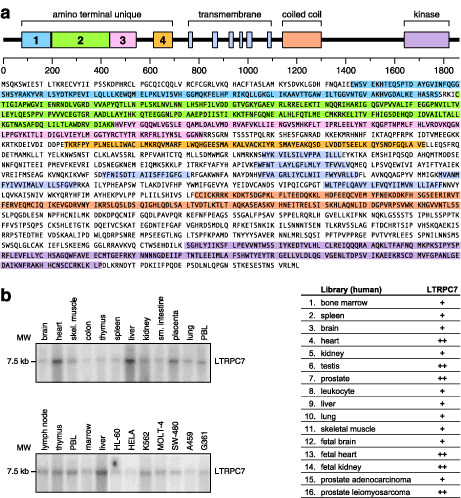
Once a partial sequence for human LTRPC7 was identified out of lymphocyte EST libraries, the corresponding EST clone was purchased and sequenced (accession number AA419407 was obtained). Partial sequences from the 5’ or 3’ ends of AA419407 were used to screen leukocyte, spleen, and kidney libraries using the GeneTrapper II method (Life Technologies) in order to extend the original sequences towards the 5’ and 3’ ends of the respective mRNA’s. Resulting clones were sequenced in both directions using standard fluorescent dideoxy sequencing techniques and partial contigs were assembled using Assembylign (Oxford Molecular, London, UK). For LTRPC7, the available coding sequence was assembled primarily from the sequences of four overlapping clones designated AS8, GT2, B5, and D2, with some sequence confirmation obtained from end sequences of many shorter clones. We also cloned the murine LTRPC7 transcript using a similar approach, and assembled a full sequence contig from two overlapping clones designated MA7 and MA5. The predicted murine LTRPC7 protein exhibited ~95% amino acid identity with the human version. Accession numbers for the murine and human LTRPC7 proteins are presently pending. Predicted proteins and hydrophobicity analyses were obtained using the Macvector program (Oxford Biotechnology), prediction of transmembrane spanning regions was performed using the TMpred program at EMBnet7, coiled coil analysis was performed using the ISREC coils server8, and BLAST alignments were obtained using the NCBI advanced BLAST server.
Northern Blotting
Multiple tissue Northern blots for human tissues and cell lines were obtained from Clontech (Palo Alto, CA), and all hybridizations were performed according to the manufacturer’s protocols. LTRPC7 Northern blots were performed using a dUTP labeled RNA probe generated from a 500 bp fragment corresponding to the most 5’ end of the available LTRPC7 coding sequence from clone AS8. The probe was generated using a T7-directed RNA probe synthesis kit from Ambion (Austin, TX).
Eukaryotic expression constructs, transfection
For the purpose of expressing LTRPC7 in eukaryotic cells, we used PCR to produce an epitope tagged expression construct from our two overlapping murine LTRPC7 clones. The LTRPC7 coding sequence was modified by removing the initiating methionine and replacing it with a sequence encoding a Kozak sequence, the FLAG tag and the additional sequence GCGGCCGCAT, and by placing a SpeI site just after the stop codon. These modifications result in an expressed protein which started with the following amino acid sequence: MGDYKDDDDKRPH followed by the murine LTRPC7 coding sequence starting at the second amino acid. This construct was expressed from the pAPuro vector which allows constitutive LTRPC7 expression from a beta-actin promotor, and from the pcDNA4/TO vector which provides tetracycline-controlled expression from a CMV promotor. The FLAG-LTRPC7/pAPuro vector was used to attempt to express LTRPC7 in several cell lines; however only very low expression was observed in transient expression experiments and no expression was ever observed in stable clones. The FLAG-LTRPC7/pCDNA4/TO construct was transfected by electroporation into HEK-293 cells expressing the tet repressor protein, and clones were selected in zeocin. Several resistant clones were selected for analysis of tetracycline-induced FLAG-LTRPC7 expression. As might be expected from our inability to express LTRPC7 using pApuro-mediated constitutive expression, all clones had low/undetectable basal expression and all clones found to express the FLAG-LTRPC7 protein subsequently exhibited growth arrest and significant toxic effects after several days of tetracycline/doxycycline induction. The clone with the highest inducible expression (c3) was chosen for subsequent electrophysiological analyses.
Immunoprecipitations and SDS/PAGE-Western blotting
Anti-FLAG immunoprecipitations were performed from lysates of 107 HEK-293 cells. Immunoprecipitated proteins were washed three times with lysis buffer, separated by SDS/PAGE using 6% polyacrylamide gels, transferred to a PVDF membrane, and analyzed by anti-FLAG immunoblotting. All procedures used standard methods.
Generation of DT-40 cells in which LTRPC7 is inducibly deficient.
Chicken LTRPC7 genomic fragments were obtained by screening lFIXII chicken genomic library using a 0.5-kb mouse LTRPC7 cDNA fragment including putative transmembrane region as a probe under a low stringent condition. The conventional targeting vectors (pLTRPC7-hisD and pLTRPC7-bsr, which allow inactivation of LTRPC7) were constructed by replacing the genomic fragment-containing exons that correspond to a part of putative transmembrane region with hisD or bsr cassette. These cassettes were flanked by 3.5 and 5.7 kb of chicken LTRPC7 genomic sequence on the 5' and 3' sides, respectively. To generate the inducible targeting vector, pLTRPC7-neo/loxP, the putative transmembrane region of LTRPC7 was inserted between two loxP sites for the vector pKSTKNEOLOXP, which has HSV thymidine kinase and loxP flanked pGK-neo. Then, the 3.5-kb fragment 5' upstream and the 2.6-kb fragment 3' downstream of the loxP flanked region were inserted.
As noted in the main text, in our initial attempt to generate DT-40 cells genetically deficient in the ltrpc7 gene, the targeting constructs, pLTRPC7-bsr and pLTRPC7-hisD, were sequentially introduced into DT-40 cells. Although the first allele targeting by using pLTRPC7-bsr resulted in success with high frequency (71%), clones harboring two targeted alleles were not obtained after several rounds of transfection with pLTRPC7-hisD. Since transfection with pLTRPC7-hisD also worked for the first allele targeting, these results suggested that inactivation of the ltrpc7 gene might lead to lethality in DT-40 cells.
Based on these results, the Cre-loxP system was utilized for disruption of the ltrpc7 gene. The expression plasmid pANMerCreMer-hyg encoding tamoxifen-regulated chimeric Cre enzyme was linearized and introduced into wild-type DT-40. Transfectants were selected in the presence of hygromycine B (2 mg/ml) and resistant clones were screened for inducible-Cre expression by Western blotting analysis. Then, pLTRPC7-neo/loxP was transfected into the clone expressing inducible-Cre, and was selected with both hygromycine B (2 mg/ml) and G418 (2 µg/ml). After confirming successful targeting by Southern blot analysis, cells were cultured in the presence of 200 nM tamoxifen to examine the potentiality of Cre-mediated recombination. Then, pTRPC7-hisD was transfected into the capable clones, and was selected with hygromycine B (2 mg/ml), G418 (2 mg/ml) and histidinol (0.5 mg/ml).

Figure S1.
LTRPC7 is a novel, ubiquitously expressed (GIF 47.5 KB)
(a) Top panel: schematic of LTRPC7 with amino terminal unique regions 1-4 (these regions are defined by their particularly high homology throughout the LTRPC family, and because the sequences between them are of variable length and level of homology in different LTRPC members), transmembrane domain regions (spans are based on TMpred and hydrophobicity analyses), coiled coil region (approximate region based on COILS output graph), and the MHCK/EEF2a kinase homology domain (from BLAST alignments). Bottom panel: predicted protein sequence of human LTRPC7. (b) Left panel: Northern blot analysis of LTRPC7 transcript expression in various human tissues and cell lines. Right panel: RT-PCR analysis of LTRPC7 transcript expression in various human tissues and cell lines. + indicates a band of the correct size was present, ++ indicates an intense band of the predicted size was present. Specificity of the PCR assay was confirmed by the cloning of partial LTRPC7 cDNA's from the kidney, spleen, and leukocyte libraries.
Rights and permissions
About this article
Cite this article
Nadler, M., Hermosura, M., Inabe, K. et al. LTRPC7 is a Mg·ATP-regulated divalent cation channel required for cell viability.Nature 411, 590–595 (2001). https://doi.org/10.1038/35079092
- Received: 05 December 2000
- Accepted: 23 April 2001
- Issue Date: 31 May 2001
- DOI: https://doi.org/10.1038/35079092