Epidermal immunization by a needle-free powder delivery technology: Immunogenicity of influenza vaccine and protection in mice (original) (raw)
Human vaccines are typically injected into muscle despite its inconsequential function in the induction of subsequent immune responses. In contrast, skin, a potent immunological induction site, is rarely used for vaccination because of its poor accessibility by needle and its poor permeability to topically applied vaccines. When special delivery systems are used, administering vaccines to the skin is very efficacious. Vaccination with a live attenuated vaccine by skin scarification led to global eradication of the deadly smallpox disease1,2. Particle-mediated DNA immunization to the skin requires 0.4–4% the DNA required for intramuscular injection3,4.
Human skin consists of an epidermis of columnar epithelium and a dermis of fibrous connective tissue. The keratinized epidermal cells with their tight conjunction form the stratum corneum that prevents molecules of 500 Da or greater to penetrate5,6. The underlying viable epidermis, with its dense network of antigen-presenting cells (Langerhans cells) and relative lack of sensory nerve endings, has long been recognized as a safe and effective target tissue for vaccination7. However, the epidermis is too thin for needle injection. Topical application of protein antigens can result in an immune response, although high antigen dose and toxic adjuvant may be required8,9,10. Here we describe a new technology, epidermal powder immunization (EI), that effectively delivers powdered vaccines to the viable epidermis by using a helium-powered, needle-free PowderJect system. EI induced serum antibody response to the influenza vaccines and provided protection against homologous and heterologous challenge in a mouse challenge model.
Powder delivery system
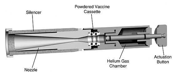
The helium-powered PowderJect device for delivering powdered vaccines has been described11,12. The device used here was a reusable research model 15 cm in length with an ‘actuation’ button, a helium gas chamber, a vaccine cassette, a nozzle and a ‘silencer’ (Fig. 1). The stainless steel gas chamber was filled with approximately 5 ml medical-grade helium gas to a pressure of 50 bar. When the device was activated, the released helium gas ruptured the membranes of the trilaminate cassette and accelerated the entrapped vaccine powders to a high speed so that the particles perforated the stratum corneum and landed in the epidermis. The helium gas was reflected off the skin and was ‘exhausted’ through the vented silencer.
Figure 1: Cross-sectional view of the re-chargeable PowderJect ND device.
Powdered vaccine is housed in the cassette. The release of 5 ml helium gas from the gas chamber propels the vaccine particles from the stationary cassette through the nozzle and into the skin. The device is ready to be used again after the gas chamber is filled with helium and a new vaccine cassette is inserted.
Comparison of EI and needle injection
Although the intramuscular route is commonly used for administering human vaccines, subcutaneous, intraperitoneal and intramuscular routes are often used interchangeably for immunizing experimental animals such as mice. To compare EI with needle injection through these conventional routes, we used a formalin-inactivated Aichi/68 influenza virus to immunize BALB/c mice (n = 8 per group). For EI, the vaccine was formulated with trehalose into a powder with microscopic particles 20–53 μm in diameter. We administered 1 mg powder containing 5 μg vaccine (total viral protein) to the shaved abdominal skin of BALB/c mice using the PowderJect ND device.
There was no bleeding, edema or any other macroscopically detectable local reactions at the vaccination sites immediately after vaccination and up to 7 days after immunization. However, measurement of transepidermal water loss (TEWL) using a TEWL meter (Cortex Technology Aps, Hadsund, Denmark) 30 minutes after vaccination showed increased water evaporation at the vaccination site, an indication of stratum corneum perforation. Erythema was not visually apparent because of the pink color of the mouse skin and the usually mild manifestation of erythema in mice.
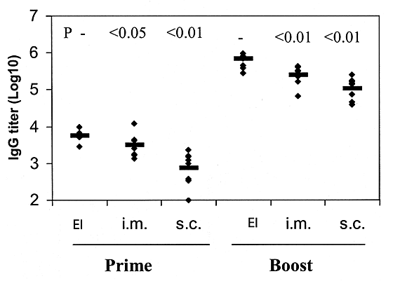
We measured antibody responses using enzyme-linked immunosorbent assay13 (ELISA). After a single immunization, EI elicited a serum immunoglobulin (Ig) G titer to influenza virus that was significantly higher than that induced by either intramuscular injection (P < 0.05, _t_-test) or subcutaneous injection (P < 0.01, _t_-test) (Fig. 2). Boosting by EI resulted in a 12,000% increase in the serum antibody titer (Fig. 2). Comparison of the sera after boosting showed that the antibody titer elicited by EI was approximately 300 and 700% higher than that induced by intramuscular and subcutaneous injection, respectively (P < 0.01, _t_-test).
Figure 2: The immunogenicity of formalin-inactivated Aichi/68 influenza virus administered via various routes.
Each mouse was vaccinated twice on days 0 and 28 with 5 μg vaccine by EI or by intramuscular (i.m) or subcutaneous needle injection (s.c.). Serum antibody titers on days 28 and 42 were determined by ELISA. Data represent IgG titers from individual mice (♦) and the mean titer (-) of eight mice receiving the same treatment. Pre-immunization sera had a titer of less than 200. Above data points (P values), EI compared with needle injection (Student's _t_-test).
Challenge experiments
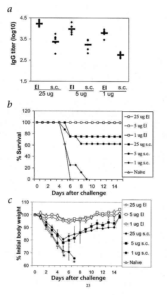
To determine whether EI elicits protective immunity, we immunized mice (n = 8 per group) with the formalin-inactivated Aichi strain of influenza and challenged them with a mouse-adapted homologous strain. The immunization consisted of a single dose of 25, 5, or 1 μg total virus protein administered by either EI or needle injection. We used the subcutaneous route for needle injection because the small musculature in mice could lead to inaccurate intramuscular injection. We measured antibody responses by ELISA 28 days after vaccination. EI elicited antibody titers that were dose-dependent. The IgG titers from the subcutaneous injection were approximately 10% of those after EI at each vaccine dose (Fig. 3_a_; P < 0.001, _t_-test).
Figure 3: Protection in the mouse influenza challenge model.
Mice (n = 8 per group) were immunized with 25, 5 or 1 μg Aichi influenza virus by either EI or subcutaneous needle injection (s.c.). a, Antibody response. Sera were collected 28 d after the immunization and assayed by ELISA. Data represent IgG titers of individual mice (♦) and the mean titer of eight mice (-). P < 0.001, EI compared with subcutaneous injection control for each dose (_t_-test). b, Protection against mortality. Mice were challenged 32 d after immunization with a mouse-adapted Aichi strain. All mice receiving EI (□) of 25, 5 or 1 μg vaccine survived the 21-day monitoring period. P = 0.14, 0.06 and < 0.01, EI compared with subcutaneous injection control for doses of 25, 5 and 1 μg, respectively (JMP survival analysis software, SAS Institute Inc., Cary, North Carolina). c, Protection against weight loss. Data represent mean percent body weight relative to that before challenge. *, less than 50% of the mice alive.
We then challenged mice with 10 minimal lethal doses (LD50) of a mouse-adapted Aichi strain 32 days after vaccination and monitored protection against mortality for 21 days. All eight naive mice died within 7 days of challenge. Delivery of 25, 5 or 1 μg vaccine by EI all conferred 100% protection against mortality, whereas subcutaneous injection of the same three vaccine doses led to only 75%, 62.5% and 0% survival, respectively (Fig. 3_b_).
We also monitored the change in body weight in these mice after challenge. Naive mice lost an average of 30% of their body weight by day 5 before succumbing to the influenza infection between days 5 and 7 (Fig. 3_c_). All mice receiving EI showed a decrease of only 5% or less in body weight during the 21-day monitoring period. The mice immunized subcutaneously, like the non-immunized control mice, lost an average of 20–30% of their body weight 5 days after challenge. At that point, the mice that survived began to regain weight (Fig. 3_c_). Five days after challenge, the body weight of the mice immunized subcutaneously was significantly less than that of mice vaccinated by EI (P < 0.001, _t_-test).
To measure protection against virus replication in the lungs, we immunized additional groups of mice (n = 4 per group) by either subcutaneous injection or EI with 5 or 1 μg influenza vaccine and boosted them on day 28. We challenged these mice with 10 LD50 infectious virus 18 days after the booster immunization, and measured virus titers in the lungs 4 days after challenge by plaque-forming assay14. All four non-immunized control mice had virus in their lungs after challenge, with an average titer of approximately 11± 0.23 × 10⁸ plaque-forming units/gram tissue. All mice receiving EI had no detectable virus in their lungs (>99.9999% clearance), indicating that EI with influenza vaccine led to accelerated clearance of virus from the lungs. Subcutaneous injection using the dose of 5 μg, but not 1 μg, led to a 53% reduction in virus titers compared with that of non-immunized control mice.
EI using trivalent human influenza vaccine
To determine whether EI could be used to deliver commercial human vaccines, we immunized BALB/c mice with the trivalent influenza vaccine manufactured for vaccination during the 1998–1999 season. We vaccinated each mouse with 10% of a human dose (1.5 μg hemagglutinin for each strain) on days 0 and 28, and measured the antibody response on days 28 and 42. We injected control mice subcutaneously with the same dose of aqueous vaccine. We challenged all mice with 10 LD50 mouse-adapted Aichi/68 influenza virus.
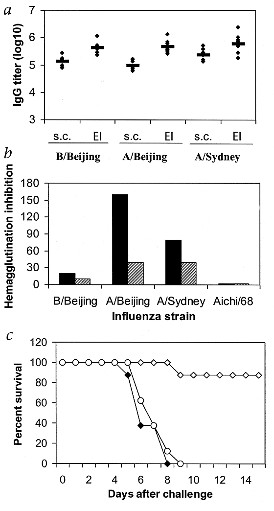
EI elicited serum IgG antibody response to all three strains of the influenza virus measured by ELISA and the titers were significantly higher than those of the subcutaneously injected control mice for each strain (Fig. 4_a_; P < 0.01, _t_-test). The hemagglutination inhibition titer in the pooled sera collected 2 weeks after boost was 200–400% higher for each strain in mice vaccinated by EI than in mice immunized by subcutaneous needle injection (Fig. 4_b_). All the naive mice and mice immunized subcutaneously died as a result of the challenge. Seven of eight mice vaccinated by EI were protected (Fig. 4_c_), although these mice lost up to 20% of their body weight 5 days after challenge (data not shown). Further study demonstrated that this heterologous protection was attributed to immunization by the A/Syndney/5/97 (H3N2) strain of the trivalent vaccine.
Figure 4: Immunogenicity of trivalent human influenza vaccine.
BALB/c mice (n = 8 per group) were vaccinated on days 0 and 28 with 10% of a human dose (1.5 μg hemagglutinin for each strain), and antibody response was determined 14 d after boost. Control mice were injected subcutaneously with the same amount of aqueous vaccine. Mice were challenged 20 d after boost with 10 LD50 Aichi/68 influenza virus. a, Serum IgG titers measured by ELISA. Data represent IgG titers of individual mice (♦) and the mean titers of eight mice (-). P < 0.001, EI compared with subcutaneous injection control (s.c.) for each strain (_t_-test). b, Hemagglutination inhibition titer, in sera pooled from eight mice. Filled bars, EI; grey bars, subcutaneous injection. Sera collected before immunization had titer less than 10. c, Survival after challenge with Aichi/68 influenza strain. ⋄, EI; ♦, subcutaneous injection; ○, naive. P < 0.01, EI compared with naive or subcutaneous injection (JMP survival analysis software).
Given the protection offered by EI against a strain isolated 30 years ago, we next explored its mechanism. Serum against the trivalent vaccine had no hemagglutination inhibition antibodies against the Aichi/68 strain. This is consistent with findings in the literature that hemagglutinin offers little or no protection to strains isolated 10 years apart15,16. Powder delivery did not seem to induce mucosal immunity, as shown by examination of the lung lavage fluid of the mice receiving EI in which only antigen-specific IgG, but not IgA, was demonstrated by ELISA. Protection against mortality but not initial infection from EI, as indicated by the weight loss of the challenged mice, seems to be the same as that afforded by the cytotoxic T cells against the conserved nucleoprotein of the influenza17,18,19. EI may result in a cytotoxic T-cell response by cytosolic deposition of small particles in the Langerhans cells. However, no cytotoxic T-cell activity to a known epitope of the nucleoprotein was detected in mice receiving EI of the formalin-inactivated PR8 strain of the influenza17. Further studies are needed to determine whether EI elicits a cytotoxic T-cell response to other epitopes of the nucleoprotein and other proteins.
Discussion
Here we evaluated a laboratory-purified influenza vaccine and commercial trivalent influenza vaccine for their immunogenicity in mice after EI. EI was effective in inducing a protective immune response in a mouse model. EI of influenza vaccine resulted in protection against mortality, weight loss and virus replication in the lungs when the mice were challenged with a homologous virus. Partial protection against mortality was also demonstrated against challenge with an influenza strain isolated 30 years ago.
The efficacy of EI is likely attributable to targeting vaccines to the immunologically potent epidermis that is richly populated with Langerhans cells7. Langerhans cells have potent antigen-processing and -presentation ability and are important in the initiation and maintenance of immune responses20,21,22. Delivering vaccines in close proximity to the Langerhans cells, sometimes directly to these cells by powder delivery, may facilitate the antigen-recognition and -uptake process.
The efficacy of EI demonstrated here is consistent with results of previous clinical studies of hepatitis B and rabies vaccines. Intradermal injection of these vaccines required only 10% of an intramuscular dose to elicit an equivalent antibody response and seroconversion rate23,24,25. The relative lack of nerve endings in the epidermis will likely make the EI safer than intradermal needle injection25. Unlike other topical application techniques6,8, EI works by forcing particles directly to the target tissue and might be used to deliver most, if not all, vaccines now in use, regardless of their particle size and chemical nature. The safety aspects of the EI (needle-free and pain-free) address the urgent need for a safe method to administer the rapidly growing number of vaccines.
Methods
Vaccines.
Commercial human influenza vaccine used for the 1998–1999 vaccination season was obtained from the Swiss Serum Institute (Bern, Switzerland). It is a split vaccine composed of A/Beijing 262/95-like, A/Sydney 5/97-like and B/Harbin 7/94-like strains. The formalin-inactivated Aichi virus (A/Aichi/68 (H3N2)) was made in our own laboratory as described26.
Vaccine powder formulation.
To prepare vaccines for EI, an appropriate amount of vaccine and trehalose solution (100 mg/ml in deionized water) were combined and plated in a glass Petri dish. The mixture was dried overnight in a desiccator (Nalgene, Rochester, New York) purged with N2 gas. The dried solid was collected, ground with a pestle and mortar, and sieved using stainless steel sieves 3 inches in diameter. Dry powders of the 20- to 53-μm size fraction were loaded in a trilaminate cassette and used to immunize mice. The trilaminate cassette is constructed of a thick ethylene vinyl acetate washer with rupture membrane heat-sealed to each side. The vaccine powders are housed between the rupture membranes. The body of the cassette has an outer diameter of 11 mm, an inner diameter of 6 mm and a height of 4 mm. The rupture membrane is a thin film (20 μm) made of semi-transparent polycarbonate. Each cassette is loaded with 1 mg powder containing 25 μg or less vaccine.
Immunization.
BALB/c mice (female, 6 weeks old; Harlan Sprague Dawley, Indianapolis, Indiana) were used. Mice were fully anesthetized by intraperitoneal injection of ketamine and xylazine, and their abdominal skin was shaved using a hair-clipper before powder delivery. For EI, the PowderJect ND device was placed against the shaved abdominal skin of the mice and the ‘actuation’ button was pressed. Control mice were injected with aqueous vaccine using a 26.5-gauge needle. The subcutaneous injection was given in the scruff of the neck and the intramuscular injection was made in the upper thigh.
References
- Fenner, F. A successful eradication campaign. Global eradication of smallpox. Rev. Infect. Dis. 4, 916–930 (1997).
Article Google Scholar - Breman, J.G. & Arita, I. The confirmation and maintenance of small eradication. N. Engl. J. Med. 303, 1263–1273 (1980).
Article CAS Google Scholar - Fynan, E.F et al. DNA vaccines: Protective immunizations by parenteral, mucosal, and gene-gun inoculations. Proc. Natl. Acad. Sci. USA 90, 11478–11482 (1993).
Article CAS Google Scholar - Swain, W.F. et al. Tolerability and immune responses in humans to a PowderJect DNA vaccine for hepatitis B. Developmental Biology: Development and Clinical Progress of DNA Vaccines (ed. Brown, F., Cichutek, K. Robertson, J.) Vol. 104, 115–119 (Karger, Basel, Switzerland, 2000).
CAS Google Scholar - Bos, J.D., Das, P.K. & Kapsenberg, M.L. in Skin Immune System (SIS) 2nd ed. (ed. Bos, J.D.) 9–15 (CRC, Boca Raton, Florida, 1997).
Google Scholar - Paul, A, Cevc, G. & Bachhawat, B.K. Transdernal immunization with an integral membrane component, gap junction protein, by means of ultradeformable drug carriers, transfersomes. Vaccine 16, 188–195 (1998).
Article CAS Google Scholar - Teunissen M.B.M., Kapsenberg, M.L. & Bos, J.D. in Skin Immune System (SIS) 2nd ed. (ed. Bos, J.D.) 59–75 (CRC, Boca Raton, Florida, 1997).
Google Scholar - Glenn, G.M., Rao, M., Matyas, G.R. & Alving, C.R. Skin immunization made possible by cholera toxin. Nature 391, 851 (1998).
Article CAS Google Scholar - Glenn, G.M. et al. Transcutaneous immunization with cholera toxin protects mice against lethal mucosal toxin challenge. J. Immunol. 161, 3211–3214 (1998).
CAS PubMed Google Scholar - Glenn, G.M., Scharton-Kersten, T., Vassell, R., Matyas, G.R. & Alving, C.R. Transcutaneous immunization with bacterial ADP-ribosylating exotoxin as antigens and adjuvants. Infect. Immun. 67, 1100–1106 (1999).
CAS PubMed PubMed Central Google Scholar - Sarphie, D.F., Varadi, A., Bellhouse, B.J. & Ashcroft, S.J.H. Transdermal delivery of powdered bovine insulin to diabetic and bob-diabetic Wistar rats. Diabetes 44, 129 (1995).
Google Scholar - Sarphie, D.F., Johnson, B., Cormier, M., Burkoth, T.L. & Bellhouse, B.J. Bioavailability following transdermal powdered delivery (TPD) of radiolabeled insulin to hairless guinea pigs. J. Controlled Release 47, 61–69 (1997).
Article CAS Google Scholar - Chen, D. et al. Evaluation of purified UspA from M. catarrhalis as a vaccine in a murine model after active immunization. Infect. Immun. 64, 1900–1905 (1996).
CAS PubMed PubMed Central Google Scholar - Novak, M., Moldoveanu, Z., Schafer, D.P., Mestechy, J. & Compans, R.W. Murine model for evaluation of protective immunity to influenza virus. Vaccine 11, 55–60 (1993).
Article CAS Google Scholar - Rimmelzwaan, G.F. et al. Influenza subtype cross-reactivities of haemagglutionation inhibiting and virus neutralizing serum antibodies induced by infection or vaccination with an ISCOM-based vaccine. Vaccine 17, 2512–2516 (1999).
Article CAS Google Scholar - Saelens, X. et al. Protection of mice against a lethal influenza virus challenge after immunization with yeast-derived secreted influenza virus hemagglutinin. Eur. J. Biochem. 260, 166–175 (1999).
Article CAS Google Scholar - Ulmer, J.B. et al. Heterologous protection against influenza by injection of DNA encoding a viral protein. Science 259, 1745–1749 (1993).
Article CAS Google Scholar - Webster, R.G. & Askonas, B.A. Cross-protection and cross-reactive cytotoxic T cells induced by influenza virus vaccines in mice. Eur. J. Immunol. 10, 396–401 (1980).
Article CAS Google Scholar - Wraith, D.C., Vessey, A.E. & Askonas, B.A. Purified influenza virus nucleoprotein protects mice from lethal infection. J. Gen. Virol. 68, 433–440 (1987).
Article CAS Google Scholar - Flohe, S.B., Bauer, C. & Moll, H. Antigen-pulsed epidermal Langerhans cells protect susceptible mice from infection with the intracellular parasite Leishmania major. Eur. J. Immunol. 28, 3800–3811 (1998).
Article CAS Google Scholar - Condon, C. et al. DNA-based immunization by in vivo transfection of dendritic cells. Nature Med. 2, 1122–1128 (1996).
Article CAS Google Scholar - Becker, Y. et al. HIV-peplotion vaccine. A novel approach to vaccination against AIDS by transepithelial transport of viral peptides to Langerhans cells for induction of cytotoxic T cells by HLA class I and CD1 molecules for long term protection. Adv. Exp. Med. Biol. 397, 97–104 (1996).
Article CAS Google Scholar - Fadda, G. et al. Efficacy of hepatitis B immunization with reduced intradermal doses. Eur. J. Immunol. 3, 176–180 (1987).
CAS Google Scholar - Bernard, K.W. et al. Preexposure immunization with intradermal human diploid cell rabies vaccine. Risk and benefits of primary and booster vaccination. J. Am. Med. Assoc. 257, 1059–1063 (1987).
Article CAS Google Scholar - Horowitz, M.M., Ershler, W.B., McKinney, W.P. & Battiola, R.J. Duration of immunity after hepatitis B vaccination: Efficacy of low-dose booster vaccine. Ann. Intern. Med. 108, 185–189 (1988).
Article CAS Google Scholar - Sheerar, M.G., Easterday, B.C. & Hinshaw, V.S. Antigenic conservation of H1N1 swine influenza viruses. J. Gen. Virol. 70, 3297–3303 (1989).
Article Google Scholar
Acknowledgements
The authors thank J. Haynes, L. Roberts, S. Umlauf and W. Swain for evaluating the manuscript.
Author information
Authors and Affiliations
- PowderJect Vaccines, 585 Science Drive, Madison, 53719, Wisconsin, USA
Dexiang Chen, Ryan L. Endres, Cherie A. Erickson, Kathleen F. Weis & Lendon G. Payne - Department of Pathobiological Sciences, School of Veterinary Medicine, University of Wisconsin, Madison, 53706, Wisconsin, USA
Martha W. McGregor & Yoshihiro Kawaoka
Authors
- Dexiang Chen
You can also search for this author inPubMed Google Scholar - Ryan L. Endres
You can also search for this author inPubMed Google Scholar - Cherie A. Erickson
You can also search for this author inPubMed Google Scholar - Kathleen F. Weis
You can also search for this author inPubMed Google Scholar - Martha W. McGregor
You can also search for this author inPubMed Google Scholar - Yoshihiro Kawaoka
You can also search for this author inPubMed Google Scholar - Lendon G. Payne
You can also search for this author inPubMed Google Scholar
Corresponding author
Correspondence toDexiang Chen.
Rights and permissions
About this article
Cite this article
Chen, D., Endres, R., Erickson, C. et al. Epidermal immunization by a needle-free powder delivery technology: Immunogenicity of influenza vaccine and protection in mice.Nat Med 6, 1187–1190 (2000). https://doi.org/10.1038/80538
- Issue Date: October 2000
- DOI: https://doi.org/10.1038/80538


