High expression of Lin28 is associated with tumour aggressiveness and poor prognosis of patients in oesophagus cancer (original) (raw)
Main
MicroRNAs (miRNAs) are a class of small nonprotein-coding RNAs that act by endogenous interference. They bind to the 3′ untranslated region of the target mRNAs, leading to translational repression or reduced stability of the mRNA (Bartel, 2004). MicroRNAs are known to have important roles in various biological processes, such as cell differentiation, cell proliferation, apoptosis, and metabolism. MicroRNAs also have emerged as central regulators of cancer (Calin and Croce, 2006). Their aberrant expression in many tumours indicates that they could function as tumour suppressors or oncogenes. Moreover, there is increasing evidence that miRNA expression is potentially useful biomarker for the diagnosis, prognosis and tailoring of therapy for patients with various cancers, but the mechanism involved in the regulation of miRNA expression is still uncertain. Recent studies have indicated that miRNA biogenesis can be regulated posttranscriptionally by _trans_-acting factors (Lee et al, 2003; Denli et al, 2004). It is reported that the biogenesis of let-7 family members, which seem to act as tumour suppressor miRNAs, is negatively regulated posttranscriptionally by Lin28 in embryonic stem cells and certain cancer cell lines (Heo et al, 2008; Newman et al, 2008; Piskounova et al, 2008; Viswanathan et al, 2008; Hagan et al, 2009).
Lin28 is a RNA-binding protein originally identified as a key regulator of developmental timing in Caenorhabditis elegans (Moss et al, 1997). Lin28 is a conserved cytoplasmic protein but is exported to the nucleus where it seems to regulate the translation or stability of mRNAs by localising them to P-bodies (Balzer and Moss, 2007). In mammals, Lin28 is widely expressed in embryonic stem cells and in early embryogenesis, but its expression is downregulated with differentiation (Moss and Tang, 2003). Recently, Lin28 was also used with three other factors (OCT4, SOX2, NANOG) to reprogramme human somatic fibroblasts to pluripotency (Yu et al, 2007). This finding suggests that Lin28 is related to stem cell function.
On the other hand, it was recently reported that Lin28 is upregulated in human tumours and functions as an oncogene promoting transformation and tumour progression (Viswanathan et al, 2009). Depletion of Lin28 and expression of let-7 suppressed bone metastasis, while Lin28 expression resulted in bone metastasis in mice implanted with breast tumour cells (Dangi-Garimella et al, 2009). Lin28B protein, a homologue of Lin28, is also overexpressed in hepatocellular carcinoma and induction of expression with exogenous Lin28B promoted cancer cell proliferation (Guo et al, 2006). Lin28B is also induced by Myc and plays an important role of Myc-dependent cellular proliferation (Chang et al, 2009). Thus, altered expression of Lin28 seems to promote tumourigenesis and can be associated with advanced malignancy.
In this study, we analysed the expression of Lin28 and Lin28B, a homologue of Lin28, in oesophageal cancer by immunohistochemistry and determined the relationship between their expression and various clinicopathological parameters including prognosis of patients. Moreover, in in vitro studies, we also examined the relationship between Lin28B expression and aggressiveness of oesophageal cancer cells using oesophageal cell lines.
Patients and Methods
Patients and tissue samples
All 161 tissue samples were obtained from patients who underwent radical oesophagectomy with lymph node dissection for thoracic oesophageal cancers between 2000 and 2006 at the Department of Gastroenterological Surgery, Graduate School of Medicine, Osaka University. Informed consent was obtained from each patient at that of surgery. Of these patients, 94 received preoperative chemotherapy followed by surgery while the remaining 67 patients underwent surgery without preoperative therapy. The preoperative chemotherapeutic regimen was cisplatin at 70 mg m–2, adriamycin at 35 mg m–2 (by rapid intravenous infusion on day 1), and 5-FU at 700 mg m–2 (by continuous intravenous infusion on day 1 through day 7) (Miyata et al, 2009). Two courses of chemotherapy were used, separated by a 4-week interval. The median duration of follow-up was 32.7 months (range, 3.1–97.9 months), and 72 patients (44.7%) died during the follow-up period.
Immunohistochemistry
A staining score of Lin28 and Lin28B was calculated based on the proportion of immunostained cancer cells to that of all cancer cells in three fields of view. The expression level was categorised as high (staining score >50%) or low (staining score ⩽50%).
For immunohistochemistry, 4 _μ_m-thick sections cut from formalin-fixed, paraffin-embedded (FFPE) tissue blocks were deparaffinised and rehydrated using xylene and serial dilutions of ethanol. Antigen retrieval was performed in 1 mmol l–1 sodium citrate buffer (pH 6.0 for Lin28, pH9.0 for Lin28B) by autoclave treatment at 121°C for 15 min, and then the sections were incubated with goat serum for 20 min to block nonspecific binding followed by incubation with the primary polyclonal rabbit antibody; anti-Lin28 (1 : 200 dilution; Proteintech, Chicago, IL, USA) at room temperature for 2 h and anti-Lin28B (1 : 50 dilution; Cell Signaling, Danvers, MA, USA) overnight at 4°C. After incubation with anti-rabbit secondary antibody (Vector Laboratories, Burlingame, CA, USA) for 20 min, the antigen–antibody complexes were visualised using VECTASTAIN ABC kit (Vector Laboratories) according to the protocol supplied by the manufacturer. The sections were counterstained with haematoxylin. All sections were examined independently by two coauthors (R Hamano and H Miyata), who were blinded to the clinical information.
Clinical and histopathological evaluation of response to chemotherapy
The clinical response to chemotherapy was categorised according to the criteria of the World Health Organization response criteria for measurable disease (Miller et al, 1981) and the Japanese Society for Esophageal Diseases (Japan Esophageal Society, 2009b). A complete response was defined as total regression of the primary tumour and its disappearance on CT scan and/or PET scan and endoscopy. A partial response (PR) was defined as >50% reduction in primary tumour size and lymph node metastasis, as confirmed by CT scan. Progressive disease (PD) was defined as >25% increase in the size of the primary tumour or the appearance of new lesions. Cases that did not meet the criteria of PR or PD were defined as stable disease.
For histopathological assessment, serial 4 _μ_m-thick tissue sections of the primary tumour and lymph nodes were cut from the surgical specimens, fixed with 10% buffered formalin, embedded in paraffin, and stained with haematoxylin and eosin. The extent of viable residual carcinoma at the primary site was assessed semiquantitatively, based on the estimated percentage of viable residual carcinoma in relation to the macroscopically identifiable tumour bed that was evaluated histopathologically. Briefly, the percentage of viable residual cancer cells within the total cancerous tissue was assessed as follows: grade 3, no viable residual tumour cells; grade 2, <2/3 residual tumour cells; grade 1b, 1/3–2/3 residual tumour cells; grade 1a, >2/3 residual tumour cells; grade 0, no significant response to chemotherapy (Miyata et al, 2009; Japan Esophageal Society, 2009a).
Cell lines and culture conditions
Five established cell lines derived from oesophageal squamous cell carcinoma (TE-1, -8, -10, -13, -15) were obtained from the Riken Cell Bank (Tsukuba, Japan). All cell lines were cultured in RPMI 1640 (Life Technologies, Gaithersburg, MD, USA) containing 10% fetal bovine serum (Sigma-Aldrich Co., St Louis, MO, USA) and 1% penicillin/streptomycin (Life Technologies Inc.), under a humidified atmosphere with 5% CO2 at 37°C.
Immunoblotting
Adherent cells grown to 50–80% confluence were washed with ice-cold phosphate-buffered saline and lysed in RIPA buffer (Thermo Fisher Scientific Inc., Waltham, MA, USA) or Sample buffer (Wako Pure Chemical Industries, Osaka, Japan) and a cocktail of phosphatase inhibitors (Thermo Fisher Scientific Inc.) on ice. Lysates were spun and the supernatant was collected. Equal amounts of cell extracts (15 _μ_g) were fractionated by SDS–PAGE gel (Bio-Rad Laboratories, Hercules, CA, USA) and transferred onto hydrophobic polyvinylidene difluoride membranes (ImmobilonP, Millipore, Bedford, MA, USA). The membranes were blocked by incubation in 5% milk followed by incubation overnight at 4°C with the primary antibodies, and then with the secondary antibodies for 1 h at room temperature. The following antibodies were used in this study; anti-actin (dilution, 1 : 1000, Sigma-Aldrich Co.), and anti-Lin28B (dilution, 1 : 1000, Abcam, Cambridge, MA, USA). Immune complexes were detected using Detection Kit (GE Healthcare, Buckinghamshire, UK).
Small interfering RNA transfection
Cells were cultured to 60–80% confluence and transfected with 1 _μ_M of small interfering RNAs (siRNAs) that targeted human Lin28B (Si-Lin28B) or negative control oligonucleotides (Applied Biosystems, Foster City, CA, USA) using siPORT NeoFX Transfection Agent (Ambion, Austin, TX, USA) according to the protocol provided by the manufacturer. After transfection, the cells were cultured for 72 h and intermediate samples were collected at 24 and 48 h and analysed by immunoblotting and MTT (3-(4,5-dimethylthiazol-2-yl)-2,5-diphenyltetrazolium bromide) assay.
MTT cell proliferation assay
The MTT assay was used to assess proliferative activity. After siRNA transfection, cells were seeded into 96-well plates at 5 × 10³ per well and incubated overnight under standard culture condition. Following incubation for 24, 48, 72, and 96 h, 10 _μ_l of MTT solution was added to each well and the plates were incubated for another 3 h at 37°C, and formazan crystals were dissolved with 100 _μ_l of 0.04 N HCl-isopropanol. The absorbance of individual wells was read at 550 nm test wavelength and 655 nm reference wavelengths using a microplate reader (Bio-Rad Laboratories). Cell proliferation activities of siRNA transfected cells and negative control transfected cells were determined by absorbance values.
Invasion assay
In vitro cell invasion was assayed using the BioCoat Matrigel Invasion Chambers (Becton Dickinson Biosciences, Sparks, MD, USA) using the procedure recommended by the manufacturer. Briefly, the transfected cells were harvested and placed in the upper chamber (2.5 × 10⁵ cells per well) in serum-free medium. After incubation at 37°C for 48 h to allow invasion of the Matrigel-coated chamber, the invaded cells on the lower surface were fixed and stained using Diff-Quik stain kit (Dade Behring Inc., Newark, DE, USA), whereas the noninvading cells on the upper surface were scraped and washed away. Finally, the number of invaded cells was counted under a microscope in nine random fields ( × 200).
RNA isolation from FFPE specimens
Total RNA was isolated from the FFPE tissue specimens using the RecoverAll Total Nucleic Acid Isolation Kit (Ambion) according to the instructions supplied by the manufacturer. Briefly, each FFPE tissue block was cut into 20-_μ_m thick pieces, and four slices were placed in a centrifuge tube. To liquefy the paraffin, 100% xylene and 100% ethanol was added to each tube. After centrifugation, the precipitated samples were air dried and treated with protease in heat blocks for 3 h at 50°C. Then, each sample was treated with isolation reagent and filtered. Each filter was treated with DNase and incubated for 30 min at room temperature. After washing the filter with washing reagents, it was treated with warmed Elution Solution and centrifuged to pass the mixture through the filter. The eluate contained the isolated RNA.
Quantitative real-time reverse transcription–PCR
The complementary DNA was synthesised from 10 ng of total RNA using the TaqMan miRNA Reverse Transcription Kit and specific stem-loop reverse transcription primers (Applied Biosystems) according to the protocol provided by the manufacturer. The reverse transcription was set at these conditions: 16°C for 30 min followed by 40°C for 30 min and 85°C for 5 min. Real-time PCR reaction was performed using TaqMan Universal PCR master mix No AmpErase UNG and TaqMan miRNA specific PCR primers (Applied Biosystems). A 20 _μ_l of the reaction product was incubated in a 96-well optical plate at 95°C for 10 min, followed by 40 cycles at 95°C for 15 s, and 60°C at 1 min using ABI PRISM 7900HT (Applied Biosystems). The miRNA expression value was expressed relative to that of RNU48 and analysed using the 2−ΔΔCt method (Livak and Schmittgen, 2001; Hamano et al, 2011).
Statistical analysis
All data are expressed as mean±s.d. The relationship between miRNA expression and each clinicopathological variable was analysed by _χ_2 test, Fisher's exact test, or Mann–Whitney _U_-test. Time to recurrence was defined as the time interval between the date of surgery and the date of diagnosis of first recurrence or last date of follow-up if recurrence was not observed. Overall survival time was censored at the date of the last follow-up if death did not occur. For survival analysis, the Kaplan–Meier method was used to assess survival time distribution according to miRNA expression level and the log-rank test was used to examine the differences between groups. A _P_-value of <0.05 denoted the presence of statistically significant difference between groups. All statistical analyses were performed with JMP ver. 8.0 software (SAS Institute Inc., Cary, NC, USA).
Results
High expression of Lin28 and Lin28B in oesophageal cancer cells
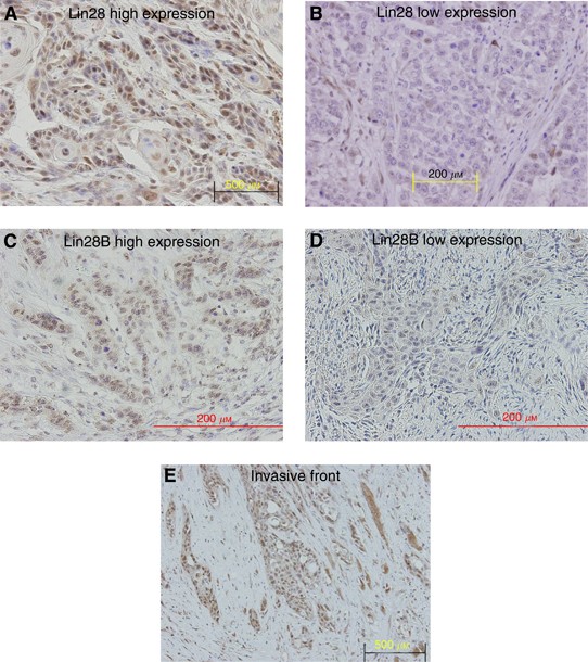
First, we determined the expression pattern of Lin28 and Lin28B in cancerous and non-cancerous tissues from patients with oesophageal cancer. Lin28 staining was predominantly detected in the nuclei of cancer cells, though cytoplasmic staining was also evident in some cancer cells (Figure 1A and B). Lin28B staining was also predominantly detected in the nuclei of cancer cells (Figure 1C and D). This finding is in agreement with the results of a previous study of Lin28 expression in the cytoplasm and its transportation to the nucleus (Balzer and Moss, 2007). The carcinoma cells at the invasive front tended show strong staining for Lin28 and Lin28B compared with those in other areas (Figure 1E). On the other hand, in non-cancerous tissue, differentiated cells were not stained although the cytoplasm of some basal cells was stained weakly for Lin28 and Lin28B.
Figure 1
Lin28 and Lin28B immunostaining in oesophageal cancer. (A) Lin28 positively stained cells. (B) Lin28 negatively stained cells. (C) Lin28B positively stained cells. (D) Lin28B negatively stained cells. (E) The carcinoma cells at the invasive front.
High expression of Lin28 is associated with poor prognosis
Tables 1 and 2 summarise the relationship between Lin28 and Lin28B expression and various clinicopathological parameters. There were no significant relationships between Lin28 expression and tumour differentiation or tumour depth. However, high expression of Lin28 correlated significantly with lymph node metastasis (_P_=0.035) and lymphatic vessel invasion (_P_=0.047). Similar to Lin28, high expression of Lin28B correlated significantly with lymph node metastasis (_P_=0.017) and lymphatic vessel invasion (_P_=0.002). Moreover, the expression of Lin28B significantly correlated with tumour depth (_P_=0.005).
Table 1 Correlation between Lin28 expression and various clinicopathological features of patients with oesophageal cancer
Table 2 Correlation between Lin28B expression and various clinicopathological features of patients with oesophageal cancer
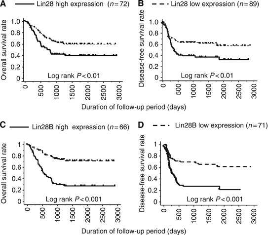
High expression of Lin28 correlated significantly with shortened survival including both overall survival and disease-free survival (Figure 2A and B). High expression of Lin28B also correlated significantly with shortened survival (Figure 2C and D). Furthermore, multivariate analysis identified Lin28B expression as an independent prognostic factor, along with the number of metastatic lymph nodes (Table 3). These results suggest that Lin28 and Lin28B may influence the malignant potential of oesophageal cancer. However, Lin28 expression did not correlate with clinical and pathological responses to preoperative chemotherapy (Table 4).
Figure 2
Correlation between Lin28/Lin28B expression and survival of 161 patients with oesophageal cancer. High expression of Lin 28 (A and B)/Lin28B (C and D) correlated with shortened survivals.
Table 3 Results of univariate and multivariate Cox's models for disease-free survival
Table 4 Relationship between Lin28 expression and response to preoperative chemotherapy in patients with oesophageal cancer
Inhibition of Lin28B expression regulates cellular behaviour
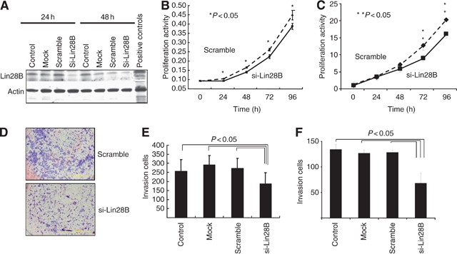
The above results suggest that Lin28 and Lin28B are potentially associated with aggressiveness of oesophageal cancer. In the next series of studies, in vitro experiments were conducted to examine the effect of these expressions on the malignant potential of oesophageal cancer cells. First, we screened several oesophageal cancer cell lines, and found that some cell lines express Lin28B, while expression of Lin28 is quite low in all cell lines examined (data not shown). Thus, to study the effects of expression of Lin28B on cellular proliferation, its expression was knocked down by transfecting si-Lin28B in TE-13 oesophageal cancer cells (Figure 3A). The proliferative activity of Lin28B-knockdown cells was significantly reduced compared with that of control cells (Figure 3B). Second, the invasion assay was conducted to assess the role of Lin28B in lymph node metastasis by invasion to lymphatics. The invasive activity of Lin28B-knockdown cells was clearly reduced compared with that of negative control cells (Figure 3D and E). In another oesophageal cancer cell line, TE-10, the reduced proliferation and invasive activity of Lin28B-knockdown cells were confirmed (Figure 3C and F).
Figure 3
Proliferative and invasive activities of Lin28B-knockdown cells. (A) Western blotting to confirm reduced Lin28B expression following transfection of si-Lin28B in TE-13. (B) Proliferative activities of Lin28B-knockdown cells and control cells in TE-13. (C) Proliferative activities of Lin28B-knockdown cells and control cells in TE-10. (D) Invasive activities of Lin28B-knockdown cells and control cells in TE-13. (E) Quantitative analysis of invasive activity in TE-13 (data are mean±s.d. of three experiments). (F) Quantitative analysis of invasive activity in TE-10 (data are mean±s.d. of three experiments).
Relationship between Lin28 expression and let-7 expression
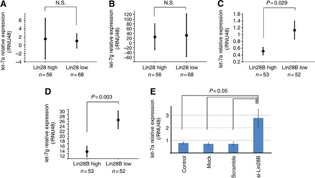
Lin28 is described as a negative regulator of let-7 biogenesis (Heo et al, 2008; Viswanathan et al, 2008). We investigated the relationship between expression of Lin28 and Lin28B and let-7 expression. We found significant relationship between let-7 expression and Lin28B expression, but not Lin28 expression in the surgical specimens of oesophageal cancer (Figure 4A–D). In vitro assay showed that let-7 was upregulated in cultured Lin28B-knockdown oesophageal cancer cells, compared with control cells (Figure 4E). This finding is consistent with the results of the previous study showing a relationship between Lin28B and let-7 (King et al, 2011).
Figure 4
Relationship between Lin28B expression and let-7 expression in oesophageal cancer. (A and B) Analysis of surgical specimens showed no significant relationship between Lin28 expression and let-7 expression, determined by real-time RT–PCR. (C and D) Analysis of surgical specimens showed significantly relationship between Lin28B expression and let-7 expression, determined by real-time RT–PCR. (E) In vitro assay using oesophageal cancer cell showed upregulation of let-7 expression in Lin28B-knockdown cells, compared with control cells.
Discussion
Lin28 is a negative regulator of let-7 family, which may act as a tumour suppressor miRNA, suggesting that Lin28 could contribute to tumourigenesis. The present study demonstrated that high expression of Lin28 and Lin28B is associated with lymph node metastasis and poor prognosis of patients with oesophageal cancers. In vitro studies confirmed that Lin28B expression was associated with aggressiveness of oesophageal cancer through increased proliferation and invasive activities in oesophageal cancer cells.
Recent studies suggest that Lin28 functions as an oncogene promoting malignant transformation and tumour progression (Viswanathan et al, 2009). Indeed, several recent reports demonstrated that Lin28 expression correlates with survival of patients with malignant diseases (Guo et al, 2006). In ovarian cancer, patients with high Lin28B expression had shorter progression-free and overall survival times than those with low Lin28B expression (Lu et al, 2009). In another recent report, high Lin28B staining intensity in stage I/II colon cancers correlated with reduced survival and increased probability of tumour recurrence (King et al, 2011). Our result of the correlation between high expression of Lin28 and Lin28B and poor prognosis of patients with oesophageal cancers is compatible with the above studies. Thus, Lin28 expression may be clinically relevant prognostic marker in various malignancies including oesophageal cancer.
The present study demonstrated that Lin28 expression is associated with tumour aggressiveness through increased proliferation of oesophageal cancer cells. One recent study demonstrated that Lin28B is necessary and sufficient for Myc-mediated let-7 repression, and that Lin28B has an important role in Myc-dependent cellular proliferation (Chang et al, 2009). Another study showed that high expression of Lin28 and Lin28B correlated with low let-7 expression and upregulation of let-7 target such as HMGA2 and Myc, and that knockdown of Lin28B expression impaired cellular proliferation (Viswanathan et al, 2009). Thus, Lin28 may increase proliferation of oesophageal cancer cells by directly inhibiting let-7 expression and subsequently upregulating HMGA2 and Myc, which are targets of let-7.
In this study, high expression of Lin28 correlated significantly with lymph node metastasis in patients with oesophageal cancers. A recent study showed that Raf kinase inhibitory protein repressed breast tumour cell intravasation and bone metastasis in a mouse model, through inhibition of mitogen-activated protein kinase, leading to decreased transcription of Lin28 and enhanced expression of let-7 by Myc (Dangi-Garimella et al, 2009). These data provided the first evidence for the roles of Lin28 and let-7 expression in tumour metastasis, in addition to the regulation of tumour growth. Another recent study demonstrated that constitutive expression of Lin28B expression in colon cancer cells confers metastatic ability by showing that mice xenografted with Lin28B expressing colon cancer cells developed much more metastasis in the liver, lung, and mesenterium compared with mice of the empty vector control group (King et al, 2011). Thus, Lin28 expression may have an important role in cancer metastasis. In the present study, Lin28B expression was significantly associated with the invasive activity of oesophageal cancer cells. The result that high expression of Lin28 correlated with lymph node metastasis in oesophageal cancer may depend on the increased invasiveness through Lin28 expression.
Several recent studies have identified the important roles of Nanog, Sox2 and Oct3/4, which are involved in reprogramming and maintenance of stem cell function, in tumour aggressiveness (Ben-Porath et al, 2008). Immunohistochemical analysis of gastric cancer (Lin et al, 2011) and colorectal cancer (Meng et al, 2010) showed that overexpression of Nanog correlated strongly with lymph node metastasis and poor prognosis of patients. In hepatocellular carcinoma, Sox2 and Oct4 were identified as independent prognostic factors with poorest prognosis in patients with tumours that co-expressed Sox2/Oct4 proteins (Huang et al, 2011). In oesophageal cancer, the expression of Oct3/4 and Sox2 proteins was reported to correlate with advanced cancer, which in turn correlated with poor clinical outcome (Wang et al, 2009). In our study, Lin28, a reprogramming factor, was associated with tumour aggressiveness and poor prognosis of patients with oesophageal cancer. Thus, reprogramming factors, which regulates stem cell like properties such as pluripotency and self-renewal in normal cells, may confer the high malignant potential of cancer cells.
In the present study, Lin28B expression correlated inversely with let-7 expression in oesophageal cancer cell line and human oesophageal cancers, although no such correlation was identified between Lin28 and let-7 expression. Several studies have indicated that Lin28/Lin28B is part of the regulatory network that also involves let-7. Lin28/Lin28B represses let-7, which itself represses Lin28/Lin28B by binding to the 3′UTR of Lin28/Lin28B transcripts, thus forming a double-negative feedback loop. A second feedback loop is that Lin28/Lin28B de-represses c-Myc by inhibiting let-7, and c-Myc transcriptionally activates Lin28/Lin28B (Chang et al, 2009; Dangi-Garimella et al, 2009). A third feedback loop involves NF-_κ_B, Lin28B, let-7 and IL-6 (Iliopoulos et al, 2009). NF-_κ_B induces Lin28B expression, leading to inhibition of let-7 and expression of the encoding IL-6 (a let-7 target). IL-6 can itself activate NF-_κ_B, resulting in a positive feedback loop. Thus, Lin28 and let-7 may form a complex feedback loop in malignant transformation. Moreover, one recent study showed that Lin28 and Lin28B function through distinct mechanisms to block let-7 processing (Piskounova et al, 2011). Further studies are required to elucidate the roles of Lin28/Lin28B and let-7 network in oesophageal cancers.
In summary, we examined in the present study the clinical significance of Lin28 and Lin28B expression in oesophageal cancer and demonstrated that high expressions of Lin28 and Lin28B are associated with lymph node metastasis and poor prognosis of patients with oesophageal cancers. Moreover, in vitro studies confirmed that Lin28B expression was associated with aggressiveness of oesophageal cancer by increasing the proliferation and invasiveness of oesophageal cancer cells. Further studies are needed to confirm the role of feedback loops including Lin28/Lin28B and let-7 in oesophageal cancer.
Change history
23 January 2013
This paper was modified 12 months after initial publication to switch to Creative Commons licence terms, as noted at publication
References
- Balzer E, Moss EG (2007) Localization of the developmental timing regulator Lin28 to mRNP complexes, P-bodies and stress granules. RNA Biol 4: 16–25
Article CAS Google Scholar - Bartel DP (2004) MicroRNAs: genomics, biogenesis, mechanism, and function. Cell 116: 281–297
Article CAS Google Scholar - Ben-Porath I, Thomson MW, Carey VJ, Ge R, Bell GW, Regev A, Weinberg RA (2008) An embryonic stem cell-like gene expression signature in poorly differentiated aggressive human tumors. Nat Genet 40: 499–507
Article CAS Google Scholar - Calin GA, Croce CM (2006) MicroRNA signatures in human cancers. Nat Rev Cancer 6: 857–866
Article CAS Google Scholar - Chang TC, Zeitels LR, Hwang HW, Chivukula RR, Wentzel EA, Dews M, Jung J, Gao P, Dang CV, Beer MA, Thomas-Tikhonenko A, Mendell JT (2009) Lin-28B transactivation is necessary for Myc-mediated let-7 repression and proliferation. Proc Natl Acad Sci USA 106: 3384–3389
Article CAS Google Scholar - Dangi-Garimella S, Yun J, Eves EM, Newman M, Erkeland SJ, Hammond SM, Minn AJ, Rosner MR (2009) Raf kinase inhibitory protein suppresses a metastasis signalling cascade involving LIN28 and let-7. EMBO J 28: 347–358
Article CAS Google Scholar - Denli AM, Tops BB, Plasterk RH, Ketting RF, Hannon GJ (2004) Processing of primary microRNAs by the microprocessor complex. Nature 432: 231–235
Article CAS Google Scholar - Guo Y, Chen Y, Ito H, Watanabe A, Ge X, Kodama T, Aburatani H (2006) Identification and characterization of lin-28 homolog B (LIN28B) in human hepatocellular carcinoma. Gene 384: 51–61
Article CAS Google Scholar - Hagan JP, Piskounova E, Gregory RI (2009) Lin28 recruits the TUTase Zcchc11 to inhibit let-7 maturation in mouse embryonic stem cells. Nat Struct Mol Biol 16: 1021–1025
Article CAS Google Scholar - Hamano R, Miyata H, Yamasaki M, Kurokawa Y, Hara J, Moon JH, Nakajima K, Takiguchi S, Fujiwara Y, Mori M, Doki Y (2011) Overexpression of miR-200c induces chemoresistance in esophageal cancers mediated through activation of the Akt signaling pathway. Clin Cancer Res 17: 3029–3038
Article CAS Google Scholar - Heo I, Joo C, Cho J, Ha M, Han J, Kim VN (2008) Lin28 mediates the terminal uridylation of let-7 precursor MicroRNA. Mol Cell 32: 276–284
Article CAS Google Scholar - Huang P, Qiu J, Li B, Hong J, Lu C, Wang L, Wang J, Hu Y, Jia W, Yuan Y (2011) Role of Sox2 and Oct4 in predicting survival of hepatocellular carcinoma patients after hepatectomy. Clin Biochem 44: 582–589
Article CAS Google Scholar - Iliopoulos D, Hirsch HA, Struhl K (2009) An epigenetic switch involving NF-kappaB, Lin28, Let-7 MicroRNA, and IL6 links inflammation to cell transformation. Cell 139: 693–706
Article CAS Google Scholar - Japan Esophageal Society (2009a) Japanese Classification of Esophageal Cancer, tenth edition: part I. Esophagus 6: 1–25
Article Google Scholar - Japan Esophageal Society (2009b) Japanese Classification of Esophageal Cancer, tenth edition: part II and III. Esophagus 6: 71–95
Article Google Scholar - King CE, Cuatrecasas M, Castells A, Sepulveda A, Lee JS, Rustgi AK (2011) Lin28b promotes colon cancer progression and metastasis. Cancer Res 71: 4260–4268
Article CAS Google Scholar - Lee Y, Ahn C, Han J, Choi H, Kim J, Yim J, Lee J, Provost P, Rådmark O, Kim S, Kim VN (2003) The nuclear RNase III Drosha initiates microRNA processing. Nature 425: 415–419
Article CAS Google Scholar - Lin T, Ding YQ, Li JM (2011) Overexpression of Nanog protein is associated with poor prognosis in gastric adenocarcinoma. Med Oncol (in press)
- Livak KJ, Schmittgen TD (2001) Analysis of relative gene expression data using real-time quantitative PCR and the 2(-Delta Delta C(T)) method. Methods 25: 402–408
Article CAS Google Scholar - Lu L, Katsaros D, Shaverdashvili K, Qian B, Wu Y, de la Longrais IA, Preti M, Menato G, Yu H (2009) Pluripotent factor lin-28 and its homologue lin-28b in epithelial ovarian cancer and their associations with disease outcomes and expression of let-7a and IGF-II. Eur J Cancer 45: 2212–2218
Article CAS Google Scholar - Meng HM, Zheng P, Wang XY, Liu C, Sui HM, Wu SJ, Zhou J, Ding YQ, Li JM (2010) Overexpression of nanog predicts tumor progression and poor prognosis in colorectal cancer. Cancer Biol Ther 9 (in press)
Article CAS Google Scholar - Miller AB, Hoogstraten B, Staquet M, Winkler A (1981) Reporting results of cancer treatment. Cancer 47: 207–214
Article CAS Google Scholar - Miyata H, Yoshioka A, Yamasaki M, Nushijima Y, Takiguchi S, Fujiwara Y, Nishida T, Mano M, Mori M, Doki Y (2009) Tumor budding in tumor invasive front predicts prognosis and survival of patients with esophageal squamous cell carcinomas receiving neoadjuvant chemotherapy. Cancer 115: 3324–3334
Article Google Scholar - Moss EG, Lee RC, Ambros V (1997) The cold shock domain protein LIN-28 controls developmental timing in C. elegans and is regulated by the lin-4 RNA. Cell 88: 637–646
Article CAS Google Scholar - Moss EG, Tang L (2003) Conservation of the heterochronic regulator Lin-28, its developmental expression and microRNA complementary sites. Dev Biol 258: 432–442
Article CAS Google Scholar - Newman MA, Thomson JM, Hammond SM (2008) Lin-28 interaction with the Let-7 precursor loop mediates regulated microRNA processing. RNA 14: 1539–1549
Article CAS Google Scholar - Piskounova E, Polytarchou C, Thornton JE, Lapierre RJ, Pothoulakis C, Hagan JP, Iliopoulos D, Gregory RI (2011) Lin28A and Lin28B inhibit let-7 MicroRNA biogenesis by distinct mechanisms. Cell 147: 1066–1079
Article CAS Google Scholar - Piskounova E, Viswanathan SR, Janas M, LaPierre RJ, Daley GQ, Sliz P, Gregory RI (2008) Determinants of microRNA processing inhibition by the developmentally regulated RNA-binding protein Lin28. J Biol Chem 283: 21310–21314
Article CAS Google Scholar - Viswanathan SR, Daley GQ, Gregory RI (2008) Selective blockade of microRNA processing by Lin28. Science 320: 97–100
Article CAS Google Scholar - Viswanathan SR, Powers JT, Einhorn W (2009) Lin28 promotes transformation and is associated with advanced human malignancies. Nat Genet 41: 843–848
Article CAS Google Scholar - Wang Q, He W, Lu C, Wang Z, Wang J, Giercksky KE, Nesland JM, Suo Z (2009) Oct3/4 and Sox2 are significantly associated with an unfavorable clinical outcome in human esophageal squamous cell carcinoma. Anticancer Res 29: 1233–1241
CAS PubMed Google Scholar - Yu J, Vodyanik MA, Smuga-Otto K, Antosiewicz-Bourget J, Frane JL, Tian S, Nie J, Jonsdottir GA, Ruotti V, Stewart R, Slukvin II, Thomson JA (2007) Induced pluripotent stem cell lines derived from human somatic cells. Science 318: 1917–1920
Article CAS Google Scholar
Author information
Authors and Affiliations
- Department of Gastroenterological Surgery, Osaka University Graduate School of Medicine, Suita, Yamadaoka 2-2, Osaka, 565-0871, Japan
R Hamano, H Miyata, M Yamasaki, K Sugimura, K Tanaka, Y Kurokawa, K Nakajima, S Takiguchi, Y Fujiwara, M Mori & Y Doki
Authors
- R Hamano
You can also search for this author inPubMed Google Scholar - H Miyata
You can also search for this author inPubMed Google Scholar - M Yamasaki
You can also search for this author inPubMed Google Scholar - K Sugimura
You can also search for this author inPubMed Google Scholar - K Tanaka
You can also search for this author inPubMed Google Scholar - Y Kurokawa
You can also search for this author inPubMed Google Scholar - K Nakajima
You can also search for this author inPubMed Google Scholar - S Takiguchi
You can also search for this author inPubMed Google Scholar - Y Fujiwara
You can also search for this author inPubMed Google Scholar - M Mori
You can also search for this author inPubMed Google Scholar - Y Doki
You can also search for this author inPubMed Google Scholar
Corresponding author
Correspondence toH Miyata.
Additional information
This work is published under the standard license to publish agreement. After 12 months the work will become freely available and the license terms will switch to a Creative Commons Attribution-NonCommercial-Share Alike 3.0 Unported License.
Rights and permissions
From twelve months after its original publication, this work is licensed under the Creative Commons Attribution-NonCommercial-Share Alike 3.0 Unported License. To view a copy of this license, visit http://creativecommons.org/licenses/by-nc-sa/3.0/
About this article
Cite this article
Hamano, R., Miyata, H., Yamasaki, M. et al. High expression of Lin28 is associated with tumour aggressiveness and poor prognosis of patients in oesophagus cancer.Br J Cancer 106, 1415–1423 (2012). https://doi.org/10.1038/bjc.2012.90
- Received: 12 October 2011
- Revised: 23 January 2012
- Accepted: 23 February 2012
- Published: 20 March 2012
- Issue Date: 10 April 2012
- DOI: https://doi.org/10.1038/bjc.2012.90


