Exploiting the defensive sugars of HIV-1 for drug and vaccine design (original) (raw)
A remarkable feature of the human immunodeficiency virus (HIV) is the dense carbohydrate (glycan) array that surrounds the exposed envelope antigens. However, the HIV genome encodes no gene products capable of synthesizing carbohydrates: its surface antigens are glycosylated entirely by host cellular enzymes. This extensive glycosylation is known to affect almost every aspect of virus biology. The folding of viral glycoproteins, the transmission of the virus and the nature of the immune response to infection are all profoundly affected by this glycosylation.
The coating of HIV with immunologically 'self' glycans (that is, those synthesized by the host cell) has a predictable effect on viral immunogenicity: antibodies against most of the available antigenic surface of HIV do not normally occur. Here we review the apparently contradictory roles of HIV glycans as both powerful adaptations for virus survival and targets for therapeutic intervention. Our understanding of the extraordinary extent to which HIV relies on the human glycosylation pathway has exposed new vulnerabilities that are now the target of both drug and vaccine design.
Structure and selection of viral carbohydrates
Synthesis and structure of gp120/gp41 carbohydrates
The types of glycan found on HIV are determined by the interactions between the three-dimensional structure of the envelope proteins and the biosynthetic environment of the cell that the virus has infected1. However, the locations of N-linked attachment sites2 (Asn-X-Ser/Thr-X, where X is not proline) and O-linked attachment sites3 are directly encoded by the viral genome. Importantly, the positioning of N-linked glycans is well conserved between both isolates and clades, particularly when compared with the high degree of variability within the viral envelope4,5.
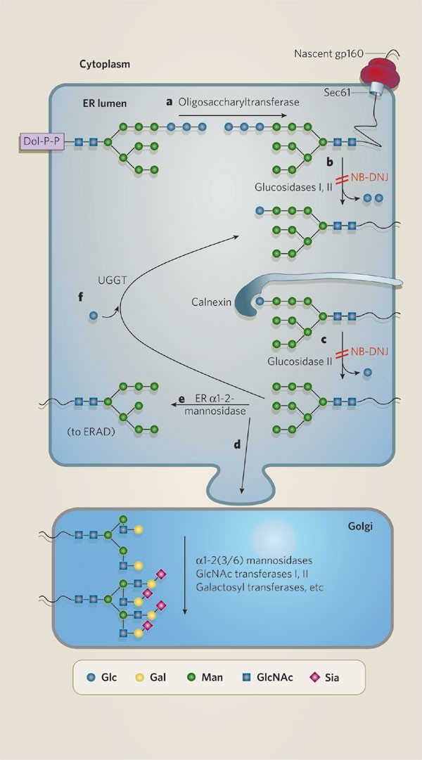
The envelope gene is translated as gp160 and later cleaved by furin proteases into gp120 and gp41. The nascent gp160 is transported by Sec61 to the endoplasmic reticulum, where N-linked glycan precursors (Glc3Man9GlcNAc2) are transferred co-translationally to the amide groups of asparagine residues. The first and second glucose residues of Glc3Man9GlcNAc2 glycans are removed by glucosidases I and II. Monoglucosylated gp160 is a substrate for the chaperones calnexin and calreticulin. While bound to calnexin/calreticulin, gp160 (re)folds through interactions with other endoplasmic reticulum chaperones and disulphide isomerases such as ERp-57. The hydrolysis of the final glucose–mannose (Glcα1-2Man) bond by glucosidase II frees gp160 from calnexin/calreticulin and allows it to exit from the endoplasmic reticulum. Misfolded glycoproteins are reglucosylated by the 'folding sensor' UDP-glucose glucosyltransferase and can rebind to calnexin/calreticulin for further refolding cycles. After release from calnexin/calreticulin, the oligomannose-bearing glycoprotein exits from the endoplasmic reticulum and moves into the Golgi, where further mannosidase trimming to Man5GlcNAc2 initiates the synthesis of complex-type glycans (Fig. 1). However, buried glycans are protected from the processing that normally occurs in the endoplasmic reticulum/Golgi, meaning that they remain as 'immature' precursor glycans6,7. In the case of gp160, the steric occlusion of closely spaced N-linked carbohydrates means that less than 50% of the envelope glycans exiting from the Golgi are properly processed8,9. A full discussion of N-linked glycosylation is beyond the scope of this review; however, the conserved functional roles of N-linked glycans in the endoplasmic reticulum10 and the diversification of these structures in the Golgi11 have been well reviewed elsewhere.
Figure 1: HIV gp160 N-linked glycosylation.
Glc3Man9GlcNAc2 is transferred from dolichol pyrophosphate (Dol-P-P) to nascent polypeptides entering the endoplasmic reticulum (ER) through Sec61 (a), and this transfer is mediated by oligo-saccharyltransferase, which recognizes N-linked glycosylation sequences (Asn-X-Ser/Thr-X) in gp160. After removal of two glucose units (b), the monoglucosylated glycan (Glc1Man9GlcNAc2) binds to calnexin or calreticulin and promotes glycoprotein folding. Hydrolysis of the final glucose–mannose bond by glucosidase II frees gp160 from calnexin (c), and it can then exit from the ER (d) and enter the Golgi, where glycoproteins undergo further processing to become complex-type glycans. Glycoproteins with prolonged residence in the ER are eventually subject to trimming by ER α1-2-mannosidase (e), which removes a terminal mannose residue from the 'middle' branch (forming Man8(B)GlcNAc2), a signal for ER-associated degradation (ERAD). Misfolded glycoproteins are reglucosylated (f) by UDP-glucose glucosyl transferase (UGGT) and can then rebind calnexin for another cycle of refolding. Inhibitors of the pathway, such as NB-DNJ, can prevent formation of N-glycan intermediate structures that are crucial for the correct folding of the glycoprotein.
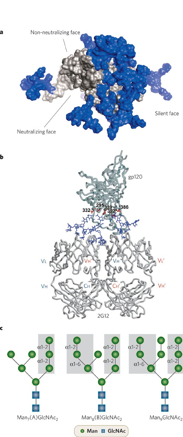
Most of the viral envelope surface is covered by carbohydrates (Fig. 2a). Structural knowledge of HIV gp120 has revealed that within this glycan shield oligomannose glycans are predominantly found in a cluster away from the receptor-binding sites and trimer interface8 (Fig. 2b, c). This distribution fits well with the 'antigenic map' of gp120 (refs 8, 12,13,14), which divides the glycoprotein into three regions: the neutralizing face, the non-neutralizing face and the heavily glycosylated silent face (Fig. 3a). Interestingly, sites with oligomannose glycans are more strongly conserved than those with complex-type carbohydrates4 and are located predominantly on the silent face. A potential explanation for the conservation of the oligomannose glycoforms is the interaction of gp120 with host mannose-specific lectins15 (as discussed below).
Figure 2: The carbohydrate shield of gp120.
a, Electron micrograph of HIV-1. Carbohydrates are stained with ruthenium red (dark), showing extensive occlusion of the antigenic surface by host-derived carbohydrates. The outer domain of gp120 is particularly rich in conserved glycosylation sites. (Image courtesy of CDC public image database.) b, The glycosylation surface of gp120 can therefore be roughly partitioned into two regions: the oligomannose glycans (green), found on the densely glycosylated outer domain, and the complex sugars (red), which are distributed on the more exposed receptor-binding sites and hypervariable loops8. The site-specific analysis of monomeric gp120 used to provide these data may not fully reflect the occlusion that occurs within a trimer, so the abundance of oligomannose glycans is likely to exceed that of the related monomer. (Molecular model of gp120 based on crystal structure of CD4-liganded core14,97, with perspective shown being that from the viral membrane. Model courtesy of M. Wormald.) c, Representation of the structure of the oligomannose glycan Man9GlcNAc2, showing the structure, glycosidic linkages and identity of the D1, D2 and D3 termini of the A, B and C arms.
Figure 3: Antigenicity and glycosylation of HIV gp120.
a, Antigenic map of monomeric HIV gp120, based on the crystal structure of gp120 (ref. 13). The neutralizing face contains the receptor-binding sites, and the non-neutralizing face contains epitopes that are exposed on free, monomeric gp120 but are hidden by adjacent gp120/gp41 subunits on the functional, trimeric form of gp120/gp41 (ref. 98). The silent face is heavily glycosylated (with N-linked glycans (blue)) and, with the known exception of the 2G12 epitope, immunosilent. b, Interaction between gp120 and IgG 2G12. The domain-exchanged configuration of 2G12 results from a non-covalent interaction between two heavy chains (red and blue) that provides an extended paratope capable of high-affinity (multivalent) interaction with gp120 sugars81. The positioning of the reducing termini of the 2G12-bound glycans is consistent with mutagenesis data that indicate that the oligomannose glycans at Asn 332 and Asn 392 of gp120 are critical for 2G12 binding. (Model of 2G12–gp120 complex reproduced, with permission, from ref. 81.) c, The Manα1-2Manα1-2Man and Manα1-2Manα1-6Man motifs recognized by 2G12 (refs 80, 99; grey shading) are found on gp120's main oligomannose glycoforms, thus minimizing the effective microheterogeneity of these glycans.
An unusual feature of gp120 is the degree to which sugar–sugar interactions are formed between neighbouring glycans16,17. For most glycoproteins, N-linked carbohydrates exhibit considerable conformational freedom beyond that of the protein18. However, the glycans of HIV are constrained within tight clusters16,17. Unusually extensive electron density is visible for many of the carbohydrates present in the crystal structure of gp120SIV, from the closely related simian immunodeficiency virus, indicating a rigid carbohydrate field stabilized by a sugar–sugar hydrogen-bond network16,17. Although not unique to HIV (for example, ordered oligomannose glycans have also been observed on the envelope glycoprotein of the Epstein–Barr virus19), this atypical clustering has direct functional, immunological and therapeutic consequences.
Biological roles of HIV glycans
Although HIV is generally specific for CD4+ cells, the type of secondary co-receptor that is bound by gp120 defines the exact tropism of the virus. HIV specific for the chemokine co-receptor CCR5 (R5 viruses) is found during early infection in most individuals, and primarily infects circulating, activated and memory CCR5+ T lymphocytes and macrophages. CXCR4-specific HIV (X4 viruses) infect a wider range of CD4+ cells, including naive T cells20. There is a direct link between glycosylation and viral tropism: a change in tropism from CCR5 to CXCR4 is closely connected with changes in N-linked glycosylation within the extended variable loops (V1/V2 and V3) of gp120 (refs 20, 21). However, beyond the positioning of glycans, it has also been established that many of the specific carbohydrate motifs found on HIV gp120 have immunological properties. For example, α2-6-linked sialic acids, which are characteristic of glycoproteins from CD4+ T cells, are known ligands for CD22, an immunosuppressive lectin found on B cells22. Bisecting _N-_acetylglucosamine residues, found on gp120 from T-cell lines23, can limit natural-killer-cell function. However, the extent to which the types of carbohydrate on HIV have important roles in vivo requires further study. The use of gp120 from infected cells rather than cultured cell lines with unrepresentative complex glycosylation will be crucial to the success of such studies.
Given that much of the antigenic surface of gp120 is mannosylated (Fig. 2), we might expect gp120 to be readily neutralized by the mannose-specific lectins of the innate immune system — notably, mannose-binding lectin (MBL). However, although MBL interacts with gp120 in vitro, and has been shown to neutralize laboratory-adapted strains of HIV, it only weakly neutralizes primary isolates24,25. Indeed, rather than serving as a target for the innate immune system, it seems that HIV uses host lectins for survival. For example, the oligomannose carbohydrates of gp120 bind to C-type lectins such as the mannose receptor, langerin and DC-SIGN (dendritic-cell-specific intercellular adhesion molecule-3-grabbing non-integrin)26,27. These lectins are expressed on dendritic cell subsets, including those located in (sub)mucosal tissue. It has been proposed that DC-SIGN+ cells (and perhaps other C-type lectin-expressing cells such as macrophages28) sequester HIV-1 particles by means of a non-infectious pathway, and subsequently present the virus to T cells across an immunological synapse. This is known as trans infection. However, the biological significance of trans infection is controversial, with recent evidence indicating that direct cis infection of dendritic-cell subsets accounts for long-term transfer of the virus to T cells29. This is consistent with evidence indicating that the productive infection of dendritic cells by HIV-1 is dependent on, or at least significantly enhanced by, the presence of DC-SIGN30. Other lectins are also present on these cells and DC-SIGN-independent transmission of HIV from dendritic cells to T cells has been documented28,31. Nonetheless, DC-SIGN/lectin-mediated early infection, whether cis or trans, represents an attractive target for antimicrobial intervention (as discussed below).
Immune tolerance
The antigenic surfaces of viruses, prokaryotes and eukaryotes are covered by 'shields' of polysaccharides or glycoconjugates such as glycolipids and glycoproteins. Much, if not all, of this carbohydrate diversity is a product of antigenic selection or co-evolution32,33. Thus the humoral immune system is highly effective at discriminating self from non-self carbohydrates, as has been revealed by microarray analysis of human serum specificities34. Classical examples of immunological discrimination of carbohydrates include the rejection of xeno-tissues expressing α-galactose epitopes or heterologous blood-group antigens (the A, B and H antigens differ only in terms of the identity of the non-reducing terminal monosaccharide).
However, in the case of viruses such as HIV, the relationship between host and pathogen is subverted by the fact that the carbohydrates on the pathogen are themselves synthesized by the host; it might seem inevitable that B-cell self tolerance will limit the scale of the anticarbohydrate response. An interesting exception may be the primary virions that establish the initial infection. These are glycosylated by the donor host and may carry non-self antigens. Indeed, HIV-1 from an AA or AO donor is susceptible to antibody-mediated, complement-dependent neutralization by BB, BO or OO host antibodies and vice versa35. It is thought that transmission of viruses is decreased between individuals (or species) with heterologous blood groups35,36. A strategy augmenting this naturally occurring phenomenon could potentially contribute to herd immunity against HIV.
Glycan microheterogeneity
About 1% of the human genome is dedicated to the diversification of glycans in the Golgi11. Analysis of gp120 N-linked glycans revealed that each contains an average of 5 (major) glycoforms8. Assuming independent variance for 25 sites, this represents a maximum of 525 different gp120 glycoforms, for any given sequence. The potential chemical diversity of a single HIV clone exceeds the genetic diversity of the entire HIV epidemic. A direct consequence of microheterogeneity is that any neutralizing anticarbohydrate agent may only be effective against a subset of a given viral clone or quasi-species.
Humoral selection of HIV carbohydrates
Numerous studies have shown that glycosylation influences the binding of antibodies against gp120 (refs 5, 37). The acquisition (or loss) of a glycosylation site can have a dramatic effect on the immunogenicity of the surrounding protein surface. For example, glycans on the variable (V1/V2 (ref. 38), V3 (ref. 39) and V4 (ref. 40)) loops of gp120 have been shown to modulate the binding of monoclonal antibodies to these regions of gp120. These 'glycan-dependent' epitopes seem to result from glycan occlusion of proximal protein epitopes, or indirect distal conformational perturbation, rather than direct antibody–carbohydrate binding.
Analysis of HIV envelope sequences indicates that highly variable regions are often adjacent to N-linked glycosylation sites. Moreover, although relatively well conserved, the positions of N-linked glycans do shift during the course of infection. This has led to the concept of the 'evolving glycan shield' of gp120, in which changes in N-linked glycosylation enhance the rate of immune evasion5. In this model, the 'dynamic shielding' of the gp120 surface by large glycans amplifies the antigenic effect of relatively small sequence changes (to the Asn-X-Ser/Thr-X motif).
A potential, related role for N-linked glycans in immune escape might be for 'silent' mutations to accumulate within a region of the protein surface that is occluded by carbohydrate. Thus a cryptic pool of variation may evolve whose antigenic exposure is effectively suppressed, until the loss of a 'protective' glycan. This process would provide for the accumulation of diversity while avoiding the fitness costs associated with individual components of that variation. The basic requirements for this process are well established: the protection of protein epitopes by N-linked glycans38,39,40 and the location of positive selection in and around shifting glycosylation sites4,5. Similar evolutionary 'capacitors' have been suggested for the transient expression of morphological diversity in fruitfly developmental genes41 and in the prion-mediated translation of normally silent portions of the yeast genome42.
Accumulation of glycans under antibody selection
The mean number of N-linked sugar sites has not changed significantly during the known course of the HIV-1 epidemic4. However, within the course of an infection, there is some evidence for a consistent accumulation of N-linked glycans after establishment of the virus in a new host43,44. Similarly, viruses isolated early on after transmission seem to have shorter variable loops and fewer glycosylation sites45,46. These observations support a model in which, early in infection and in the absence of antibody-mediated selection, glycosylation sites are dispensed with in favour of replicative efficiency, and only return when the virus is subject to mounting humoral neutralization pressure. However, this phenomenon is not universal, and is proposed to be more prevalent in some viral subtypes than others45,47,48. Furthermore, longitudinal studies have reported this effect in only some cases47,49, indicating that infection-specific factors determine the rate of glycan accumulation.
The most detailed analysis so far sampled the diversity of glycosylation-site mutations within viral quasi-species over time. Divergent envelope sequences (that is, those of quasi-species that had spread most from the inoculum sequence) had acquired extra N-linked glycosylation sites, whereas non-divergent ones had not50. Interestingly, within separate, divergent quasi-species there was a remarkable degree of convergent evolution at glycosylation hotspots, suggesting an unexpected constraint on the potential diversity of HIV. The convergent evolution of glycosylation sites may have important implications for both vaccine design and antiviral therapeutic lectins: viruses resistant to such therapy would revert to a sensitive phenotype upon passage to an uninfected host.
Therapeutics to exploit the glycan shield of HIV
Inhibition of glycan biosynthesis prevents infectious virion formation
Several classes of drug are known to inhibit key stages of mammalian glycan biosynthesis. Notable examples are imino sugars, such as _N_-butyldeoxynojirimycin (NB-DNJ), which can inhibit the trimming of Glc3Man9GlcNAc2 to Glc1Man9GlcNAc2 in the endoplasmic reticulum and prevent entry of its carrier glycoprotein into the calnexin/calreticulin folding cycle. NB-DNJ treatment leads to structural alterations in gp120 (ref. 51), and viruses expressed in the presence of this drug fail to undergo productive post-receptor-binding rearrangements or fusion with the target-cell membrane51,52,53.
Interestingly, despite its inhibition of a host process, NB-DNJ is moderately well tolerated in humans54,55. It is already used at low concentrations for the treatment of glycolipid storage disorders56, although at the higher concentrations needed for antiviral activity NB-DNJ causes unwanted inhibition of disaccharidases in the intestine54,55. The molecular basis for the enhanced sensitivity of viral, but not host, glycoproteins to glucosidase inhibitors remains unknown and warrants further investigation. Potential explanations may involve the link between lectin-mediated retention of glucosylated gp120 in the endoplasmic reticulum and gp120's complex disulphide bond network that is required for productive infection by T-cell thiol-reactive proteins57,58. Alternatively, the sensitivity of gp120 to glucosidase inhibition may simply be an example of the proposed correlation between N-glycosylation density and calnexin/calreticulin dependence10.
Lectin-mediated HIV neutralization
The glycans of gp120 are attractive targets for exogenous lectins that prevent viral infection or transmission. The primary mechanisms of these lectins are competitive inhibition of the association of gp120 with DC-SIGN59 or disruption of receptor-induced conformational changes and/or inhibition of membrane fusion60. A wide range of plant, animal and microbial lectins have been assayed for efficacy against HIV. Many of these lectins bind to the mannose residues of gp120, but other monosaccharides (such as _N_-acetylglucosamine, galactose and fucose) are also candidates. Lectin specificity for HIV, and not for host proteins, seems to rely on the unusual density of the sugars found on gp120 and their specificity for terminal residues, such as mannose, that are not normally observed in mammalian proteins61.
In the absence of an effective vaccine for HIV/AIDS (acquired immunodeficiency syndrome), microbicidal lectins are promising complements to traditional barrier protection; at least one, cyanovirin, has shown efficacy in vivo62. Cyanovirin is a small bacterial lectin with a specificity for α1-2-linked mannose residues, similar to that of the antibody 2G12 (see below). Although systemic use of antiviral lectins has yet to be investigated, their ability to prevent cell–cell virus transfer63 could make them a valuable therapeutic tool. The more general specificity of lectins, compared with that of traditional small-molecule inhibitors (such as those targeting the viral reverse transcriptase and protease64), makes them potentially powerful antivirals. Many independent mutations65,66 are required for viral escape. However, the development of viable therapeutic lectins is not without challenges, and the mucosal or systemic immunogenicity of these foreign antigens and the risk of cross-reactivity with host carbohydrates both need to be addressed61.
The future of anticarbohydrate therapeutics
HIV has evolved to exploit the human glycosylation machinery. Although this yields considerable advantages for the virus, the sheer extent of this glycosylation provides a window of discrimination between host and virus. Of course, it is possible that HIV could evolve to dispense with its glycosylation under drug or lectin selection. However, because glucosidases I and II are host enzymes, viral mutants that escape the drugs targeting these proteins might be less likely to arise. It has also been argued that escape from the selection pressure targeted at HIV glycosylation would remove a major defensive strength from the virus67. Immune escape from antiviral therapy, although possible, might be bought at a significant cost to the virus's inherent ability to evade immunity. Moreover, as described above, in the absence of selection, identical glycans might convergently evolve (that is, reappear) in a new host. Thus, at an epidemiological level, resistance may be limited by the selection pressures that drive the formation of the glycan shield of gp120.
Vaccines to exploit the glycan shield of HIV
Neutralization of HIV by a carbohydrate-binding antibody
Most antibodies against HIV-1 are either directed against non-neutralizing epitopes (for example, those found on monomeric gp120 but not on functional trimers; Fig. 3a) or exert a selection pressure that HIV rapidly evades through changes in its envelope sequence68,69. Similarly, most immunogens based on the envelope proteins of HIV elicit a narrow antibody response, specific only for non-neutralizing epitopes or those that are poorly conserved between strains70,71. Nonetheless, a few antibodies, isolated from infected individuals, do exhibit a potent neutralizing activity against a broad range of HIV isolates68,72,73,74,75,76,77,78. Remarkably, given the generally poor immunogenicity of heterogeneous, self glycans outlined above, one of these broadly neutralizing antibodies, IgG1 2G12, binds directly and exclusively to the carbohydrates of gp120 (refs 15, 75, 79,80,81; Fig. 3b).
The structure and specificity of the 2G12 epitope
The 2G12 epitope is formed in the dense cluster of oligomannose glycans found on the outer domain of gp120 (refs 15, 79). The antibody itself adopts a unique domain-exchanged configuration, with the variable (VH) domains of the two heavy chains crossing over at the junction between the first constant (CH1) domain and the VH domain to form a non-standard I-shaped antibody81. The antibody-binding fragment (Fab) accommodates Manα1-2Man residues, provided by the non-reducing termini of Man6–9GlcNAc2 glycans, along the extended paratope formed by adjacent VH domains. The 2G12 structure, combined with studies of its epitope and specificity (Fig. 3b, c), reveals how this antibody is capable of high-affinity interaction with a broad range of viral isolates (41%, with low activity against clade C, owing to the absence of a required key glycosylation site in gp120 (refs 15, 76)). The glycan cluster is conserved between isolates, and the carbohydrate-recognition motif is conserved between glycoforms. This conservation is consistent with the hypothesis that the 2G12 epitope is maintained by the convergent evolution of glycosylation sites driven by neutralizing antibody selection43. The clustering of self sugars in a non-self manner provides the basis for immunological discrimination15,79.
Progress and obstacles for synthetic mannose vaccines against HIV
Chemical synthesis directed towards molecular mimics of the 2G12 epitope have yielded antigens that can interact with 2G12 (refs 80, 82,83,84,85,86,87). The advantage of the synthetic approach is the potential variation in design for antigen optimization and presentation. However, one potential disadvantage of the chemical approaches reported so far for 2G12 is the degree of internal flexibility evident within the compounds. Thus a unique biophysical feature of gp120 (the inter-glycan hydrogen-bond network figure) is not replicated17. The high affinity of 2G12 for gp120 derives at least in part from the lowered entropic penalty normally associated with the binding of unconstrained glycans81. The inherent flexibility of synthetic vaccines may limit the affinity maturation of any cognate antibody or B-cell receptor, not just 2G12. Thus, in this limited case, specific antigenicity for 2G12 is also a measure of immunogenicity.
The seemingly unique status of 2G12-like antibodies presents a serious challenge to the synthetic strategy88. If 2G12-like antibodies are hardly ever elicited by what is probably their optimal cognate epitope (the silent face of gp120), then how can mimics of the same structure be expected to be any more effective? Implicit in this challenge are two assumptions that warrant inspection. The first is that all protective anti-oligomannose antibodies against gp120 would need to resemble the highly mutated, domain-exchanged configuration of the 2G12 Fab. The second is that the best immunogen for a given epitope is that with the closest homology.
It is tempting to assume that the unusual nature of the 2G12 epitope15,79 and the specific structural basis for its recognition81 are related. However, this does not exclude the possibility of alternative modes of recognition of gp120 glycans. For example, it would be sterically possible for two Fab arms to bind to widely spaced carbohydrates within a single gp120 monomer, or for the trimeric form of gp120 to accommodate binding of Fabs across different monomeric subunits. Provided that the immunological constraints that seem to have driven the formation of 2G12 (discrimination from self glycosylation, high avidity and minimal microheterogeneity79) are observed, it may be possible for alternative structural determinants to be included in a gp120-mimetic immunogen. Moreover, the tendency for convergent evolution43,50 of oligomannose glycosylation sites suggests that an oligomannose vaccine might have the unusual property of being assisted by the HIV-resistance mechanisms that normally protect the virus from antibody neutralization.
Convergent approaches towards anticancer and HIV vaccine design
The challenge of eliciting high-affinity antibodies against glycans that are fundamentally self units is not without precedent. Elevated or altered expression of self glycolipids (such as Globo-H, GM2 and GM3) and glycoproteins (including the TN, sialyl-TN (STN), and sialyl-LewisA (SLeA), SLeX and SLeY epitopes) is associated with many cancers89. The concept of clustering of normally self antigens to improve immunogenicity, and the importance of carrier peptides such as keyhole limpet haemocyanin90, has been investigated, along with strategies such as immunization with peptide mimetics of glycan structures91 (an approach that has also been explored for HIV92). Although these approaches have yet to translate into clinical vaccines (see page 1000), it has been established that high-titre, class-switched antibodies against self carbohydrate epitopes can be elicited93,94. This provides an important rational justification for research into carbohydrate vaccines against HIV-1. Constructs, carriers and adjuvants that prove successful for cancer antigens should be considered for use in HIV vaccine development.
Drawing clues from nature to develop a carbohydrate vaccine for HIV-1
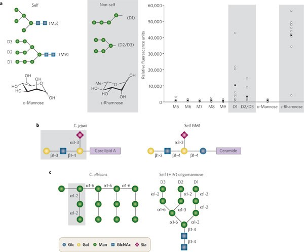
An interesting feature of the humoral anticarbohydrate repertoire is the inherent specificity against oligosaccharide motifs that constitute the 2G12 epitope. For example, the serum antibody recognition of the D1 and D2/D3 termini is similar to the specificity of 2G12 itself 34,80 (Fig. 4a). However, unlike 2G12, serum anti-oligomannose antibodies do not bind strongly to these structures when part of the larger self (Man5–9GlcNAc2) structures. So self motifs are antigenic when out of context.
Figure 4: Self/non-self discrimination and antigenic mimicry.
a, The right panel shows the extent of binding of human sera (n = 10, open circles; mean values are shown as filled boxes) to a glycan microarray, as determined using fluorescently labelled antihuman antibodies34. The discrimination between self mannosides and non-self mannosides (grey shading) is closely regulated, presenting a challenge to vaccine design. The carbohydrates that bind 2G12 — Manα1-2Manα1-2Manα1-3Man (compound D1, left panel) and (Manα1-2Manα1-6)(Manα1-2Manα1-3)Man (compound D2/D3) — are naturally antigenic. These antigenic structures are nonetheless immunosilent in the context of a self glycan. The immunological discrimination is evident at an atomic level: changing the optical configuration and substitution at a single carbon position changes the antigenicity of the monosaccharide D-mannose, compared with L-rhamnose. Thus L-rhamnose may be a powerful antigenic monosaccharide for inclusion in synthetic 'mannose' vaccines. b, Carbohydrate mimicry between C. jejuni and self ganglioside GM1. Autoimmune neuropathies, including Guillain–Barré syndrome, are a direct consequence of this molecular mimicry100. c, Carbohydrate mimicry between the mannan of C. albicans and the oligomannose glycans of gp120.
The ability to overcome self tolerance to carbohydrate structures — the goal of both anticancer and HIV vaccines — is already exhibited by some pathogenic antigens. Infection with Campylobacter jejuni elicits IgG antibodies against the bacterial lipo-oligosaccharides that also bind identical structures found on the gangliosides of peripheral nervous tissue95 (resulting in Guillain–Barré syndrome; Fig. 4b). The role of immunogenic carriers (for example, core lipid A) in breaking immune tolerance to a self antigen might be explored in the design of oligomannose vaccines. Thus, a component of pathogenesis in one disease (in this case Guillain–Barré) might be translated into a tool for vaccine design against another (HIV).
The search for immunogens that can elicit 2G12-like antibodies should include those pathogens whose mannose epitopes already seem to elicit a strong antimannose response. HIV is not the only pathogen to include the Manα1-2Man motif in its surface. Several such pathogens are endemic to the human population; some, such as Candida albicans, are particularly associated with HIV-1 infection96 (Fig. 4c). The B-cell clone that was the ancestor of 2G12 may have initially recognized mannose-type structures on a pathogen other than HIV. A conceptually related approach is to chemically modify the glycan structures of the 2G12 epitope to include antigenic carbohydrates. Again, microarray analysis of serum antibodies indicates that rhamnose (6-deoxymannose), although structurally related to mannose, is strongly antigenic34. Introduction of antigenic motifs into normally immunosilent epitopes might help to break the immune silence of HIV carbohydrates.
Perspective and future directions
Immune selection against HIV-1 has driven the evolution of a dense array of N-linked carbohydrate sites43,50. Although the targeting of a host cell property presents challenges in the form of either toxicity (in the case of lectins and glycosylation inhibitors) or autoimmunity (in the case of vaccines), the extensive and unusual nature of HIV glycosylation may provide a window of discrimination between the function and structure of human glycans and those of HIV. From the perspective of vaccine design, it is known that the human immune system is capable of providing at least one solution to the recognition of self glycans on HIV. In the broadest sense, research towards a carbohydrate vaccine for HIV must address the question of how or why some antigens are able to elicit an immune response to carbohydrate structures with self components. Observations from autoimmunity suggest that the answer to this enigma may already exist in nature: the challenge is to find a solution specific to HIV.


