Protective astrogenesis from the SVZ niche after injury is controlled by Notch modulator Thbs4 (original) (raw)
- Letter
- Published: 24 April 2013
- Dominic Luciano2,3,
- Rebecca Jo1,2,
- Khadar Abdi2,
- Patricia Paez-Gonzalez2,
- Huaxin Sheng4,
- David S. Warner4,
- Chunlei Liu5,6,
- Cagla Eroglu2,3,7 &
- …
- Chay T. Kuo1,2,3,7,8
Nature volume 497, pages 369–373 (2013)Cite this article
- 18k Accesses
- 258 Citations
- 40 Altmetric
- Metrics details
Subjects
Abstract
Postnatal/adult neural stem cells (NSCs) within the rodent subventricular zone (SVZ; also called subependymal zone) generate doublecortin (Dcx)+ neuroblasts that migrate and integrate into olfactory bulb circuitry1,2. Continuous production of neuroblasts is controlled by the SVZ microenvironmental niche3,4. It is generally thought that enhancing the neurogenic activities of endogenous NSCs may provide needed therapeutic options for disease states and after brain injury. However, SVZ NSCs can also differentiate into astrocytes. It remains unclear whether there are conditions that favour astrogenesis over neurogenesis in the SVZ niche, and whether astrocytes produced there have different properties compared with astrocytes produced elsewhere in the brain5. Here we show in mice that SVZ-generated astrocytes express high levels of thrombospondin 4 (Thbs4)6,7, a secreted homopentameric glycoprotein, in contrast to cortical astrocytes, which express low levels of Thbs4. We found that localized photothrombotic/ischaemic cortical injury initiates a marked increase in Thbs4hi astrocyte production from the postnatal SVZ niche. Tamoxifen-inducible nestin-creER tm 4 lineage tracing demonstrated that it is these SVZ-generated Thbs4hi astrocytes, and not Dcx+ neuroblasts, that home-in on the injured cortex. This robust post-injury astrogenic response required SVZ Notch activation modulated by Thbs4 via direct Notch1 receptor binding and endocytosis to activate downstream signals, including increased Nfia transcription factor expression important for glia production8. Consequently, Thbs4 homozygous knockout mice (_Thbs4_KO/KO) showed severe defects in cortical-injury-induced SVZ astrogenesis, instead producing cells expressing Dcx migrating from SVZ to the injury sites. These alterations in cellular responses resulted in abnormal glial scar formation after injury, and significantly increased microvascular haemorrhage into the brain parenchyma of _Thbs4_KO/KO mice. Taken together, these findings have important implications for post-injury applications of endogenous and transplanted NSCs in the therapeutic setting, as well as disease states where Thbs family members have important roles9,10.
This is a preview of subscription content, access via your institution
Access options
Subscribe to this journal
Receive 51 print issues and online access
$199.00 per year
only $3.90 per issue
Buy this article
- Purchase on SpringerLink
- Instant access to the full article PDF.
USD 39.95
Prices may be subject to local taxes which are calculated during checkout
Additional access options:
Figure 1: SVZ generation of Thbs4hi astrocytes.
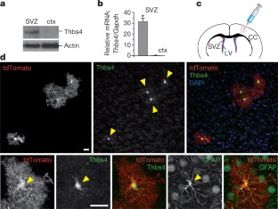
The alternative text for this image may have been generated using AI.
Figure 2: Thbs4hi astrocyte production after photothrombotic cortical injury.
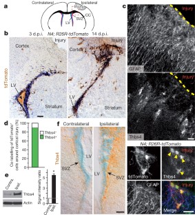
The alternative text for this image may have been generated using AI.
Figure 3: Notch signalling and regulation of injury-induced SVZ astrogenesis.
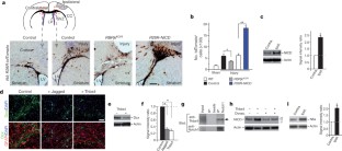
The alternative text for this image may have been generated using AI.
Figure 4: SVZ astrogenesis defects in Thbs4 mutant mice after cortical injury.
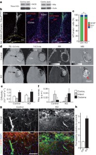
The alternative text for this image may have been generated using AI.
References
- Kriegstein, A. & Alvarez-Buylla, A. The glial nature of embryonic and adult neural stem cells. Annu. Rev. Neurosci. 32, 149–184 (2009)
Article CAS Google Scholar - Kelsch, W., Sim, S. & Lois, C. Watching synaptogenesis in the adult brain. Annu. Rev. Neurosci. 33, 131–149 (2010)
Article CAS Google Scholar - Ihrie, R. A. & Alvarez-Buylla, A. Lake-front property: a unique germinal niche by the lateral ventricles of the adult brain. Neuron 70, 674–686 (2011)
Article CAS Google Scholar - Paez-Gonzalez, P. et al. Ank3-dependent SVZ niche assembly is required for the continued production of new neurons. Neuron 71, 61–75 (2011)
Article CAS Google Scholar - Molofsky, A. V. et al. Astrocytes and disease: a neurodevelopmental perspective. Genes Dev. 26, 891–907 (2012)
Article CAS Google Scholar - Lawler, J. et al. Identification and characterization of thrombospondin-4, a new member of the thrombospondin gene family. J. Cell Biol. 120, 1059–1067 (1993)
Article CAS Google Scholar - Eroglu, C. The role of astrocyte-secreted matricellular proteins in central nervous system development and function. J. Cell Commun. Signal. 3, 167–176 (2009)
Article Google Scholar - Deneen, B. et al. The transcription factor NFIA controls the onset of gliogenesis in the developing spinal cord. Neuron 52, 953–968 (2006)
Article CAS Google Scholar - Adams, J. C. & Lawler, J. The thrombospondins. Cold Spring Harb. Perspect. Biol. 3, a009712 (2011)
Article Google Scholar - Liauw, J. et al. Thrombospondins 1 and 2 are necessary for synaptic plasticity and functional recovery after stroke. J. Cereb. Blood Flow Metab. 28, 1722–1732 (2008)
Article CAS Google Scholar - Kuo, C. T. et al. Postnatal deletion of Numb/Numblike reveals repair and remodeling capacity in the subventricular neurogenic niche. Cell 127, 1253–1264 (2006)
Article CAS Google Scholar - Frolova, E. G. et al. Thrombospondin-4 regulates vascular inflammation and atherogenesis. Circ. Res. 107, 1313–1325 (2010)
Article CAS Google Scholar - Thored, P. et al. Persistent production of neurons from adult brain stem cells during recovery after stroke. Stem Cells 24, 739–747 (2006)
Article CAS Google Scholar - Givogri, M. I. et al. Notch signaling in astrocytes and neuroblasts of the adult subventricular zone in health and after cortical injury. Dev. Neurosci. 28, 81–91 (2006)
Article CAS Google Scholar - Li, L. et al. Focal cerebral ischemia induces a multilineage cytogenic response from adult subventricular zone that is predominantly gliogenic. Glia 58, 1610–1619 (2010)
Article Google Scholar - Maxwell, K. A. & Dyck, R. H. Induction of reproducible focal ischemic lesions in neonatal mice by photothrombosis. Dev. Neurosci. 27, 121–126 (2005)
Article CAS Google Scholar - Aguirre, A., Rubio, M. E. & Gallo, V. Notch and EGFR pathway interaction regulates neural stem cell number and self-renewal. Nature 467, 323–327 (2010)
Article ADS CAS Google Scholar - Morrison, S. J. et al. Transient Notch activation initiates an irreversible switch from neurogenesis to gliogenesis by neural crest stem cells. Cell 101, 499–510 (2000)
Article CAS Google Scholar - Han, H. et al. Inducible gene knockout of transcription factor recombination signal binding protein-J reveals its essential role in T versus B lineage decision. Int. Immunol. 14, 637–645 (2002)
Article CAS Google Scholar - Carlén, M. et al. Forebrain ependymal cells are Notch-dependent and generate neuroblasts and astrocytes after stroke. Nature Neurosci. 12, 259–267 (2009)
Article Google Scholar - Meng, H., Zhang, X., Hankenson, K. D. & Wang, M. M. Thrombospondin 2 potentiates notch3/jagged1 signaling. J. Biol. Chem. 284, 7866–7874 (2009)
Article CAS Google Scholar - Yamamoto, S., Charng, W. L. & Bellen, H. J. Endocytosis and intracellular trafficking of Notch and its ligands. Curr. Top. Dev. Biol. 92, 165–200 (2010)
Article CAS Google Scholar - Namihira, M. et al. Committed neuronal precursors confer astrocytic potential on residual neural precursor cells. Dev. Cell 16, 245–255 (2009)
Article CAS Google Scholar - Moseley, M. E., de Crespigny, A. J., Roberts, T. P., Kozniewska, E. & Kucharczyk, J. Early detection of regional cerebral ischemia using high-speed MRI. Stroke 24, I60–I65 (1993)
CAS PubMed Google Scholar - Patel, M. R., Edelman, R. R. & Warach, S. Detection of hyperacute primary intraparenchymal hemorrhage by magnetic resonance imaging. Stroke 27, 2321–2324 (1996)
Article CAS Google Scholar - Daneman, R., Zhou, L., Kebede, A. A. & Barres, B. A. Pericytes are required for blood-brain barrier integrity during embryogenesis. Nature 468, 562–566 (2010)
Article ADS CAS Google Scholar - Faulkner, J. R. et al. Reactive astrocytes protect tissue and preserve function after spinal cord injury. J. Neurosci. 24, 2143–2155 (2004)
Article CAS Google Scholar - Robel, S., Berninger, B. & Gotz, M. The stem cell potential of glia: lessons from reactive gliosis. Nature Rev. Neurosci. 12, 88–104 (2011)
Article CAS Google Scholar - Aboody, K., Capela, A., Niazi, N., Stern, J. H. & Temple, S. Translating stem cell studies to the clinic for CNS repair: current state of the art and the need for a Rosetta Stone. Neuron 70, 597–613 (2011)
Article CAS Google Scholar - Alcantara Llaguno, S. et al. Malignant astrocytomas originate from neural stem/progenitor cells in a somatic tumor suppressor mouse model. Cancer Cell 15, 45–56 (2009)
Article Google Scholar - Scheffler, B. et al. Phenotypic and functional characterization of adult brain neuropoiesis. Proc. Natl Acad. Sci. USA 102, 9353–9358 (2005)
Article ADS CAS Google Scholar - Christopherson, K. S. et al. Thrombospondins are astrocyte-secreted proteins that promote CNS synaptogenesis. Cell 120, 421–433 (2005)
Article CAS Google Scholar - Macia, E. et al. Dynasore, a cell-permeable inhibitor of dynamin. Dev. Cell 10, 839–850 (2006)
Article CAS Google Scholar - Platel, J. C. et al. NMDA receptors activated by subventricular zone astrocytic glutamate are critical for neuroblast survival prior to entering a synaptic network. Neuron 65, 859–872 (2010)
Article CAS Google Scholar - McDowell, K. A. et al. Reduced cortical BDNF expression and aberrant memory in Carf knock-out mice. J. Neurosci. 30, 7453–7465 (2010)
Article CAS Google Scholar - Lee, J. K. et al. Photochemically induced cerebral ischemia in a mouse model. Surg. Neurol. 67, 620–625 (2007)
Article Google Scholar - Johnson, G. A., Cofer, G. P., Gewalt, S. L. & Hedlund, L. W. Morphologic phenotyping with MR microscopy: the visible mouse. Radiology 222, 789–793 (2002)
Article Google Scholar - Basser, P. J., Mattiello, J. & LeBihan, D. MR diffusion tensor spectroscopy and imaging. Biophys. J. 66, 259–267 (1994)
Article CAS Google Scholar - Li, W., Wu, B. & Liu, C. Quantitative susceptibility mapping of human brain reflects spatial variation in tissue composition. Neuroimage 55, 1645–1656 (2011)
Article Google Scholar
Acknowledgements
We thank D. Melton (Harvard) for R26R-NICD mice; T. Honjo (Kyoto) for RBPjk-flox mice; F. Wang for R26R-tdTomato mice; E. Rawlins (Cambridge) for Foxj1-creER t2 mice; B. Deneen (B.C.M.) and S. Singh for discussions; G. Lyons, R. Andersen, P. Heine, D. Fromme and S. Collins for project assistance; Duke Flow Cytometry Facility for help with FACS; W. Li and Duke Center for In vivo microscopy/brain imaging for MRI analyses; and T. Lechler, A. West and B. Hogan for comments on manuscript. This work was supported by National Biomedical Technology Resource Center Grant P41 RR005959 (C.L.) of P41 EB015897 to Duke Center for In Vivo Microscopy; George and Jean Brumley Endowment, Sontag Foundation, David and Lucile Packard Foundation, March of Dimes, and NIH Director’s New Innovator Award 1 DP2 OD004453-01 (C.T.K.).
Author information
Authors and Affiliations
- Department of Pediatrics, George and Jean Brumley Neonatal-Perinatal Research Institute, Duke University School of Medicine, Durham, North Carolina 27710, USA,
Eric J. Benner, Rebecca Jo & Chay T. Kuo - Department of Cell Biology, Duke University School of Medicine, Durham, North Carolina 27710, USA,
Dominic Luciano, Rebecca Jo, Khadar Abdi, Patricia Paez-Gonzalez, Cagla Eroglu & Chay T. Kuo - Department of Neurobiology, Duke University School of Medicine, Durham, North Carolina 27710, USA,
Dominic Luciano, Cagla Eroglu & Chay T. Kuo - Department of Anesthesiology, Duke University School of Medicine, Durham, North Carolina 27710, USA,
Huaxin Sheng & David S. Warner - Department of Radiology, Brain Imaging and Analysis Center, Duke University School of Medicine, Durham, North Carolina 27710, USA,
Chunlei Liu - Department of Radiology, Duke University School of Medicine, Durham, North Carolina 27710, USA,
Chunlei Liu - Duke Institute for Brain Sciences, Duke University School of Medicine, Durham, North Carolina 27710, USA,
Cagla Eroglu & Chay T. Kuo - Preston Robert Tisch Brain Tumor Center, Duke University School of Medicine, Durham, North Carolina 27710, USA,
Chay T. Kuo
Authors
- Eric J. Benner
- Dominic Luciano
- Rebecca Jo
- Khadar Abdi
- Patricia Paez-Gonzalez
- Huaxin Sheng
- David S. Warner
- Chunlei Liu
- Cagla Eroglu
- Chay T. Kuo
Contributions
E.J.B. performed injury and biochemical experiments; D.L. performed gene expression and live-imaging experiments; K.A. performed in vivo immunoprecipitation experiments; P.P.-G. performed SVZ antibody staining and analyses; R.J., H.S. and D.S.W. assisted with injuries and their analyses; C.L. performed MRI scanning and quantitative analyses; C.E. provided reagents and experimental insight; C.T.K. performed transplantations and conceived the project. E.J.B., D.L. and R.J. assembled figures and C.T.K. wrote the paper. All authors discussed results and commented on the manuscript.
Corresponding author
Correspondence toChay T. Kuo.
Ethics declarations
Competing interests
The authors declare no competing financial interests.
Supplementary information
PowerPoint slides
Rights and permissions
About this article
Cite this article
Benner, E., Luciano, D., Jo, R. et al. Protective astrogenesis from the SVZ niche after injury is controlled by Notch modulator Thbs4.Nature 497, 369–373 (2013). https://doi.org/10.1038/nature12069
- Received: 10 January 2012
- Accepted: 13 March 2013
- Published: 24 April 2013
- Issue date: 16 May 2013
- DOI: https://doi.org/10.1038/nature12069
This article is cited by
Editorial Summary
Stem-cell-mediated recovery from brain injury
The subventricular zone (SVZ) of the rodent brain is known to host precursor cells that can generate neurons or glia, depending on the microenvironment. The question arises, might activating this niche following brain injury play a role in tissue repair? Here, Chay Kuo and colleagues identify a specific population of SVZ-generated astrocytes in mice that increases in number post-injury. These activated astrocytes migrate to the site of injury, unlike their cortex-generated counterparts. This robust post-injury response requires Notch signalling, and when this population of astrocytes is disrupted tissue recovery is compromised.