WDR62 is associated with the spindle pole and is mutated in human microcephaly (original) (raw)
- Letter
- Published: 03 October 2010
- Maryam Khurshid1 na1,
- Julie Désir2 na1,
- Ofélia P Carvalho1,
- James J Cox1,
- Gemma Thornton1,
- Rizwana Kausar3,
- Muhammad Ansar3,
- Wasim Ahmad3,
- Alain Verloes4,
- Sandrine Passemard4,5,
- Jean-Paul Misson6,
- Susan Lindsay7,
- Fanni Gergely8,
- William B Dobyns9,
- Emma Roberts10,
- Marc Abramowicz2 &
- …
- C Geoffrey Woods1
Nature Genetics volume 42, pages 1010–1014 (2010)Cite this article
- 4240 Accesses
- 274 Citations
- Metrics details
Subjects
Abstract
Autosomal recessive primary microcephaly (MCPH) is a disorder of neurodevelopment resulting in a small brain1,2. We identified WDR62 as the second most common cause of MCPH after finding homozygous missense and frame-shifting mutations in seven MCPH families. In human cell lines, we found that WDR62 is a spindle pole protein, as are ASPM and STIL, the MCPH7 and MCHP7 proteins3,4,5. Mutant WDR62 proteins failed to localize to the mitotic spindle pole. In human and mouse embryonic brain, we found that WDR62 expression was restricted to neural precursors undergoing mitosis. These data lend support to the hypothesis that the exquisite control of the cleavage furrow orientation in mammalian neural precursor cell mitosis, controlled in great part by the centrosomes and spindle poles, is critical both in causing MCPH when perturbed and, when modulated, generating the evolutionarily enlarged human brain6,7,8,9.
This is a preview of subscription content, access via your institution
Access options
Subscribe to this journal
Receive 12 print issues and online access
$259.00 per year
only $21.58 per issue
Buy this article
- Purchase on SpringerLink
- Instant access to the full article PDF.
USD 39.95
Prices may be subject to local taxes which are calculated during checkout
Additional access options:
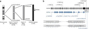
Figure 1: A summary of the linkage strategy used to define the MCPH2 region and mutations found in WDR62 in MCPH2 families.
The alternative text for this image may have been generated using AI.
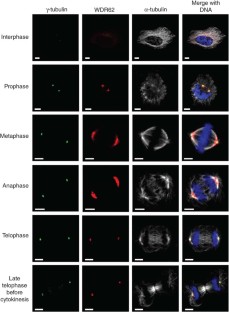
Figure 2: Subcellular localization of WDR62 throughout the cell cycle.
The alternative text for this image may have been generated using AI.
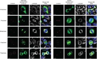
Figure 3: Overexpression of WDR62-GFP wild type and c.1313G>A (p.Arg438His) mutant constructs in HeLa cells.
The alternative text for this image may have been generated using AI.
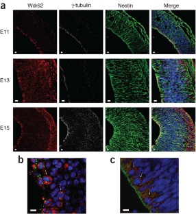
Figure 4: Endogenous expression pattern of WDR62 in human and mouse embryonic brain.
The alternative text for this image may have been generated using AI.
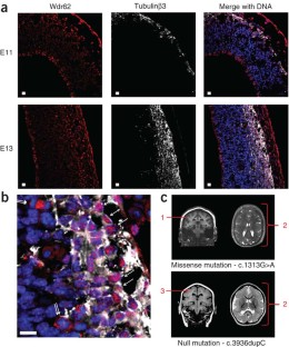
Figure 5: Wdr62 expression in newborn, newly arrived cortical neurons in the developing cerebral cortex and brain imaging from two individuals with WDR62 mutations.
The alternative text for this image may have been generated using AI.
References
- Mochida, G.H. & Walsh, C.A. Genetic basis of developmental malformations of the cerebral cortex. Arch. Neurol. 61, 637–640 (2004).
Google Scholar - Woods, C.G., Bond, J. & Enard, W. Autosomal recessive primary microcephaly (MCPH): a review of clinical, molecular, and evolutionary findings. Am. J. Hum. Genet. 76, 717–728 (2005).
Google Scholar - Bond, J. et al. ASPM is a major determinant of cerebral cortical size. Nat. Genet. 32, 316–320 (2002).
Google Scholar - Pfaff, K.L. et al. The zebra fish cassiopeia mutant reveals that SIL is required for mitotic spindle organization. Mol. Cell. Biol. 27, 5887–5897 (2007).
Google Scholar - Bond, J. & Woods, C.G. Cytoskeletal genes regulating brain size. Curr. Opin. Cell Biol. 18, 95–101 (2006).
Google Scholar - Fish, J.L., Kosodo, Y., Enard, W., Pääbo, S. & Huttner, W.B. Aspm specifically maintains symmetric proliferative divisions of neuroepithelial cells. Proc. Natl. Acad. Sci. USA 103, 10438–10443 (2006).
Google Scholar - Fish, J.L., Dehay, C., Kennedy, H. & Huttner, W.B. Making bigger brains–the evolution of neural-progenitor-cell division. J. Cell Sci. 121, 2783–2793 (2008).
Google Scholar - Marthiens, V. & ffrench-Constant, C. Adherens junction domains are split by asymmetric division of embryonic neural stem cells. EMBO Rep. 10, 515–520 (2009).
Google Scholar - Thornton, G.K. & Woods, C.G. Primary microcephaly: do all roads lead to Rome? Trends Genet. 25, 501–510 (2009).
Google Scholar - Roberts, E. et al. The second locus for autosomal recessive primary microcephaly (MCPH2) maps to chromosome 19q13.1–13.2. Eur. J. Hum. Genet. 7, 815–820 (1999).
Google Scholar - Jackson, A.P. et al. Identification of microcephalin, a protein implicated in determining the size of the human brain. Am. J. Hum. Genet. 71, 136–142 (2002).
Google Scholar - Bond, J. et al. A centrosomal mechanism involving CDK5RAP2 and CENPJ controls brain size. Nat. Genet. 37, 353–355 (2005).
Google Scholar - Kumar, A., Girimaji, S.C., Duvvari, M.R. & Blanton, S.H. Mutations in STIL, encoding a pericentriolar and centrosomal protein, cause primary microcephaly. Am. J. Hum. Genet. 84, 286–290 (2009).
Google Scholar - Passemard, S. et al. Primary autosomal recessive microcephaly. GeneReviews. (University of Washington, Seattle, WA, 2009) <http://www.ncbi.nlm.nih.gov/pubmed/20301772>.
- Roberts, E. et al. Autosomal recessive primary microcephaly: an analysis of locus heterogeneity and phenotypic variation. J. Med. Genet. 39, 718–721 (2002).
Google Scholar - Mardis, E.R. New strategies and emerging technologies for massively parallel sequencing: applications in medical research. Genome Med. 1, 40 (2009).
Google Scholar - Field, M. et al. Mutations in the BRWD3 gene cause X–linked mental retardation associated with macrocephaly. Am. J. Hum. Genet. 81, 367–374 (2007).
Google Scholar - Nousiainen, M., Silljé, H.H., Sauer, G., Nigg, E.A. & Körner, R. Phosphoproteome analysis of the human mitotic spindle. Proc. Natl. Acad. Sci. USA 103, 5391–5396 (2006).
Google Scholar - Malik, R. et al. Quantitative analysis of the human spindle phosphoproteome at distinct mitotic stages. J. Proteome Res. 8, 4553–4563 (2009).
Google Scholar - do Carmo Avides, M., Tavares, A. & Glover, D.M. Polo kinase and Asp are needed to promote the mitotic organizing activity of centrosomes. Nat. Cell Biol. 3, 421–424 (2001).
Google Scholar - Caspi, M. et al. LIS1 missense mutations: variable phenotypes result from unpredictable alterations in biochemical and cellular properties. J. Biol. Chem. 278, 38740–38748 (2003).
Google Scholar - Gardette, R., Courtois, M. & Bisconte, J.C. Prenatal development of mouse central nervous structures: time of neuron origin and gradients of neuronal production. A radioautographic study. J. Hirnforsch. 23, 415–431 (1982).
Google Scholar - Uylings, H.B. Development of the cerebral cortex in rodents and man. Eur. J. Morphol. 38, 309–312 (2000).
Google Scholar - Bystron, I., Blakemore, C. & Rakic, P. Development of the human cerebral cortex: Boulder Committee revisited. Nat. Rev. Neurosci. 9, 110–122 (2008).
Google Scholar - Attardo, A., Calegari, F., Haubensak, W., Wilsch-Brauninger, W.M. & Huttner, W.B. Live imaging at the onset of cortical neurogenesis reveals differential appearance of the neuronal phenotype in apical versus basal progenitor progeny. PLoS ONE 3, e2388 (2008).
Google Scholar - Noctor, S.C., Martinez-Cerdeno, V. & Kriegstein, A.R. Distinct behaviors of neural stem and progenitor cells underlie cortical neurogenesis. J. Comp. Neurol. 508, 28–44 (2008).
Google Scholar - O'Rahilly, R. & Müller, F. Developmental Stages in Human Embryos: Revised and New Measurements. Cells Tissues Organs 192, 73–84 (2010).
Google Scholar - Bilgüvar, K. et al. Whole-exome sequencing identifies recessive WDR62 mutations in severe brain malformations. Nature. published online, doi:10.1038/nature09327 (22 August 2010).
- Barr, F.A. & Gruneberg, U. Cytokinesis: placing and making the final cut. Cell 131, 847–860 (2007).
Google Scholar - Barr, A.R., Kilmartin, J.V. & Gergely, F. CDK5RAP2 functions in centrosome to spindle pole attachment and DNA damage response. J. Cell Biol. 189, 23–39 (2010).
Google Scholar - Götz, M. & Huttner, W.B. The cell biology of neurogenesis. Nat. Rev. Mol. Cell Biol. 6, 777–788 (2005).
Google Scholar - Kosodo, Y. et al. Asymmetric distribution of the apical plasma membrane during neurogenic divisions of mammalian neuroepithelial cells. EMBO J. 23, 2314–2324 (2004).
Google Scholar
Acknowledgements
The authors would like to thank the research families for their participation in this project and the Wellcome Trust, Medical Research Council, Action Research and the Higher Education Commission of Pakistan for funding (to A.K.N., M.K., O.P.C., J.J.C., G.T. and E.R.). J.D. was supported by the Belgian Kids' Fund. M.A. was supported by grants from the Fonds Erasme and the Belgian Fonds de la Recherche Scientifique Médicale (FRSM). We thank S. Strollo for expert technical assistance. We thank the Medical Research Council (MRC)-Wellcome Trust Human Developmental Biology Resource (HDBR), Newcastle for providing the human tissue for the expression studies.
Author information
Author notes
- Adeline K Nicholas, Maryam Khurshid and Julie Désir: These authors contributed equally to this work.
Authors and Affiliations
- Department of Medical Genetics, Cambridge Institute for Medical Research, University of Cambridge, Cambridge, UK
Adeline K Nicholas, Maryam Khurshid, Ofélia P Carvalho, James J Cox, Gemma Thornton & C Geoffrey Woods - Department of Medical Genetics, Hôpital Erasme and Institut de Recherche Interdisciplinaire en Biologie Humaine et Moléculaire, Université libre de Bruxelles (IRIBHM), ULB, Brussels, Belgium
Julie Désir & Marc Abramowicz - Department of Biochemistry, Faculty of Biological Sciences, Quaid-i-Azam University, Islamabad, Pakistan
Rizwana Kausar, Muhammad Ansar & Wasim Ahmad - Department of Genetics, Robert Debré University Hospital, Paris, France
Alain Verloes & Sandrine Passemard - Department of Child Neurology, Assistance publique-Hôpitaux de Paris (AP-HP) Robert Debré University Hospital, Paris, France
Sandrine Passemard - University of Liège Medical School and Department of Pediatrics, La Citadelle University Hospital, Liège, Belgium
Jean-Paul Misson - Institute of Human Genetics, Newcastle University, International Centre for Life, Central Parkway, Newcastle upon Tyne, UK
Susan Lindsay - Cancer Research UK Cambridge Research Institute, Li Ka Shing Centre, Cambridge, UK
Fanni Gergely - Department of Human Genetics, University of Chicago, Chicago, Illinois, USA
William B Dobyns - Microcephaly and Neurogenesis research group, Leeds Institute of Molecular Medicine, St. James's University Hospital, Leeds, UK
Emma Roberts
Authors
- Adeline K Nicholas
- Maryam Khurshid
- Julie Désir
- Ofélia P Carvalho
- James J Cox
- Gemma Thornton
- Rizwana Kausar
- Muhammad Ansar
- Wasim Ahmad
- Alain Verloes
- Sandrine Passemard
- Jean-Paul Misson
- Susan Lindsay
- Fanni Gergely
- William B Dobyns
- Emma Roberts
- Marc Abramowicz
- C Geoffrey Woods
Contributions
The following authors contributed to the design of the study: A.K.N., M.K., J.J.C., F.G., E.R., M. Abramowicz and C.G.W. The following authors generated experimental data: A.K.N., M.K., J.D., O.P.C., G.T., R.K., M. Ansar, F.G., W.B.D., E.R. and C.G.W. Reagents were contributed by R.K., M. Abramowicz, W.A., A.L., S.P., J.-P.M., S.L., M. Abramowicz and C.G.W. The paper was written by A.K.N., M.K., O.P.C., J.J.C., W.A., S.L., F.G., W.B.D. and C.G.W.
Corresponding authors
Correspondence toMarc Abramowicz or C Geoffrey Woods.
Ethics declarations
Competing interests
The authors declare no competing financial interests.
Supplementary information
Rights and permissions
About this article
Cite this article
Nicholas, A., Khurshid, M., Désir, J. et al. WDR62 is associated with the spindle pole and is mutated in human microcephaly.Nat Genet 42, 1010–1014 (2010). https://doi.org/10.1038/ng.682
- Received: 27 May 2010
- Accepted: 10 September 2010
- Published: 03 October 2010
- Issue date: November 2010
- DOI: https://doi.org/10.1038/ng.682
This article is cited by
WDR62-deficiency Causes Autism-like Behaviors Independent of Microcephaly in Mice
- Dan Xu
- Yiqiang Zhi
- Zhiheng Xu
Neuroscience Bulletin (2022)
Novel phenotype and genotype spectrum of WDR62 in two patients with associated primary autosomal recessive microcephaly
- Hajar Aryan
- Shaghayegh Zokaei
- Ali Reza Tavasoli
Irish Journal of Medical Science (1971 -) (2022)
Molecular evolutionary analysis of human primary microcephaly genes
- Nashaiman Pervaiz
- Hongen Kang
- Amir Ali Abbasi
BMC Ecology and Evolution (2021)
The journey of Zika to the developing brain
- Francesca Rombi
- Richard Bayliss
- Sharon Yeoh
Molecular Biology Reports (2020)
Primary microcephaly with an unstable genome
- Shibin Xu
- Xingxuan Wu
- Xingzhi Xu
Genome Instability & Disease (2020)