A culture system to study oligodendrocyte myelination processes using engineered nanofibers (original) (raw)
- Article
- Published: 15 July 2012
- Michelle K Leach3,
- Stephanie A Redmond1,2,
- S Y Christin Chong1,2,
- Synthia H Mellon4,
- Samuel J Tuck5,
- Zhang-Qi Feng3,
- Joseph M Corey3,5,6 &
- …
- Jonah R Chan1,2
Nature Methods volume 9, pages 917–922 (2012)Cite this article
- 9047 Accesses
- 380 Citations
- 43 Altmetric
- Metrics details
Subjects
Abstract
Current methods for studying central nervous system myelination necessitate permissive axonal substrates conducive to myelin wrapping by oligodendrocytes. We have developed a neuron-free culture system in which electron-spun nanofibers of varying sizes substitute for axons as a substrate for oligodendrocyte myelination, thereby allowing manipulation of the biophysical elements of axonal-oligodendroglial interactions. To investigate axonal regulation of myelination, this system effectively uncouples the role of molecular (inductive) cues from that of biophysical properties of the axon. We use this method to uncover the causation and sufficiency of fiber diameter in the initiation of concentric wrapping by rat oligodendrocytes. We also show that oligodendrocyte precursor cells display sensitivity to the biophysical properties of fiber diameter and initiate membrane ensheathment before differentiation. The use of nanofiber scaffolds will enable screening for potential therapeutic agents that promote oligodendrocyte differentiation and myelination and will also provide valuable insight into the processes involved in remyelination.
This is a preview of subscription content, access via your institution
Access options
Subscribe to this journal
Receive 12 print issues and online access
$259.00 per year
only $21.58 per issue
Buy this article
- Purchase on SpringerLink
- Instant access to the full article PDF.
USD 39.95
Prices may be subject to local taxes which are calculated during checkout
Additional access options:
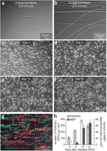
Figure 1: Temporal and spatial behavior of OPCs on polystyrene fibers are similar to that of OPCs on live axons.
The alternative text for this image may have been generated using AI.
Figure 2: Fiber diameter is sufficient to initiate myelination.

The alternative text for this image may have been generated using AI.
Figure 3: Quantification of fiber diameter threshold and size preference for ensheathment and wrapping.

The alternative text for this image may have been generated using AI.
Figure 4: The majority of the cells in the cultures are capable of ensheathing and wrapping fibers above a minimum threshold.

The alternative text for this image may have been generated using AI.
References
- Colello, R.J. & Pott, U. Signals that initiate myelination in the developing mammalian nervous system. Mol. Neurobiol. 15, 83–100 (1997).
Article CAS Google Scholar - Fruttiger, M., Calver, A.R. & Richardson, W.D. Platelet-derived growth factor is constitutively secreted from neuronal cell bodies but not from axons. Curr. Biol. 10, 1283–1286 (2000).
Article CAS Google Scholar - Jiang, F., Frederick, T.J. & Wood, T.L. IGF-I synergizes with FGF-2 to stimulate oligodendrocyte progenitor entry into the cell cycle. Dev. Biol. 232, 414–423 (2001).
Article CAS Google Scholar - Nave, K.A. & Trapp, B.D. Axon-glial signaling and the glial support of axon function. Annu. Rev. Neurosci. 31, 535–561 (2008).
Article CAS Google Scholar - Nave, K.A. Myelination and support of axonal integrity by glia. Nature 468, 244–252 (2010).
Article CAS Google Scholar - Friede, R.L. Control of myelin formation by axon caliber (with a model of the control mechanism). J. Comp. Neurol. 144, 233–252 (1972).
Article CAS Google Scholar - Voyvodic, J.T. Target size regulates calibre and myelination of sympathetic axons. Nature 342, 430–433 (1989).
Article CAS Google Scholar - de Waegh, S.M., Lee, V.M. & Brady, S.T. Local modulation of neurofilament phosphorylation, axonal caliber, and slow axonal transport by myelinating Schwann cells. Cell 68, 451–463 (1992).
Article CAS Google Scholar - Garcia, M.L. et al. NF-M is an essential target for the myelin-directed “outside-in” signaling cascade that mediates radial axonal growth. J. Cell Biol. 163, 1011–1020 (2003).
Article CAS Google Scholar - Garcia, M.L. et al. Phosphorylation of highly conserved neurofilament medium KSP repeats is not required for myelin-dependent radial axonal growth. J. Neurosci. 29, 1277–1284 (2009).
Article CAS Google Scholar - Waxman, S.G. & Bennett, M.V. Relative conduction velocities of small myelinated and non-myelinated fibres in the central nervous system. Nat. New Biol. 238, 217–219 (1972).
Article CAS Google Scholar - Rosenberg, S.S., Kelland, E.E., Tokar, E., De la Torre, A.R. & Chan, J.R. The geometric and spatial constraints of the microenvironment induce oligodendrocyte differentiation. Proc. Natl. Acad. Sci. USA 105, 14662–14667 (2008).
Article CAS Google Scholar - Gertz, C.C. et al. Accelerated neuritogenesis and maturation of primary spinal motor neurons in response to nanofibers. Dev. Neurobiol. 70, 589–603 (2010).
Article CAS Google Scholar - Leach, M.K., Feng, Z.Q., Tuck, S.J. & Corey, J.M. Electrospinning fundamentals: optimizing solution and apparatus parameters. J. Vis. Exp. 47, 2494 (2011).
Google Scholar - Bullock, P.N. & Rome, L.H. Glass micro-fibers: a model system for study of early events in myelination. J. Neurosci. Res. 27, 383–393 (1990).
Article CAS Google Scholar - Howe, C.L. Coated glass and vicryl microfibers as artificial axons. Cells Tissues Organs 183, 180–194 (2006).
Article CAS Google Scholar - Chan, J.R. et al. NGF controls axonal receptivity to myelination by Schwann cells or oligodendrocytes. Neuron 43, 183–191 (2004).
Article CAS Google Scholar - Chong, S.Y. et al. Neurite outgrowth inhibitor Nogo-A establishes spatial segregation and extent of oligodendrocyte myelination. Proc. Natl. Acad. Sci. USA 109, 1299–1304 (2011).
Article Google Scholar - Ruit, K.G., Elliott, J.L., Osborne, P.A., Yan, Q. & Snider, W.D. Selective dependence of mammalian dorsal root ganglion neurons on nerve growth factor during embryonic development. Neuron 8, 573–587 (1992).
Article CAS Google Scholar - Remahl, S. & Hildebrand, C. Changing relation between onset of myelination and axon diameter range in developing feline white matter. J. Neurol. Sci. 54, 33–45 (1982).
Article CAS Google Scholar - Kleitman, N., Wood, P.M. & Bunge, R.P. in Culturing Nerve Cells (eds. Banker, G. & Goslin, K.) Ch. 14, 337–377 (MIT Press, 1991).
Google Scholar - Fanarraga, M.L., Griffiths, I.R., Zhao, M. & Duncan, I.D. Oligodendrocytes are not inherently programmed to myelinate a specific size of axon. J. Comp. Neurol. 399, 94–100 (1998).
Article CAS Google Scholar - Colognato, H., Ramachandrappa, S., Olsen, I.M. & ffrench-Constant, C. Integrins direct Src family kinases to regulate distinct phases of oligodendrocyte development. J. Cell Biol. 167, 365–375 (2004).
Article CAS Google Scholar - Spiegel, I. & Peles, E. A novel method for isolating Schwann cells using the extracellular domain of Necl1. J. Neurosci. Res. 87, 3288–3296 (2009).
Article CAS Google Scholar - Schwab, M.E. & Schnell, L. Region-specific appearance of myelin constituents in the developing rat spinal cord. J. Neurocytol. 18, 161–169 (1989).
Article CAS Google Scholar - Corey, J.M. et al. The design of electrospun PLLA nanofiber scaffolds compatible with serum-free growth of primary motor and sensory neurons. Acta Biomater. 4, 863–875 (2008).
Article CAS Google Scholar - Grimes, M.L. et al. Endocytosis of activated TrkA: evidence that nerve growth factor induces formation of signaling endosomes. J. Neurosci. 16, 7950–7964 (1996).
Article CAS Google Scholar - Lewallen, K.A. et al. Assessing the role of the cadherin/catenin complex at the Schwann cell-axon interface and in the initiation of myelination. J. Neurosci. 31, 3032–3043 (2011).
Article CAS Google Scholar
Acknowledgements
We thank W. Stallcup for the rabbit anti-PDGFRα antibody; M.L. Wong for sectioning nanofiber cultures for electron microscopy analysis at the W.M. Keck Foundation Advanced Microscopy Laboratory at UCSF; R. Langen and M. Isas for assistance, advice and support with the electron microscopy; the other members of the Chan laboratory and the MS Research Group at UCSF for encouragement, advice and insightful discussions. This work was supported by the US National Multiple Sclerosis Society Career Transition Award (TA 3008A2/T), the Harry Weaver Neuroscience Scholar Award (JF 2142-A2/T) and the US National Institutes of Health/National Institute of Neurological Disorders and Stroke (NS062796-02) to J.R.C.
Author information
Authors and Affiliations
- Department of Neurology, University of California, San Francisco (UCSF), San Francisco, California, USA
Seonok Lee, Stephanie A Redmond, S Y Christin Chong & Jonah R Chan - Program in Neuroscience, UCSF, San Francisco, California, USA
Seonok Lee, Stephanie A Redmond, S Y Christin Chong & Jonah R Chan - Department of Biomedical Engineering, University of Michigan, Ann Arbor, Michigan, USA
Michelle K Leach, Zhang-Qi Feng & Joseph M Corey - Department of Obstetrics, Gynecology and Reproductive Sciences, UCSF, San Francisco, California, USA
Synthia H Mellon - Department of Neurology, University of Michigan, Ann Arbor, Michigan, USA
Samuel J Tuck & Joseph M Corey - Geriatric Research, Education and Clinical Center, Veterans Affairs Ann Arbor Healthcare Center, Ann Arbor, Michigan, USA
Joseph M Corey
Authors
- Seonok Lee
- Michelle K Leach
- Stephanie A Redmond
- S Y Christin Chong
- Synthia H Mellon
- Samuel J Tuck
- Zhang-Qi Feng
- Joseph M Corey
- Jonah R Chan
Contributions
S.L., M.K.L., S.A.R., S.Y.C.C. and J.R.C. performed experiments. S.L., M.K.L., S.H.M., S.J.T., Z.-Q.F., J.M.C. and J.R.C. provided reagents. S.L., M.K.L., S.A.R., S.Y.C.C., S.H.M., S.J.T., Z.-Q.F., J.M.C. and J.R.C. provided intellectual contributions. S.L. and J.R.C. analyzed the data and wrote the paper.
Corresponding authors
Correspondence toJoseph M Corey or Jonah R Chan.
Ethics declarations
Competing interests
The authors declare no competing financial interests.
Supplementary information
Rights and permissions
About this article
Cite this article
Lee, S., Leach, M., Redmond, S. et al. A culture system to study oligodendrocyte myelination processes using engineered nanofibers.Nat Methods 9, 917–922 (2012). https://doi.org/10.1038/nmeth.2105
- Received: 16 April 2012
- Accepted: 22 June 2012
- Published: 15 July 2012
- Issue date: September 2012
- DOI: https://doi.org/10.1038/nmeth.2105