Simultaneous imaging of morphological plasticity and calcium dynamics in dendrites (original) (raw)
- Protocol
- Published: 22 November 2006
Nature Protocols volume 1, pages 1859–1864 (2006) Cite this article
- 721 Accesses
- 18 Citations
- Metrics details
Abstract
The structure and function of the nervous system are intricately connected. To investigate their relationship it is essential to image neuronal structure and function simultaneously with high spatio-temporal resolution. For this purpose, we describe here a two-step strategy. First, to visualize neurons and their entire dendritic arborization in neuronal tissue, we use ballistic delivery or single-cell electroporation of a fluorescent calcium indicator and a red fluorescent dye. Second, dual wavelength wide-field fluorescence microscopy or confocal microscopy enables imaging structural plasticity of dendrites (including filopodia and spines) and calcium dynamics together. We routinely apply this strategy to developing neurons in live tissue, but mature neurons can also be loaded and imaged as described. For labeling cells and setting up imaging equipment, approximately 2 h are required.
This is a preview of subscription content, access via your institution
Access options
Subscribe to this journal
Receive 12 print issues and online access
$259.00 per year
only $21.58 per issue
Buy this article
- Purchase on SpringerLink
- Instant access to the full article PDF.
USD 39.95
Prices may be subject to local taxes which are calculated during checkout
Additional access options:
Figure 1: Single cell electroporation.
The alternative text for this image may have been generated using AI.
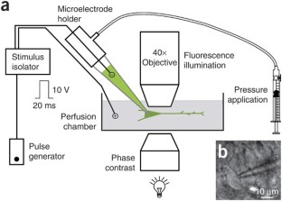
Figure 2: Setup for the consecutive imaging of two wavelengths using wide-field microscopy and a CCD camera.

The alternative text for this image may have been generated using AI.
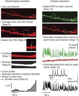
Figure 3: Analysis of morphological plasticity and calcium dynamics.
The alternative text for this image may have been generated using AI.
Figure 4: Imaging structural plasticity and calcium dynamics in dendrites of a CA3 pyramidal neuron from the neonatal rat hippocampus.

The alternative text for this image may have been generated using AI.
References
- Feng, G. et al. Imaging neuronal subsets in transgenic mice expressing multiple spectral variants of GFP. Neuron 28, 41–51 (2000).
Article CAS Google Scholar - Denk, W., Strickler, J.H. & Webb, W.W. Two-photon laser scanning fluorescence microscopy. Science 248, 73–76 (1990).
Article CAS Google Scholar - Engert, F. & Bonhoeffer, T. Dendritic spine changes associated with hippocampal long-term synaptic plasticity. Nature 399, 66–70 (1999).
Article CAS Google Scholar - Maletic-Savatic, M., Malinow, R. & Svoboda, K. Rapid dendritic morphogenesis in CA1 hippocampal dendrites induced by synaptic activity. Science 283, 1923–1927 (1999).
Article CAS Google Scholar - Matsuzaki, M., Honkura, N., Ellis-Davies, G.C. & Kasai, H. Structural basis of long-term potentiation in single dendritic spines. Nature 429, 761–766 (2004).
Article CAS Google Scholar - Lohmann, C., Finski, A. & Bonhoeffer, T. Local calcium transients regulate the spontaneous motility of dendritic filopodia. Nature Neurosci. 8, 305–312 (2005).
Article CAS Google Scholar - Haas, K., Sin, W.C., Javaherian, A., Li, Z. & Cline, H.T. Single-cell electroporation for gene transfer in vivo. Neuron 29, 583–591 (2001).
Article CAS Google Scholar - Rathenberg, J., Nevian, T. & Witzemann, V. High-efficiency transfection of individual neurons using modified electrophysiology techniques. J. Neurosci. Meth. 126, 91–98 (2003).
Article Google Scholar - Kettunen, P. et al. Imaging calcium dynamics in the nervous system by means of ballistic delivery of indicators. J. Neurosci. Meth. 119, 37–43 (2002).
Article CAS Google Scholar - Lohmann, C., Demas, J., Morgan, J.L. & Wong, R.O.L. in Imaging in Neuroscience and Development: A Laboratory Manual Chapter 21, p. 171–183 (CSHL Press, Cold Spring Harbor, New York, 2004).
Google Scholar - Portera-Cailliau, C., Pan, D.T. & Yuste, R. Activity-regulated dynamic behavior of early dendritic protrusions: evidence for different types of dendritic filopodia. J. Neurosci. 23, 7129–7142 (2003).
Article CAS Google Scholar - Lohmann, C., Myhr, K.L. & Wong, R.O. Transmitter-evoked local calcium release stabilizes developing dendrites. Nature 418, 177–181 (2002).
Article CAS Google Scholar - Lohmann, C. & Wong, R.O. Regulation of dendritic growth and plasticity by local and global calcium dynamics. Cell Calcium 37, 403–409 (2005).
Article CAS Google Scholar - O'Brien, J.A., Holt, M., Whiteside, G., Lummis, S.C.R. & Hastings, M.H. Modifications to the hand-held gene gun: improvements for in vitro biolistic transfection of organotypic neuronal tissue. J. Neurosci. Meth. 112, 57–64 (2001).
Article CAS Google Scholar
Acknowledgements
The Calistic technique was developed in collaboration with J. Demas, P. Kettunen, W.B. Gan and R.O.L. Wong.
Author information
Authors and Affiliations
- Max-Planck Institute of Neurobiology, Am Klopferspitz 18, Planegg-Martinsried, 82152, Germany
Susanne B Lang, Tobias Bonhoeffer & Christian Lohmann
Authors
- Susanne B Lang
- Tobias Bonhoeffer
- Christian Lohmann
Corresponding author
Correspondence toChristian Lohmann.
Ethics declarations
Competing interests
The authors declare no competing financial interests.
Rights and permissions
About this article
Cite this article
Lang, S., Bonhoeffer, T. & Lohmann, C. Simultaneous imaging of morphological plasticity and calcium dynamics in dendrites.Nat Protoc 1, 1859–1864 (2006). https://doi.org/10.1038/nprot.2006.267
- Published: 22 November 2006
- Issue date: November 2006
- DOI: https://doi.org/10.1038/nprot.2006.267
This article is cited by
Structural plasticity upon learning: regulation and functions
- Pico Caroni
- Flavio Donato
- Dominique Muller
Nature Reviews Neuroscience (2012)
Single-cell electroporation
- Manyan Wang
- Owe Orwar
- Stephen G. Weber
Analytical and Bioanalytical Chemistry (2010)