A protocol for dissecting Drosophila melanogaster brains for live imaging or immunostaining (original) (raw)
- Protocol
- Published: 07 December 2006
Nature Protocols volume 1, pages 2110–2115 (2006)Cite this article
- 13k Accesses
- 337 Citations
- 1 Altmetric
- Metrics details
Abstract
This protocol describes a basic method for dissection and immunofluorescence staining of the Drosophila brain at various developmental stages. The Drosophila brain has become increasingly useful for studies of neuronal wiring and morphogenesis in combination with techniques such as the 'mosaic analysis with a repressible cell marker' (MARCM) system, where single neurons can be followed in live and fixed tissues for high-resolution analysis of wild-type or genetically manipulated cells. Such high-resolution anatomical study of the brain is also important in characterizing the organization of neural circuits using genetic tools such as GAL4 enhancer trap lines, as Drosophila has been intensively used for studying the neural basis of behavior. Advantages of fluorescence immunostaining include compatibility with multicolor labeling and confocal or multiphoton imaging. This brain dissection and immunofluorescence staining protocol requires approximately 2 to 6 d to complete.
This is a preview of subscription content, access via your institution
Access options
Subscribe to this journal
Receive 12 print issues and online access
$259.00 per year
only $21.58 per issue
Buy this article
- Purchase on SpringerLink
- Instant access to the full article PDF.
USD 39.95
Prices may be subject to local taxes which are calculated during checkout
Additional access options:
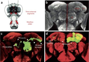
Figure 1: Visualizing neurons in the Drosophila brain.
The alternative text for this image may have been generated using AI.
References
- Lee, T. & Luo, L. Mosaic analysis with a repressible cell marker for studies of gene function in neuronal morphogenesis. Neuron 22, 451–461 (1999).
Article CAS PubMed Google Scholar - Lee, T. & Luo, L. Mosaic analysis with a repressible cell marker (MARCM) for Drosophila neural development. Trends Neurosci. 24, 251–254 (2001).
Article CAS PubMed Google Scholar - Wu, J.S. & Luo, L. A protocol for mosaic analysis with a repressible cell marker (MARCM) in Drosophila. Nat. Prot. (doi:10.1038/nprot.2006.320).
- Wong, A.M., Wang, J.W. & Axel, R. Spatial representation of the glomerular map in the Drosophila protocerebrum. Cell 109, 229–241 (2002).
Article CAS PubMed Google Scholar - Wagh, D.A. et al. Bruchpilot, a protein with homology to ELKS/CAST, is required for structural integrity and function of synaptic active zones in Drosophila. Neuron 49, 833–844 (2006).
Article CAS PubMed Google Scholar - Crittenden, J.R., Sloulakis, E.M.C., Han, K.-A., Kalderon, D. & Davis, R.L. Tripartite mushroom body architecture revealed by antigenic markers. Learn. Mem. 5, 38–51 (1998).
Article CAS PubMed PubMed Central Google Scholar - Reuter, J.E. et al. A mosaic genetic screen for genes necessary for Drosophila mushroom body neuronal morphogenesis. Development 130, 1203–1213 (2003).
Article CAS PubMed Google Scholar - Jefferis, G.S.X.E., Marin, E.C., Stocker, R.F. & Luo, L. Target neuron prespecification in the olfactory map of Drosophila. Nature 414, 204–208 (2001).
Article CAS PubMed Google Scholar - Jefferis, G.S.X.E., Marin, E.C., Watts, R.J. & Luo, L. Development of neuronal connectivity in Drosophila antennal lobes and mushroom bodies. Curr. Opin. Neurobiol. 12, 80–86 (2002).
Article CAS PubMed Google Scholar
Acknowledgements
We thank members of our laboratory for their helpful comments on this protocol. Research in our lab has been generously supported by grants from the US National Institutes of Health, and more recently from the Howard Hughes Medical Institute, for which L.L. is an investigator.
Author information
Authors and Affiliations
- Department of Biological Sciences, Howard Hughes Medical Institute, Neurosciences Program, Stanford University, Stanford, 94305-5020, California, USA
Joy S Wu & Liqun Luo
Corresponding author
Correspondence toLiqun Luo.
Ethics declarations
Competing interests
The authors declare no competing financial interests.
Rights and permissions
About this article
Cite this article
Wu, J., Luo, L. A protocol for dissecting Drosophila melanogaster brains for live imaging or immunostaining.Nat Protoc 1, 2110–2115 (2006). https://doi.org/10.1038/nprot.2006.336
- Published: 07 December 2006
- Issue date: November 2006
- DOI: https://doi.org/10.1038/nprot.2006.336