Caspase cleavage of vimentin disrupts intermediate filaments and promotes apoptosis (original) (raw)
Introduction
Apoptosis or programmed cell death is a genetically regulated cellular suicide response that results in a series of distinctive morphological events including membrane blebbing, cellular rounding, nuclear condensation/fragmentation and subsequent encapsulation of these fragments into ‘apoptotic bodies’ that are degraded by neighboring cells.1 Caspases are a conserved family of cysteine proteases with aspartate substrate specificity that play a crucial role in the execution of apoptosis by cleaving and modifying the activity/function of diverse intracellular proteins.2,3 Indeed, caspase proteolysis of several proteins, including Gas2, gelsolin, fodrin, the nuclear lamins, keratins 18 and 19, and β-catenin, has been implicated in the dramatic cytoskeletal reorganization that occurs during apoptotic cell death.4,5,6,7,8,9,10,11,12,13,14 For instance, caspase cleavage of Gas2 and gelsolin disrupts actin filaments and directly promotes many of the morphological manifestations of apoptosis.4,5 In addition, several structurally related intermediate filament proteins, including the lamins (A, B and C) and cytokeratins 18 and 19, are cleaved by caspase-6 at a conserved aspartate residue in their respective L1-2 linker domains.8,9,10,11,12,13 Caspase cleavage of the lamins is functionally linked to the disassembly of the nucleus during apoptosis.10 Clearly, the identity of additional cytoskeletal caspase targets and the functional significance of their proteolysis has yet to be elucidated.
In an effort to delineate the molecular mechanisms by which caspases induce apoptosis, we have recently developed a novel expression cloning strategy to systematically identify cDNAs encoding caspase substrates.15,16 Using this approach, we report here the isolation of the intermediate filament protein vimentin as a downstream caspase target. We demonstrate that vimentin is cleaved by multiple caspases at distinct sites, including Asp85 by caspases-3 and -7 and Asp259 by caspase-6, both in vitro and during apoptosis. We also show that caspase proteolysis of vimentin has two important functional consequences: (i) the irreversible disruption of intermediate filaments; and (ii) the generation of a pro-apoptotic amino-terminal cleavage product (amino acids 1–85) that serves to amplify the cell death signal.
Results
Isolation of vimentin as a caspase-3 substrate by small pool expression cloning
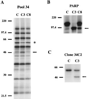
We have recently used a novel in vitro expression cloning strategy to systematically identify cDNAs encoding putative caspase substrates that are proteolyzed during apoptotic cell death.15,16 In the current screen, we incubated 35S-labeled protein pools derived from small pools of a human heart cDNA library (48 cDNAs/pool) with recombinant caspases-3 and -8. As shown in Figure 1A, 35S-labeled protein pool 34 contained a 57 kDa protein (indicated by asterisk) that was specifically cleaved by caspase-3 (C3) into an approximately 48 kDa fragment (indicated by arrow); this proteolytic fragment was not observed when protein pool 34 was incubated with control buffer (C) or caspase-8 (C8). The proteolytic activity of caspase-3 (C3) and caspase-8 (C8) was verified by incubating these proteases with 35S-labeled poly(ADP-ribose) polymerase (PARP), a well characterized caspase substrate:18 caspase-3 (C3) and, to a lesser extent, caspase-8 (C8) cleaved PARP into its signature 85 kDa fragment (Figure 1B). cDNA pool 34 was then progressively subdivided into smaller pools and the corresponding 35S-labeled protein pools were re-examined as above. As shown in Figure 1C, a single cDNA encoding a 57 kDa protein that was cleaved by caspase-3 (C3) into an approximately 48 kDa fragment was isolated. Sequence analysis of this cDNA revealed that it was human vimentin, an abundant type III intermediate filament protein that is expressed in mesenchymal tissues and in a broad spectrum of carcinomas.19,20
Figure 1
Isolation of vimentin as a caspase-3 substrate in vitro by small pool expression cloning. (A) 35S-labeled protein pool 34 contains a 57 kDa protein (indicated by asterisk) that is specifically cleaved into an approximately 48 kDa fragment (indicated by arrow) by caspase-3 (C3), but not by buffer control (C) or caspase-8 (C8). (B) Specific proteolysis of 35S-labeled PARP into its characteristic 85 kDa cleavage fragment (indicated by arrow) by caspase-3, and to a lesser extent, by caspase-8. (C) Isolation of a single cDNA from pool 34 that encodes a 57 kDa protein that is cleaved by caspase-3 into the previously noted 48 kDa fragment. Sequence analysis of this cDNA revealed that it is human vimentin. Small pool expression cloning was performed as described previously15,16 and in Materials and Methods. The molecular mass of markers is indicated at the left of each panel
Vimentin is proteolyzed at several sites including Asp85 and Asp259 by multiple caspases in vitro
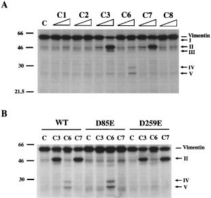
Having isolated vimentin by virtue of its sensitivity to proteolysis by caspase-3 in vitro, we next wanted to determine whether vimentin was cleaved by other caspases in vitro. As demonstrated in Figure 2A, vimentin was most efficiently proteolyzed by caspases-3, -6 and -7 into distinct patterns of proteolytic products in vitro (indicated by arrows and designated I-V); the proteolytic activity of each caspase was verified by incubation with a known 35S-labeled substrate (data not shown). Caspase-3 (C3) preferentially cleaved vimentin to generate fragments II (the above-noted 48 kDa product) and III (fragments I, IV and V were minor products); fragment II was also the most abundant product resulting from vimentin cleavage by caspase-7 (C7). In contrast, caspase-6 (C6) cleaved vimentin most efficiently into fragments I, IV and V (fragment II was less efficiently produced by caspase-6 than by caspases-3 or -7). To determine vimentin's major caspase cleavage sites in vitro, we examined its sequence and identified a potential caspase-3 cleavage site (DXXD motif) that would generate fragment II (DSVD85-F in the non-helical amino-terminus) and a potential caspase-6 site (V/IXXD motif) that would produce fragments IV and V (IDVD259-V in the L1-2 domain).21,22 We next selectively altered these aspartic acid residues by site-directed mutagenesis to create two mutant vimentin constructs: D85E and D259E. As shown in Figure 2B, incubation of 35S-labeled D85E with caspases-3 and -7 failed to generate proteolytic fragment II that was observed when 35S-labeled wild-type (WT) or D259E mutant were incubated with these caspases. Moreover, 35S-labeled D259E was resistant to proteolysis by caspase-6 at this site: fragments IV and V produced by caspase-6 cleavage of wild-type or D85E vimentin were not observed upon incubation of D259E with this protease. Taken together, these studies indicate that vimentin is preferentially cleaved by caspases-3 and -7 at Asp85 and by caspase-6 at Asp259 in vitro; the identity of vimentin's additional caspase cleavage site(s) has yet to be determined. Interestingly, vimentin's caspase-6 cleavage site (Asp259) in its L1-2 linker domain corresponds precisely to the caspase cleavage sites previously reported in the structurally related intermediate filament proteins, the lamins and cytokeratins 18 and 19.8,9,10,11,12,13
Figure 2
Vimentin is proteolyzed at several sites including Asp85 and Asp259 by multiple caspases in vitro. (A) Human vimentin is preferentially cleaved by caspases-3, -6 and -7 to yield distinct proteolytic cleavage patterns in vitro (proteolytic fragments are indicated by arrows and designated I-V). 35S-labeled human vimentin was incubated with control buffer (C), 2.5 or 25 ng of caspases-1 (C1), -2 (C2), -3 (C3), -6 (C6), -7 (C7), or -8 (C8) for 1 h at 37°C, and the cleavage reactions were separated by SDS–PAGE and visualized by autoradiography as detailed in Materials and Methods. (B) vimentin mutant D85E is resistant to proteolysis by caspases-3 and -7 at this site, while vimentin D259E is resistant to cleavage by caspase-6 at the latter site. Mutant vimentin constructs D85E and D259E were made by site-directed mutagenesis as described in Materials and Methods. 35S-labeled wild-type (WT), D85E or D259E vimentin were incubated with control buffer (C) or 25 ng of caspases-3 (C3), -6 (C6) or -7 (C7) for 1 h at 37°C and analyzed as above. The molecular mass of markers in kDa is indicated at the left of each panel
Vimentin is specifically cleaved by caspases during the induction of apoptosis in vivo
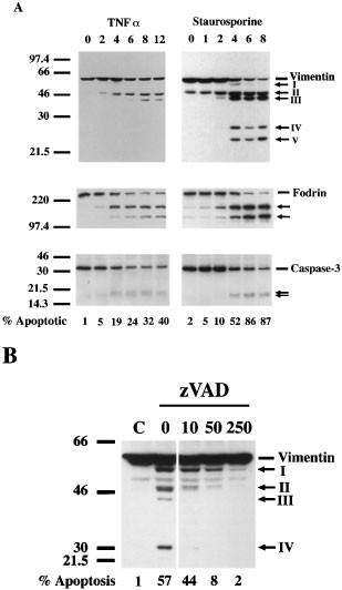
To determine whether vimentin is proteolyzed by caspases during apoptosis in vivo, we treated HeLa cells with TNF-α/cycloheximide or staurosporine for variable time periods and examined cell lysates for evidence of vimentin cleavage by immunoblotting. As shown in Figure 3A, vimentin was rapidly proteolyzed into an approximately 48 kDa fragment (indicated by arrow and designated II) within 2 h of treating cells with either TNF-α/cycloheximide or staurosporine. This cleavage occurred early during the induction of apoptosis when fewer than 10% of treated cells had condensed or fragmented nuclei indicative of apoptotic cell death (the percentage of cells with apoptotic nuclei at each time point is indicated at the bottom of Figure 3A). Additional vimentin fragments (indicated by arrows and labeled I, III-V) were subsequently generated during the induction of apoptosis by these stimuli; fragments I, IV and V were observed only after prolonged exposure of immunoblots from TNF-α treated cells (data not shown). Vimentin was also cleaved into similar sized fragments in Jurkat cells induced to undergo apoptosis by treatment with etoposide or anti-Fas mAb (data not shown). Importantly, the vimentin cleavage products observed during the induction of apoptosis in vivo were of the identical (fragments I-III) or similar (fragments IV and V) size as the corresponding fragments generated by caspase cleavage of vimentin in vitro (data not shown), thereby strongly suggesting that caspases are responsible for its apoptotic cleavage. Further support for this notion comes from the observation that vimentin's cleavage during apoptosis coincided closely with that of the well characterized caspase cytoskeletal target α-fodrin6,7 and with pro-caspase-3 proteolytic activation (manifested as a reduction in proenzyme intensity and/or processing into its p20 and p17 subunits) (Figure 3A). Moreover, as demonstrated in Figure 3B, the broad-spectrum caspase-inhibitor zVAD-fmk antagonized staurosporine-induced vimentin proteolysis and apoptotic cell death (as indicated by diminished nuclear fragmentation). Of note, the various vimentin cleavage products differed in their sensitivity to inhibition by zVAD-fmk: fragment III was completely inhibited by 10 μM, fragment IV by 50 μM, while fragments I and II were almost completely inhibited by 250 μM. Fragment V was faintly observed after prolonged exposure of immunoblots only in staurosporine-treated cells that had not received any zVAD-fmk (data not shown). Taken together, these results indicate unequivocally that vimentin is proteolytically cleaved by multiple caspases (with different sensitivities to inhibition by zVAD-fmk) acting at distinct time points during the execution of apoptotic cell death.
Figure 3
Vimentin is specifically cleaved by caspases during the induction of apoptosis in vivo. (A) Vimentin is rapidly proteolyzed during TNF-α and staurosporine-induced apoptosis into multiple fragments of similar size to those generated in vitro by caspase cleavage; the time course of vimentin's apoptotic cleavage is similar to that of α-fodrin and pro-caspase-3. HeLa cells were treated with 10 ng/ml TNF-α and 1 μg/ml cycloheximide for 0–12 h or with 1 μM staurosporine for 0–8 h. (B) zVAD-fmk inhibits staurosporine-induced vimentin proteolysis. HeLa cells were preincubated with zVAD-fmk (0–250 μM) for 2 h, and then treated for an additional 4 h with 1 μM staurosporine. Cell lysates were analyzed by immunoblotting and the percentage of apoptotic cells (indicated at the bottom) was determined by nuclear morphology as detailed in Materials and Methods. The molecular mass in kDa is indicated at the left of each panel
Caspase cleavage of vimentin disrupts intermediate filaments during apoptosis
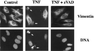
To assess the functional consequences of vimentin's proteolysis by caspases, we examined the cellular distribution of vimentin by indirect immunofluorescence during the induction of apoptosis. As shown in Figure 4, vimentin in untreated, control HeLa cells (left-hand, upper panel) was organized into an extensive network of cytoplasmic intermediate filaments. However, after 3 h of treatment with TNF-α and cycloheximide, vimentin intermediate filaments were dismantled into punctate or granular aggregates in many cells (indicated by arrows, middle upper panel). This striking cytoskeletal event coincided temporally with nuclear fragmentation (indicated by arrows, middle lower panel): under these conditions, all cells with disrupted vimentin filaments were clearly apoptotic by nuclear morphology. The caspase inhibitor zVAD-fmk antagonized vimentin intermediate filament disruption and nuclear fragmentation induced by TNF-α (right-hand, upper and lower panels, respectively), thereby confirming the critical role that caspases play in vimentin filament disassembly.
Figure 4
Caspase cleavage of vimentin disrupts intermediate filaments during apoptosis. TNF-α induces the rearrangement of vimentin intermediate filaments into punctate, granular aggregates (indicated by arrows); this cytoskeletal event coincides temporally with the induction of nuclear fragmentation (indicated by arrows). The caspase inhibitor zVAD-fmk blocks both vimentin filament disruption and nuclear fragmentation. HeLa cells were untreated (control, left-hand panels) or preincubated with DMSO (middle panels) or 250 μM zVAD-fmk (right-hand panels) for 1 h, and then treated for an additional 3 h (DMSO) or 5 h (zVAD-fmk) with 10 ng/ml TNF-α and 1 μg/ml cycloheximide. Vimentin intermediate filaments (upper panels) and nuclear morphology (lower panels) were visualized as detailed in Materials and Methods
Caspase proteolysis of vimentin at Asp85 generates a pro-apoptotic amino-terminal product that amplifies the cell death signal
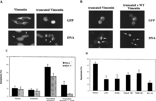
Because caspases initially cleave vimentin at Asp85 during the induction of apoptosis, we postulated that the amino-terminal cleavage product corresponding to amino acids 1–85 (‘truncated’ vimentin) might contribute to the execution of apoptosis. As shown in Figure 5A, GFP-tagged vimentin assembles into extensive intermediate filaments surrounding normal nuclei, while GFP-tagged truncated vimentin is assembly incompetent and induces cellular rounding, cytosolic shrinkage and nuclear condensation/fragmentation. Indeed, GFP-tagged truncated vimentin often forms granular aggregates in cells that resemble those seen in TNF-α-treated cells (see Figure 5B, left-hand, upper panel); cells bearing such aggregates commonly have apoptotic nuclear morphology (Figure 5B, left-hand, lower panel). Moreover, truncated vimentin interferes with the assembly of wild-type vimentin into intermediate filaments in co-transfection experiments (Figure 5B, right-hand, upper panel). As demonstrated in Figure 5C, transient expression of GFP-truncated vimentin but not full-length vimentin induces apoptosis in vimentin-positive HeLa cells and in vimentin-negative MCF-7 cells (which also lack caspase-3). Interestingly, apoptosis induced by truncated vimentin requires caspases: the broad spectrum caspase inhibitor zVAD-fmk antagonizes truncated vimentin-induced cell death. To delineate which caspases might play a role in apoptosis induced by truncated vimentin, we co-transfected HeLa cells with truncated vimentin and a variety of anti-apoptotic cDNAs. As shown in Figure 5D, dominant negative caspase-8 and dominant negative FADD, specific inhibitors of caspase-8,23 CrmA (an inhibitor of caspases-1 and -824), p35 (a non-selective caspase inhibitor) and Bcl-xL antagonize cell death induced by truncated vimentin. Hence, truncated vimentin induces apoptosis in cells by a caspase-dependent mechanism, and caspase-8 may be particularly important in mediating its pro-apoptotic actions.
Figure 5
Truncated vimentin interferes with intermediate filament assembly and induces apoptosis by a caspase-dependent mechanism. (A) HeLa cells transiently transfected with GFP-vimentin assemble into intermediate filaments surrounding normal nuclei (left-hand, upper and lower panels), while GFP-truncated vimentin is assembly incompetent and induces cellular rounding, cytosolic shrinkage and nuclear condensation/fragmentation (right-hand, upper and lower panels). (B) Vimentin-negative MCF-7 cells were transiently transfected with GFP-truncated vimentin alone or co-transfected with GFP-truncated vimentin and GFP-wild-type vimentin (1 μg each). Truncated vimentin often forms granular aggregates in MCF-7 cells (left-hand, upper panel) and interferes with wild-type vimentin's assembly into intermediate filaments (right-hand, upper panel); MCF-7 cells containing granular aggregates often have apoptotic nuclei (lower panels). (C) HeLa and MCF-7 cells were transfected with 1 μg of pEGFP-N1 plasmid containing empty vector, full-length or truncated vimentin (the latter in the presence or absence of 100 μM zVAD-fmk). Twenty-four hours later, GFP-positive cells were scored for apoptosis by nuclear morphology as detailed in ‘Materials and Methods’. (D) HeLa cells were co-transfected with 0.4 μg of pEGFP-N1 plasmid containing truncated vimentin and 1.2 μg of various anti-apoptotic cDNAs (p35, CrmA, caspase-8 dominant negative (Casp-8 DN), FADD dominant negative (FADD DN), or Bcl-xL). GFP-positive cells were scored for apoptosis as above. The data in (C) and (D) represent the mean of at least triplicate experiments±S.D.
Discussion
We have demonstrated that vimentin, an abundant type III intermediate filament protein, is rapidly proteolyzed by multiple caspases at several sites during apoptosis induced by diverse stimuli. Vimentin is initially cleaved at Asp85 (DSVD85-F) by a caspase-3/-7-like protease and subsequently at additional sites, including Asp259 (IDVD259-V) which is cleaved by a caspase-6-like protease. Importantly, vimentin is proteolyzed at Asp85 during the induction of apoptosis when the vast majority (>90%) of cells are still viable; cleavage at this site coincides temporally with caspase-3 proteolytic activation and cleavage of the caspase cytoskeletal target α-fodrin.6,7 Consequently, vimentin proteolysis precedes the dramatic reorganization of the cytoskeleton that typifies apoptotic cell death and could therefore play a role in its execution.
Although other studies have indicated that vimentin is cleaved in response to certain apoptotic stimuli,25,26 the present study provides a more detailed characterization of the molecular mechanisms underlying its apoptotic cleavage. Moreover, our results differ from those of Morishima who reported that vimentin is first cleaved at Asp259 by a caspase-8-like protease and subsequently at Asp85 by caspase-3-like protease.27 Although the explanation for this discrepancy is unclear, we have demonstrated unambiguously that Asp259 in vimentin is readily cleaved by caspase-6 in vitro but not by limited amounts (2.5 or 25 ng) of caspase-8. In contrast, Morishima shows only that caspase-8 (amount unspecified) cleaves Asp259 in vitro. Because isoleucine in the P4 substrate recognition sequence at Asp259 (IDVD259-V) is likely to be well tolerated by either caspase-6 or -8,21,22 we postulate that although caspase-8 may be able to cleave Asp259 in vitro (particularly when caspase-8 is present at high concentrations), caspase-6 is the primary caspase responsible for its proteolysis at this site.
Vimentin, then, can be added to the list of intermediate filament proteins, including the nuclear lamins and cytokeratins 18 and 19, which are cleaved by caspases during apoptotic cell death.8,9,10,11,12,13 Like these other intermediate filament proteins, vimentin is cleaved by caspase-6 or a caspase-6-like protease at a conserved motif in its L1-2 domain (IDVD in vimentin and VEV/ID in the lamins and cytokeratins). Caspase-6 cleavage of the nuclear lamins at the corresponding site directly contributes to the apoptotic destruction of the nucleus: selective inhibition of lamin proteolysis by introducing a cleavage-resistant mutant lamin into cells delays chromatin condensation and DNA fragmentation.10 However, introduction of the corresponding vimentin D259E mutant construct into MCF-7 cells (which lack vimentin) did not confer protection against apoptotic cell death (M Trivedi and V Cryns, unpublished data), likely reflecting the sensitivity of this mutant vimentin protein to caspase cleavage at additional sites. Indeed, vimentin's apoptotic cleavage at multiple sites outside the L1-L2 linker domain distinguishes it from the nuclear lamins.
We have also demonstrated that caspase cleavage of vimentin has important functional consequences, including the dismantling of vimentin intermediate filaments into granular aggregates that bear a striking resemblance to those generated by caspase proteolysis of cytokeratins.12,28 Although phosphorylation of vimentin at Ser55 leads to filament disassembly during mitosis,29 our finding that the apoptotic dismantling of vimentin filaments is zVAD-fmk-inhibitable indicates that caspases play an essential role in this process, independent of any potential phosphorylation events. Given the evidence that several intermediate filament proteins (some with different patterns of tissue expression) are caspase substrates, caspase dismantling of the intermediate filament network is a central theme in the execution of apoptosis. It seems likely that the proteolytic disassembly of this rigid cytosolic network may facilitate many of the morphological manifestations of apoptosis, including cellular rounding and packaging the debris of dying cells into apoptotic bodies.
Importantly, we have demonstrated for the first time that caspase proteolysis of vimentin plays an active role in the execution of apoptosis. Specifically, the initial cleavage of vimentin at Asp85 by a caspase-3-like protease during the induction of apoptosis generates an amino-terminal cleavage product (truncated vimentin) that is sufficient to induce apoptosis. Truncated vimentin is unable to assemble into intermediate filaments; instead, it often forms granular aggregates that closely resemble those seen in apoptotic cells and/or assumes a diffuse cytoplasmic pattern of staining. Truncated vimentin also disrupts the assembly of wild-type vimentin into intermediate filaments, a finding that is consistent with the observation that synthetic peptides derived from vimentin's amino-terminus interfere with intermediate filament assembly.30 Although we have yet to detect truncated vimentin directly in apoptotic cells, we postulate that this proteolytic fragment is likely to be short-lived, perhaps reflecting proteasomal degradation of this fragment that is prone to aggregation. Nevertheless, given the highly dynamic nature of vimentin filaments that are constantly undergoing subunit assembly and disassembly (t1/2 approximately 5 min),31 even a short-lived inhibitor of filament assembly would likely have profound cytoskeletal effects. Furthermore, we have also demonstrated that apoptosis induced by truncated vimentin is dependent on caspase activity, since non-specific caspase inhibitors (zVAD-fmk, p35 and CrmA) antagonize truncated vimentin-induced cell death. Caspase-8 may be a particularly important mediator of truncated vimentin-induced cell death: specific inhibitors of caspase-8 (dominant negative forms of caspase-8 or FADD) partially block apoptosis induced by truncated vimentin. These findings indicate that caspase cleavage of vimentin amplifies the cell death signal by generating an amino-terminal proteolytic product that, in turn, activates more caspases, thereby creating a positive feedback loop. A similar positive feedback loop has been described for other caspase substrates such as the kinase MEKK-1.32 Additional studies will be needed to clarify the mechanism(s) by which truncated vimentin activates caspases and whether the granular aggregates of vimentin participate in this process.
Materials and Methods
Cell lines and reagents
HeLa and MCF-7 cells were grown in DMEM (Life Technologies, Inc.) with 10% heat-inactivated fetal calf serum. z-Val-Ala-Asp(OMe)-fmk (zVAD-fmk) was purchased from Enzyme Systems Products and human recombinant tumor necrosis factor (TNF)-α from R&D Systems. Bisbenzimide (Hoescht #33258) and staurosporine were obtained from Sigma.
Small pool expression cloning
A human heart cDNA library in the pcDNA3 vector (Invitrogen) was subdivided into small pools (48 clones/pool); these pools were screened for cDNAs encoding putative caspase substrates as we have described previously except that recombinant caspase-3 (50 ng) and -8 (100 ng) were used instead of apoptotic extracts.15,16
Proteolytic cleavage of human vimentin in vitro
35S-labeled human vimentin was prepared by in vitro transcription/translation of pcDNA3-vimentin using the TnT T7 Quick Coupled Transcription/Translation system (Promega) according to the manufacturer's instructions. 35S-labeled vimentin was then incubated with control buffer, 2.5 or 25 ng of recombinant caspases-1, -2, -3, -6, -7, or -8 for 1 h at 37°C and the cleavage reaction products analyzed as detailed previously.6,17 The catalytic activity of each caspase was confirmed by incubation with a known 35S-labeled substrate.
Determination of vimentin's caspase cleavage sites in vitro by site-directed mutagenesis
Two mutant human vimentin constructs specifically altered at potential caspase cleavage sites, D85E and D259E, were made using the QuickChange Site-Directed Mutagenesis kit (Stratagene) according to the manufacturer's instructions with the following oligonucleotide primers: 5′-GGACTCGGTGGAATTCTCGCTGGCCG-3′ and 5′-CGGCCAGCGAGAATTCCACCGAGTCC-3′ (D85E) and 5′-CCAAATCGATGTGGAGGTTTCCAAGCCTGACC-3′ and 5′-GGTCAGGCTTGGAAACCTCCACATCGATTTGG-3′ (D259E). Mutations were verified by automated DNA sequencing of both strands. These mutant vimentin constructs were then 35S-labeled, incubated with 25 ng of caspases-3, -6 or -7, and analyzed as above.
Induction and quantitation of apoptosis
HeLa cells were grown on glass coverslips and were treated with 10 ng/ml TNF-α and 1 μg/ml cycloheximide for 0, 2, 4, 6, 8, or 12 h or with 1 μM staurosporine for 0, 1, 2, 4, 6, or 8 h. For inhibitor studies, HeLa cells were preincubated with zVAD-fmk (0, 10, 50 or 250 μM) for 2 h, and then treated for an additional 4 h with 1 μM staurosporine. Following treatment, cells were fixed in 100% methanol for 2 min at −20°C and nuclei were stained with 10 μg/ml Hoescht #33258 for 30 min. The percentage of apoptotic cells was determined by scoring for fragmented/condensed nuclei as visualized by fluorescence microscopy. For each treatment condition, at least 200 nuclei were scored; all experiments were performed in triplicate.
Immunoblotting
Whole cell lysates were prepared and examined by immunoblotting as described previously 6 using the following antibodies: vimentin mAb V9 (Sigma, 1 : 1000 dilution), α-fodrin mAb 1622 (Chemicon, 1 : 1000 dilution) and caspase-3 mAb (Transduction Laboratories, 1 : 1000 dilution).
Indirect immunofluorescence
HeLa cells were grown to approximately 50% confluence on glass coverslips: control cells were untreated, while other cells were preincubated with vehicle (DMS0) or 250 μM zVAD-fmk for 1 h prior to treatment with 10 ng/ml TNF α and 1 μg/ml cycloheximide for an additional 3 h (vehicle-treated) or 5 h (zVAD-fmk-treated). Cells were subsequently fixed in 100% methanol for 2 min at −20°C, washed in PBS, and incubated with vimentin V9 mAb (1 : 20 dilution in PBS) for 2 h at 37°C. After washing in PBS, cells were incubated with fluorescein-conjugated goat affinity purified Ab to mouse IgG (ICN Pharmaceuticals, 1 : 20 dilution in PBS) and 10 μg/ml Hoescht #33258 for 30 m at 37°C, and then again washed in PBS and mounted on glass slides with Fluoromount-G (Southern Biotechnology Associates). Vimentin intermediate filaments and nuclear morphology were visualized by fluorescence microscopy.
Construction of GFP-tagged vimentin cDNAs
Full-length and truncated (encoding amino acids 1–85) vimentin cDNAs were PCR amplied from wild-type human vimentin with the following oligonucleotide primers: 5′-GGCCGAATTTCCCATGTCCACCAGGTCCGTG-3′ and 5′-GGCCGGATCCCCTTCAAGGTCATCGTGATG-3′ (full-length) or 5′-GGCCGAATTTCCCATGTCCACCAGGTCCGTG-3′ and 5′-GGCCGGATCCAAGTCCTGCAG-3′ (truncated). The PCR products were then digested with _Eco_RI and _Bam_HI and cloned into pEGFP-N1 (Clontech) digested with the same two enzymes. The sequence of the resulting full-length and truncated vimentin constructs fused at their C-termini with GFP was confirmed by automated DNA sequencing.
Transfection experiments
HeLa and MCF-7 cells were plated on glass cover slips and transfected with 1 μg of pEGFP-N1 plasmid containing empty vector, truncated or full-length vimentin using Lipofectamine reagent (Life Technologies) according to the manufacturer's instructions. Twenty-four hours later, cells were fixed in 2.5% glutaraldehyde and nuclei stained with Hoescht #33258 as above. The percentage of transfected cells with apoptotic nuclear changes was determined by fluorescence microscopy in triplicate experiments. To determine whether truncated vimentin interferes with assembly of wild-type vimentin into intermediate filaments, MCF-7 cells were co-transfected with pEGFP-N1-truncated vimentin and pEGFP-N1-wild-type vimentin (1 μg of each plasmid), and transfected cells were analyzed as above. In other co-transfection experiments, HeLa cells were transfected with 0.4 μg of pEGFP-N1 plasmid containing truncated vimentin and 1.2 μg of various anti-apoptotic cDNAs, and the percentage of transfected cells that were apoptotic was determined as above. The caspase-8 dominant negative cDNA was kindly provided by Drs. I Sanchéz and J Yuan.
Abbreviations
zVAD-fmk:
z-Val-Ala-Asp(OMe)-fmk
TNF:
tumor necrosis factor
PARP:
poly(ADP-ribose) polymerase
kDa:
kilodaltons
SDS–PAGE:
sodium dodecyl sulfate-polyacrylamide gel electrophoresis
mAb:
monoclonal antibody
PCR:
polymerase chain reaction
GFP:
green fluorescent protein
References
- Steller H . 1995 Mechanisms and genes of cellular suicide Science 267: 1445–1449
Article CAS Google Scholar - Cryns VL, Yuan J . 1998 Proteases to die for Genes Dev. 12: 1551–1570
Article CAS Google Scholar - Thornberry NA, Lazebnik Y . 1998 Caspases: enemies within Science 281: 1312–1316
Article CAS Google Scholar - Brancolini C, Benedetti M, Schneider C . 1995 Microfilament reorganization during apoptosis: the role of Gas2, a possible substrate for ICE-like proteases EMBO J. 14: 5179–5190
Article CAS Google Scholar - Kothakota S, Azuma T, Reinhard C, Klippel A, Tang J, Chu K, McGarry TJ, Kirschner MW, Koths K, Kwiatkowski DJ, Williams LT . 1997 Caspase-3-generated fragment of gelsolin: effector of morphological change in apoptosis Science 278: 294–298
Article CAS Google Scholar - Cryns VL, Bergeron L, Zhu H, Li H, Yuan J . 1996 Specific cleavage of α-fodrin during Fas- and tumor necrosis factor-induced apoptosis is mediated by an interleukin-1β-converting enzyme/Ced-3 protease distinct from the poly(ADP-ribose) polymerase protease J. Biol. Chem. 271: 31277–31282
Article CAS Google Scholar - Wang K, Posmantur R, Nath R, McGinnis K, Whitton M, Talanian R, Glantz S, Morrow J . 1998 Simultaneous degradation of αII- and βII-spectrin by caspase 3 (CPP32) in apoptotic cells J. Biol. Chem. 273: 22490–22497
Article CAS Google Scholar - Lazebnik YA, Takahashi A, Moir RD, Goldman RD, Poirier GG, Kaufmann SH, Earnshaw WC . 1995 Studies of the lamin proteinase reveal multiple parallel biochemical pathways during apoptotic execution Proc. Natl. Acad. Sci. USA 92: 9042–9046
Article CAS Google Scholar - Orth K, Chinnaiyan AM, Garg M, Froelich CJ, Dixit VM . 1996 The CED-3/ICE-like protease Mch2 is activated during apoptosis and cleaves the death substrate lamin A J. Biol. Chem. 271: 16443–16446
Article CAS Google Scholar - Rao L, Perez D, White E . 1996 Lamin proteolysis facilitates nuclear events during apoptosis J. Cell. Biol. 135: 1441–1455
Article CAS Google Scholar - Takahashi A, Alnemri ES, Lazebnik YA, Fernandes-Alnemri T, Litwack G, Mori RD, Goldman RD, Poirier GG, Kaufmann SH, Earnshaw WC . 1996 Cleavage of lamin A by Mch2α but not CPP32: multiple interleukin 1β-converting enzyme-related proteases with distinct substrate recognition properties are active in apoptosis Proc. Natl. Acad. Sci. USA 93: 8395–8400
Article CAS Google Scholar - Caulín C, Salvesen GS, Oshima RG . 1997 Caspase cleavage of keratin 18 and reorganization of intermediate filaments during epithelial cell apoptosis J. Cell. Biol. 138: 1379–1394
Article Google Scholar - Ku N-O, Liao J, Omary MB . 1997 Apoptosis generates stable fragments of human type I keratins J. Biol. Chem. 272: 33197–33203
Article CAS Google Scholar - Brancolini C, Lazarevic D, Schneider C . 1997 Dismantling cell-cell contacts during apoptosis is coupled to a caspase-dependent proteolytic cleavage of beta-catenin J. Cell. Biol. 139: 759–771
Article CAS Google Scholar - Lustig KD, Stukenberg T, McGarry T, King RW, Cryns VL, Mead P, Zon L, Yuan J, Kirschner MW . 1997 Small pool expression screening: a novel strategy for the identification of genes involved in cell cycle control, apoptosis and early development Methods Enzymol. 283: 83–99
Article CAS Google Scholar - Cryns VL, Byun Y, Rana A, Mellor H, Lustig KD, Ghanem L, Parker PJ, Kirschner MW, Yuan J . 1997 Specific proteolysis of the kinase protein kinase C-related kinase 2 by caspase-3 during apoptosis: Identification by a novel small pool expression cloning strategy J. Biol. Chem. 272: 29449–29453
Article CAS Google Scholar - Li H, Bergeron L, Cryns V, Pasternack M, Zhu H, Shi L, Greenberg A, Yuan J . 1997 Activation of caspase-2 in apoptosis J. Biol. Chem. 272: 21010–21017
Article CAS Google Scholar - Lazebnik YA, Kaufmann SH, Desnoyers S, Poirier GG, Earnshaw WC . 1994 Cleavage of poly(ADP-ribose) polymerase by a proteinase with properties like ICE Nature 371: 346–347
Article CAS Google Scholar - Ferrari S, Battini R, Kaczmarek L, Rittling S, Calabretta B, de Riel J, Philiponis V, Wei J, Baserga R . 1986 Coding sequence and growth regulation of the human vimentin gene Mol. Cell. Biol. 6: 3614–3620
Article CAS Google Scholar - Fuchs E, Weber K . 1994 Intermediate filaments: structure, dynamics, function, and disease Annu. Rev. Biochem. 63: 345–382
Article CAS Google Scholar - Thornberry N, Rano T, Peterson E, Rasper D, Timkey T, Garcia-Calvo M, Houtzager V, Nordstrom P, Roy S, Vaillancourt J, Chapman K, Nicholson D . 1997 A combinatorial approach defines specificities of members of the caspase family and granzyme B. Functional relationships established for key mediators of apoptosis J. Biol. Chem. 272: 17907–17911
Article CAS Google Scholar - Talanian RV, Quinlan C, Trautz S, Hackett MC, Mankovich JA, Banach D, Ghayur T, Brady KD, Wong WW . 1997 Substrate specificities of caspase family proteases J. Biol. Chem. 272: 9677–9682
Article CAS Google Scholar - Sánchez I, Xu C-J, Juo P, Kakizaka A, Blenis J, Yuan J . 1999 Caspase-8 is required for cell death induced by expanded polyglutamine repeats Neuron 22: 623–633
Article Google Scholar - Zhou Q, Snipas S, Orth K, Muzio M, Dixit VM, Salvesen GS . 1997 Target protease specificity of the viral serpin CrmA. Analysis of five caspases J. Biol. Chem. 272: 7797–7800
Article CAS Google Scholar - van Engeland M, Kuijpers H, Ramaekers F, Reutelingsperger C, Schutte B . 1997 Plasma membrane alterations and cytoskeletal changes in apoptosis Exp. Cell. Res. 235: 421–430
Article CAS Google Scholar - Hashimoto M, Inoue S, Ogawa S, Conrad C, Muramatsu M, Shackelford D, Masliah E . 1998 Rapid fragmentation of vimentin in human skin fibroblasts exposed to tamoxifen: A possible involvement of caspase-3 Biochem. Biophys. Res. Commun. 247: 401–406
Article CAS Google Scholar - Morishima N . 1999 Changes in nuclear morphology during apoptosis correlate with vimentin cleavage by different caspases located either upstream or downstream of Bcl-2 action Genes to Cells 4: 401–414
Article CAS Google Scholar - MacFarlane M, Merrison W, Dinsdale D, Cohen GM . 2000 Active caspases and cleaved cytokeratins are sequestered into cytoplasmic inclusions in TRAIL-induced apoptosis J. Cell. Biol. 148: 1239–1254
Article CAS Google Scholar - Chou Y-H, Bischoff JR, Beach D, Goldman RD . 1990 Intermediate filament reorganization during mitosis is mediated by p34cdc2 phosphorylation of vimentin Cell 62: 1063–1071
Article CAS Google Scholar - Hofmann I, Herrmann H . 1992 Interference in vimentin assembly in vitro by synthetic peptides derived from the vimentin head domain J. Cell. Sci. 101: 687–700
CAS PubMed Google Scholar - Yoon M, Moir RD, Prahlad V, Goldman RD . 1998 Motile properties of vimentin intermediate filament networks in living cells J. Cell. Biol. 143: 147–157
Article CAS Google Scholar - Cardone MH, Salvesen GS, Widmann C, Johnson G, Frisch SM . 1997 The regulation of anoikis: MEKK-1 activation requires cleavage by caspases Cell 90: 315–320
Article CAS Google Scholar
Acknowledgements
We are indebted to Dr. R Talanian for providing the recombinant caspases used in this study, to Drs. I Sanchéz and J Yuan for the caspase-8 dominant negative cDNA, and to Drs. Harris Perlman and Z Oltvai for their critical reading of the manuscript. This work was supported in part by Mentored Clinical Scientist Development Award K08-CA01752 (to VL Cryns) and R01 AR41836 (to KJ Green) from the NIH, by institutional research grants to Northwestern University from the Howard Hughes Medical Institute and the American Cancer Society (to VL Cryns), and by the Elizabeth Boughton Trust (to VL Cryns).
Author information
Authors and Affiliations
- Department of Medicine, Center for Endocrinology, Metabolism and Molecular Medicine, Northwestern University Medical School, Chicago, 60611, IL, USA
Y Byun, F Chen, R Chang, M Trivedi & V L Cryns - Department of Pathology, Northwestern University Medical School, Chicago, 60611, IL, USA
K J Green
Authors
- Y Byun
You can also search for this author inPubMed Google Scholar - F Chen
You can also search for this author inPubMed Google Scholar - R Chang
You can also search for this author inPubMed Google Scholar - M Trivedi
You can also search for this author inPubMed Google Scholar - K J Green
You can also search for this author inPubMed Google Scholar - V L Cryns
You can also search for this author inPubMed Google Scholar
Corresponding author
Correspondence toV L Cryns.
Additional information
Edited by Y Lazebnik
Rights and permissions
About this article
Cite this article
Byun, Y., Chen, F., Chang, R. et al. Caspase cleavage of vimentin disrupts intermediate filaments and promotes apoptosis.Cell Death Differ 8, 443–450 (2001). https://doi.org/10.1038/sj.cdd.4400840
- Received: 17 July 2000
- Revised: 30 November 2000
- Accepted: 19 December 2000
- Published: 22 June 2001
- Issue Date: 01 May 2001
- DOI: https://doi.org/10.1038/sj.cdd.4400840
