Proapoptotic Bcl-2 family member Bim is involved in the control of mast cell survival and is induced together with Bcl-XL upon IgE-receptor activation (original) (raw)
Introduction
Mast cells are important effector and regulatory cells in inflammatory reactions. Upon activation, mast cells release and secrete inflammatory mediators, such as histamine, proteases, prostaglandin, leukotrienes and cytokines (e.g. TNF-α). Mast cells originate from hemopoietic stem cells in the bone marrow and are dispersed throughout most tissues. Although mast cells have been studied mostly in the context of allergic diseases, it is now evident that these cells also play a critical role in host defense against certain pathogens.1 Moreover, it has been demonstrated that mast cells can influence the onset and/or disease severity of chronic inflammatory reactions, for example in certain autoimmune diseases.2
In healthy individuals, the numbers of tissue mast cells are kept at a constant level, but during inflammation, such as allergy,3 allergic asthma4 or rheumatoid arthritis,5 their numbers are increased. When the inflammation has resolved, mast cell numbers decline, and there is evidence that apoptotic cell death is involved in this process. The regulation of mast cell homeostasis has not been studied in any great detail and the molecular mechanisms involved in mast cell apoptosis still need to be deciphered.
Mammals have two distinct apoptosis signaling pathways6 that converge upon activation of aspartate-specific cysteine proteases (caspases) which demolish cells by cleaving vital structural proteins and by activating latent enzymes that degrade DNA.7 The Bcl-2 family of prosurvival and proapoptotic proteins are critical regulators of cell survival7 and the balance between the two subgroups determines whether a cell will survive or undergo apoptosis. The prosurvival Bcl-2 family members include Bcl-2, Bcl-XL, Bcl-w, A1, Mcl-1 and Boo/Diva and these proteins all share three or four regions of homology, called Bcl-2 homology (BH) regions. The proapoptotic family members can be further divided into two subgroups, the Bax/Bak-like proteins and the BH3-only proteins. The Bax/Bak subgroup consists of Bax, Bak, Bok/Mtd, Bcl-xS, Bcl-GL and Bfk and these proteins share two or three BH regions and, surprisingly, at least in the case of Bax also extensive structural similarity with their prosurvival relatives.7 The BH3-only proteins include Bik/Blk/Nbk, Bad, Hrk/DP5, Bid, Bim/Bod, Noxa, Bmf and Puma/Bbc3 and they share with each other and the Bcl-2 family only the BH3 region.8 The BH3 domain is essential for the binding of these proapoptotic proteins to the antiapoptotic Bcl-2 family members and for inducing apoptosis.8 Genetic and biochemical experiments have indicated that the BH3-only proteins initiate programmed cell death and stress-induced apoptosis and that the Bax/Bak-like proteins play an essential role in cell killing further downstream.9, 10
Knowledge about the role of Bcl-2 family members in the regulation of mast cell survival and apoptosis is limited. It has been shown that Bcl-2 and Bcl-XL are expressed in mast cells,11, 12, 13 but very little is known about their function in these cells. It has been reported that _bax_−/− and _bcl_-2−/− mice have abnormal numbers of mast cells in certain tissues but no general difference with wt animals were observed.14 We have previously shown that the Bcl-2 family member A1 is critical for the prosurvival stimuli triggered by crosslinking of the high-affinity IgE receptor, Fc_ɛ_RI.15
In this study, we provide the first evidence for an essential function of the proapoptotic BH3-only protein Bim in mast cells and that its expression can be regulated by Fc_ɛ_RI activation.
Results
Bim is an inducer of cytokine withdrawal-induced apoptosis in mast cells
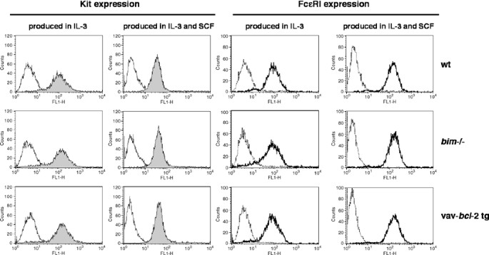
To examine whether Bim has a function in the regulation of growth factor withdrawal-induced mast cell apoptosis, we compared the survival of _in vitro_-developed bone marrow-derived mast cells (BMMC) from _bim_−/−16 and wt mice. In order to further study the role of Bcl-2 family members in the regulation of mast cell survival and apoptosis, mast cells were also generated from vav-_bcl_-2 transgenic mice, which overexpress Bcl-2 in all hemopoietic cells, including mast cells.17 The mast cells were produced in either IL-3, or in the presence of both IL-3 and stem cell factor (SCF). The BMMC developed from the wt, _bim_−/− and vav-_bcl_-2 transgenic mice all had typical mast cell morphology and staining of granula by toluidine blue. Flow cytometric analysis demonstrated that the different genotypes expressed the SCF-receptor Kit and the IgE-receptor Fc_ɛ_RI to similar extent (Figure 1).
Figure 1
Wt, _bim_−/− and vav-_bcl_-2 transgenic BMMC express the receptors Kit and Fc_ɛ_RI to similar extent. By flow cytometric analysis, the receptor expression was analyzed on BMMC produced in IL-3 and on BMMC produced in a combination of IL-3 plus SCF. Left panel: filled peaks=Kit expression, empty peaks=isotype control. Right panel: thick line=Fc_ɛ_RI expression, dotted line=isotype control. The results shown are representative of three independent experiments
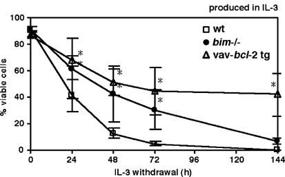
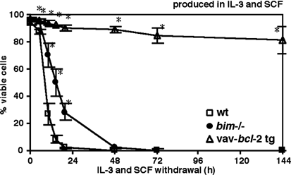
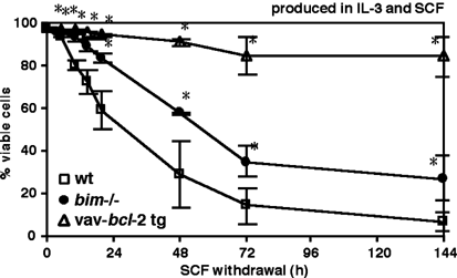
It has previously been demonstrated that mast cells undergo apoptosis as a result of growth factor withdrawal.18, 19 In our experiments, the wt, _bim_−/− and vav-_bcl_-2 transgenic mast cells were deprived of either IL-3, IL-3 and SCF, or of SCF alone. Cell death was determined by staining with propidium iodide followed by flow cytometric analysis. After 48 h of IL-3 deprivation from mast cells produced in IL-3 alone, only 13% wt cells had survived, whereas 42% of the _bim_−/− cells were still alive (Figure 2). After 15 h of growth factor (IL-3 plus SCF) deprivation of mast cells produced in both IL-3 and SCF, only 8% of the wt cells remained alive, whereas 50% of the _bim_−/− cells were still alive (Figure 3). When only SCF was withdrawn from BMMC produced in IL-3 plus SCF, 29% of the wt BMMC remained alive after 48 h, while 57% of the _bim_−/− mast cells had survived (Figure 4). Higher concentrations of IL-3 delayed the cell death, but did not rescue the cells from undergoing apoptosis (data not shown). The results demonstrate for the first time that Bim is involved in the induction of mast cell death following growth factor withdrawal. Our results also show that IL-3 alone is not sufficient to promote survival of mast cells produced in both IL-3 plus SCF.
Figure 2
Bim deficiency and _bcl_-2 transgene expression inhibit IL-3 withdrawal-induced mast cell death. Wt, _bim_−/− and vav-_bcl_-2 transgenic BMMC produced in IL-3 were deprived of IL-3, and cell viability was analyzed at the time points indicated. Cell viability was determined by propidium iodide staining and flow cytometric analysis. Data are presented as the mean ±S.D. (_n_=4). Significant differences compared to wt were obtained as indicated (*P<0.05)
Figure 3
Bim deficiency and _bcl_-2 transgene expression inhibit mast cell death induced by removal of IL-3 and SCF. Wt, _bim_−/− and vav-_bcl_-2 transgenic BMMC produced in IL-3 and SCF were deprived of both IL-3 and SCF, and cell viability was analyzed at the time points indicated as described in Figure 2. Data are presented as the mean±S.D. (_n_=6). Significant differences compared to wt were obtained as indicated (*P<0.05)
Figure 4
Bim deficiency and _bcl_-2 transgene expression inhibit SCF withdrawal-induced mast cell death. Wt, _bim_−/− and vav-_bcl_-2 transgenic BMMC produced in IL-3 and SCF were deprived of SCF, and cell viability was analyzed at the time points indicated as described in Figure 2. Data are presented as the mean±S.D. (_n_=4). Significant differences compared to wt were obtained as indicated (*P<0.05)
We also investigated the effect of overexpressing the prosurvival gene bcl-2 on growth factor withdrawal-induced mast cell apoptosis. Overexpression of Bcl-2 protected mast cells even more potently against cytokine deprivation-induced apoptosis than did loss of Bim (Figures 2, 3 and 4). Hence, additional Bcl-2 inhibitable death inducers besides Bim, most likely other BH3-only proteins, may be involved in the mast cell death triggered by growth factor withdrawal.
When growth factors were withdrawn from the vav-_bcl_-2 transgenic BMMC that had been produced in both IL-3 and SCF, there was an almost complete rescue from cell death even after 6 days, the latest time point studied (Figures 3 and 4). When IL-3 was withdrawn from the BMMC produced in only IL-3, overexpression of Bcl-2 also provided substantial protection from apoptosis, but not as much as for the cells produced in both IL-3 and SCF (Figure 2).
An interesting observation was that mast cells produced in both IL-3 and SCF are much more sensitive to growth factor withdrawal than BMMC produced in IL-3 alone (Figures 2 and 3). For wt mast cells produced in IL-3, it took 3 days of growth factor withdrawal before most cells were dead (Figure 2). However, when wt mast cells that had been produced in both IL-3 plus SCF were starved of their growth factors, almost all cells were already dead after 20 h (Figure 3). Together with the observation that upon cytokine withdrawal Bcl-2 overexpression can almost completely rescue BMMC produced in IL-3 and SCF but only partially rescue those grown in IL-3, these results indicate that there are significant differences in the mechanisms regulating apoptosis following growth factor withdrawal, depending on whether mast cells have been produced in IL-3 alone or by the combined stimulation with IL-3 and SCF.
The prosurvival effect of IgE-receptor activation is not due to inactivation of Bim
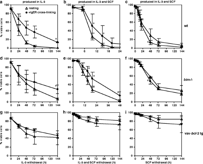
We also investigated whether inhibition of Bim could be involved in activation-induced survival of mast cells. In order to examine this, wt, _bim_−/− and vav-_bcl_-2 transgenic BMMC were deprived of growth factors with or without prior IgE-receptor crosslinking. If inactivation of Bim was essential for the prosurvival stimuli induced by IgE-receptor crosslinking, one would expect no additional prosurvival effect in Fc_ɛ_RI-stimulated _bim_−/− mast cells compared to nonstimulated _bim_−/− cells. However, both the wt and the _bim_−/− mast cells obtained a clear prosurvival stimulus upon Fc_ɛ_RI activation, indicating that inhibition of Bim alone cannot be the sole prosurvival activity of Fc_ɛ_RI stimulation (Figure 5a,b,d and e).
Figure 5
Absence of Bim does not affect the prosurvival stimulus from IgE-receptor crosslinking. In the experiments shown (a–i) wt, _bim_−/− or vav-_bcl_-2 transgenic BMMC were activated by crosslinking of the Fc_ɛ_RI by IgE, followed by withdrawal of growth factors. (a, d and g) BMMC produced in IL-3 were deprived of IL-3, with or without IgE-receptor activation prior to growth factor withdrawal. (b, e and h) BMMC produced in IL-3 and SCF were activated and deprived of IL-3 and SCF. (c, f and i) BMMC produced in IL-3 and SCF were activated, and SCF was withdrawn. In all experiments, cell viability was analyzed as described in Figure 2. Data are presented as the mean±S.D. (_n_=4–6). Significant effects compared to resting cells were obtained as indicated (*P<0.05)
Fc_ɛ_RI stimulation does not promote survival in SCF-deprived or Bcl-2 overexpressing mast cells
As already described above, the mechanisms of growth factor withdrawal-induced BMMC death depends on the cytokines in which they have been produced. Similarly, we found that the method of mast cell production affected Fc_ɛ_RI activation-induced BMMC survival. When only SCF was withdrawn from BMMC produced in IL-3 plus SCF, no prosurvival effect was obtained as a result of IgE-receptor crosslinking (Figure 5c,f and i). This differed from the experiments in which both growth factors (IL-3 plus SCF) were withdrawn from mast cells (Figure 5b and e), and from the experiments in which IL-3 was withdrawn from mast cells produced in IL-3 alone (Figure 5a,d and g). In addition, we found that vav-_bcl_-2 transgenic mast cells produced in IL-3 plus SCF did not survive better upon cytokine withdrawal when activated through their Fc_ɛ_RI compared to cells not stimulated through the Fc_ɛ_RI (Figure 5h and i).
Bim and Bcl-XL proteins are upregulated upon IgE-receptor crosslinking
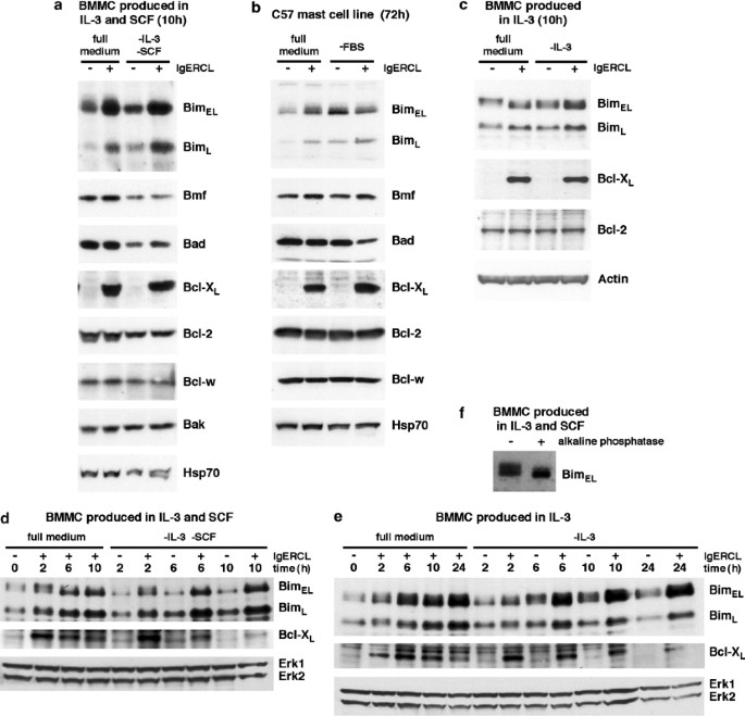
We next examined the expression of several pro- and antiapoptotic proteins of the Bcl-2 family in BMMC and in the C57 mast cell line (Figure 6). The C57 mast cell line was used to compare primary growth factor-dependent mast cells with a growth factor-independent cell line. Cell lysates were prepared from resting and Fc_ɛ_RI-activated BMMC grown for 10 h in the presence or absence of IL-3 and SCF (Figure 6a). The C57 mast cell line is growth factor independent, therefore these cells were deprived of serum instead of growth factors, and the cells were harvested and lysed 72 h after activation and/or serum deprivation (Figure 6b). All of the Bcl-2 family members analyzed were readily detectable in BMMC and C57 cells (Figure 6). These included the prosurvival proteins Bcl-XL, Bcl-2 and Bcl-w, the proapoptotic multi BH domain protein Bak and the proapoptotic BH3-only proteins Bim, Bmf and Bad.
Figure 6
Crosslinking of Fc_ɛ_RI by IgE increases the levels of Bim and Bcl-XL proteins. (a) Wt BMMC produced in IL-3 and SCF were activated by IgE-receptor crosslinking, deprived of IL-3 and SCF, or subjected to both stimuli. After 10 h, the levels of different Bcl-2 family members were analyzed by Western blotting using specific polyclonal or monoclonal antibodies. (b) C57, a cytokine-independent mast cell line, was stimulated by IgE-receptor crosslinking and/or deprived of FBS. After 72 h, protein levels were analyzed as in (a). (c) Wt BMMC produced in IL-3 were activated by IgE-receptor crosslinking and/or deprived of IL-3. After 10 h, protein levels were analyzed as in (a). (d and e) BMMC produced in IL-3 plus SCF or in IL-3 alone were activated by IgE-receptor crosslinking and/or deprived of growth factors for varying periods of time. Protein levels were analyzed as in (a). (f) Cell lysates from BMMC produced in IL-3 plus SCF were treated with alkaline phosphatase, and then analyzed by Western blotting as in (a). The Probing with anti-Hsp70, anti-Actin, or anti-Erk Abs was used as control for the amount of proteins loaded. The results shown are representative of two independent experiments
In agreement with our previous mRNA expression analysis,15 we could detect upregulation of Bcl-XL protein levels upon IgE-receptor crosslinking, both in BMMC and in C57 cells (Figure 6). We also observed that withdrawal of IL-3 and SCF caused a shift in migration of the Bcl-XL and Bcl-2 bands on the Western gel (Figure 6a). The Bcl-XL and Bcl-2 proteins were found as distinct, slow-migrating bands in lysates from the growth factor-deprived cells, whereas lysates from cells grown in the presence of cytokines also contained faster-migrating proteins (Figure 6a). This shift in migration could be due to differences in phosphorylation status. Both Bcl-XL and Bcl-2 are known to be phosphorylated and dephosphorylated, and it has been reported that this can affect their function.20, 21
It has been reported that Bim protein levels are increased upon growth factor withdrawal in several cell types.22, 23 Surprisingly, we found that both BimEL and BimL were strongly increased following IgE-receptor activation (Figure 6). We also analyzed the expression of the Bcl-2 family members Bim, Bcl-XL and Bcl-2 in BMMC produced in IL-3. A similar expression pattern could be observed in BMMC produced in IL-3 as for those produced in IL-3 plus SCF (Figure 6c). Bim and Bcl-XL were induced after IgE-receptor activation, whereas Bcl-2 levels were unaffected. This is the first observation that FcR activation can lead to upregulation of a proapoptotic BH3-only protein.
We further analyzed the kinetics of the IgE-receptor activation-induced upregulation of Bim and Bcl-XL. Both Bim and Bcl-XL were upregulated 2 h after IgE-receptor crosslinking (Figure 6d and e). This was observed in the BMMC produced in IL-3 plus SCF as well as in the cells produced in IL-3 alone.
Growth factor deprivation changed both the level and the migration pattern of the Bim protein. In BMMC grown in the presence of IL-3 and SCF, BimEL consists of proteins migrating with slightly varying mobilities on SDS-PAGE giving rise to a blurry band (Figure 6a). However, following deprivation of IL-3 and SCF, only fast-migrating BimEL was found. As for Bcl-XL and Bcl-2, also the Bim protein has been reported to be a target for phosphorylation24 and this is thought to regulate its degradation through the proteasome pathway.25, 26 In order to analyze whether the differences in migration mobility of BimEL on the Western gel is due to differences in phosphorylation status of BimEL, cell lysates from BMMC cultured in IL-3 plus SCF were treated with alkaline phosphatase before loading onto a Western gel. This treatment abolished the slower migrating portion of the BimEL band (Figure 6f), demonstrating that this upper band represents a phosphorylated form of BimEL.
Discussion
Bim has been shown to be involved in the regulation of apoptosis in several cell types, including B lymphocytes, mature T lymphocytes, thymocytes, monocytes, granulocytes, osteoclasts and neurons.16, 23, 26, 27, 28 Our studies demonstrate for the first time that Bim is involved in the induction of mast cell death following growth factor withdrawal. Since we only observed a delay in the induction of death in _bim_−/− mast cells compared to wt cells, and not a total rescue, other factor(s) in addition to Bim most likely are involved in cytokine deprivation-induced mast cell death. It is possible that other BH3-only proteins expressed in mast cells play an important role in this cell death and this needs to be investigated further by using knockout mice lacking, for example, Bad, Blk, Puma or Bid. Puma is of particular interest since it has recently been shown that Puma plays a critical role in cytokine withdrawal-induced apoptosis of thymocytes and myeloid progenitors.29, 30 The finding that overexpression of Bcl-2 protected mast cells even more potently against cytokine withdrawal-induced apoptosis than did loss of Bim provides further evidence that other Bcl-2 inhibitable death inducers besides Bim, most likely other BH3-only proteins, are involved in the mast cell death caused by growth factor deprivation.
In our experiments, we could observe some differences depending on how the BMMC were produced – in IL-3 alone or by costimulation of IL-3 plus SCF. For example, we found that Bcl-2 protected more potently from growth factor deprivation-induced apoptosis in BMMC produced in IL-3 plus SCF compared to in BMMC produced in IL-3 alone. This may indicate that in mast cells Bcl-2 can compensate more efficiently for SCF-receptor signaling than for IL-3 signaling. A possible explanation for this finding may be that Bcl-2 noninhibitable death inducers contribute to the growth factor withdrawal-induced death of mast cells produced in only IL-3. Alternatively, Bcl-2 may be more efficiently neutralized in mast cells produced in IL-3 alone compared to those produced in IL-3 plus SCF.
We also observed that the BMMC produced in both IL-3 plus SCF proliferated much more rapidly than did the BMMC produced in IL-3 alone (data not shown). This is consistent with what has been observed by others.31 This may indicate that increased level of cytokine stimulation and increased cell cycling render mast cells (and perhaps also other cell types) more dependent on growth signals.
Another interesting finding was that mast cells produced in both IL-3 plus SCF undergo apoptosis much more rapidly upon growth factor withdrawal than does BMMC produced in IL-3 alone. A similar rapid decrease in mast cell viability upon SCF withdrawal from cells maintained in SCF has been reported earlier.19 Taken together, our results demonstrate that depending on whether mast cells have been produced in IL-3 alone or by the combined stimulation with IL-3 and SCF, the cells differ significantly in their response to growth factor deprivation.
It has been reported that crosslinking of the high-affinity IgE-receptor Fc_ɛ_RI elicits a prosurvival response in mast cells.15, 32 We have previously shown that mRNA levels for the prosurvival genes A1 and Bcl-XL are increased following IgE-receptor crosslinking and that A1 is critical for the activation-induced survival of mast cells.15 We investigated whether inhibition of Bim function is the most critical prosurvival stimulus elicited by IgE-receptor crosslinking. Our results do not support this hypothesis, but instead indicate that inhibition of Bim alone cannot be the sole prosurvival activity of Fc_ɛ_RI stimulation. Since Fc_ɛ_RI stimulation did not further increase the survival of cytokine-deprived mast cells overexpressing Bcl-2, it is likely that this antiapoptotic signal prevents activation of certain BH3-only proteins, perhaps Puma or Bad, since they have been implicated in growth factor withdrawal-induced apoptosis.29, 30, 33
Not only were there differences in the mechanisms regulating apoptosis following growth factor withdrawal depending on the cytokines used for cell growth but we could also observe differences in the IgE-receptor activation-induced prosurvival stimuli. When BMMC produced in both IL-3 plus SCF were IgE-receptor activated followed by withdrawal of SCF alone, the mast cells did not survive better than did mast cells that had not been Fc_ɛ_RI stimulated. In contrast, IgE-receptor stimulation elicited a prosurvival activity in mast cells deprived of both IL-3 and SCF. Moreover, in Bcl-2-overexpressing mast cells produced in IL-3 plus SCF, there was no prosurvival response from IgE-receptor activation compared to resting mast cells, when depriving the cells of cytokines following the Fc_ɛ_RI stimulation. The protection of the cells may, in these experiments, already be maximal due to the overexpression of Bcl-2, which could explain why no further prosurvival effect is seen after Fc_ɛ_RI activation.
In a previous study, we analyzed the mRNA levels of several Bcl-2 family members in resting and IgE-receptor-activated cells, and found that the prosurvival members A1 and Bcl-XL were upregulated.15 However, at the protein level, it has not yet been thoroughly analyzed which Bcl-2 family members might be involved in the regulation of apoptosis following IgE-receptor crosslinking or growth factor withdrawal. It has previously been reported that SCF withdrawal leads to downregulation of the Bcl-2 and Bcl-XL protein levels in human mast cells.13 In some other cell types (e.g. lymphoid or neuronal cells), the proapoptotic BH3-only protein Bim has been reported to be upregulated upon growth factor withdrawal,22, 23, 34 but the expression of Bim has not yet been studied in mast cells. In this study, we found that the prosurvival Bcl-XL and, to our surprise, the proapoptotic Bim proteins were both strongly upregulated following IgE-receptor activation. This is the first time that it has been shown that FcR activation can result in upregulation of a proapoptotic BH3-only protein. We have recently shown that activation of the T-cell receptor induces upregulation of Bim and that this is critical for the deletion of autoreactive thymocytes.27 The signaling pathways responsible for Fc_ɛ_RI-mediated upregulation of Bim needs to be further investigated. Mast cell activation through Fc_ɛ_RI has been reported to stimulate the Akt signaling pathway.35 Activation of Akt has in several cell types been shown to result in phosphorylation, and hence inactivation, of forkhead transcription factors, such as FOXO3a.36 Since FOXO3a, when in an active state, is considered as an important transcription factor for Bim,22 the observed Fc_ɛ_RI-mediated upregulation of Bim was somewhat unexpected. Studies on other transcription factors besides forkhead transcription factors might shed light on signaling molecules that are responsible for the upregulation of Bim following IgE-receptor activation.
The finding that both prosurvival and proapoptotic members of the Bcl-2 protein family are upregulated in response to IgE-receptor activation might explain why this type of activation in some experimental settings results in decreased cell death, while no prosurvival effect is seen in other conditions. Cell fate is likely to be determined by the balance between the prosurvival and the proapoptotic molecules.
An interesting observation was that deprivation of IL-3 and SCF from mast cells can alter the phosphorylation state of several Bcl-2 family members, including Bim, Bcl-2 and Bcl-XL. Bim becomes dephosphorylated following withdrawal of IL-3 and SCF, while Bcl-2 and Bcl-XL appear to be phosphorylated. Several previous studies have demonstrated that the phosphorylation status can affect the function of these proteins.20, 21, 25
Although Bim is clearly an initiator of cytokine deprivation-induced mast cell death, our results indicate that other proteins are also involved in this process. Since overexpression of Bcl-2 protected mast cells against cytokine withdrawal more potently than loss of Bim, other Bcl-2 inhibitable proteins, quite likely other BH3-only proteins, are probably activated by growth factor deprivation. Our expression analysis shows that neither Bmf nor Bad protein levels are increased upon IL-3/SCF deprivation, but these proteins might still be involved in this process if their proapoptotic activity is activated by a post-translational mechanism. Moreover, there exist several other BH3-only proteins that remain to be studied for their role in regulating mast cell apoptosis.
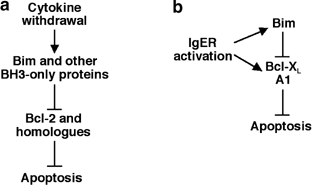
Collectively, our results show that Bim, together with other Bcl-2 inhibitable proteins, is involved in the regulation of apoptosis and survival of mast cells. In Figure 7a, a model is presented on how cytokine withdrawal could lead to mast cell apoptosis by activating Bim and other BH3-only proteins, thereby inhibiting the prosurvival effect of Bcl-2 and other prosurvival homologs. We have also found that IgE-receptor crosslinking upregulates the protein levels of both prosurvival and proapoptotic Bcl-2 family members, namely, Bcl-XL and Bim. Considering these results and previously published data regarding the involvement of A1 in IgE-receptor activation-induced mast cell survival,15 we present a model for how the fate of mast cells following IgE-receptor crosslinking could be fine-tuned by prosurvival and proapoptotic Bcl-2 family members (Figure 7b). The balance between proapoptotic and prosurvival members of the Bcl-2 protein family depends on the amounts of different family members present in a cell. Circumstances that could affect the prosurvival/proapoptotic balance include whether the proteins are in active or inactive forms, the subcellular localization of these proteins and the mode of interaction between the different family members.37
Figure 7
Two possible models for the role of Bim in the regulation of mast cell survival. (a) A mechanism by which cytokine withdrawal induces mast cells to undergo apoptosis could be that Bim and activation of other BH3-only proteins hinder prosurvival Bcl-2 homologues from performing their antiapoptotic function. (b) Crosslinking of the Fc_ɛ_RI by IgE leads to upregulation of both prosurvival and proapoptotic members of the Bcl-2 family. The balance between prosurvival and proapoptotic proteins following IgE-receptor activation of mast cells might determine if the net result of the receptor activation is increased survival or apoptosis
Materials and Methods
Mice
The mice used were Bim-deficient (266/266del) mice backcrossed for more than 12 generations onto a C57BL/6J genetic background,16 vav-_bcl_-2-69 transgenic mice, expressing human Bcl-2 under control of the vav promoter in all hemopoietic cells, on an inbred C57BL/6J genetic background17 and wt C57BL/6J mice (from the Walter and Eliza Hall Institute mouse breeding facility, Kew, Australia). All experiments with animals were performed according to the guidelines of the Royal Melbourne Hospital Research Foundation Animal Ethics Committee.
Mast cell cultures
BMMC were obtained by culturing mouse bone marrow cells in RPMI medium (Gibco) supplemented with 10% heat-inactivated fetal bovine serum (FBS) (TRACE Scientific), 50 _μ_M 2-ME, 50 _μ_g/ml penicillin, 25 _μ_g/ml streptomycin and 1000 U/ml of IL-3 (produced by X63/0 cells transfected with a mouse IL-3 expression construct). Alternatively, BMMC were produced in medium supplemented with 1000 U/ml IL-3 and 25 ng/ml SCF (rmSCF, produced in Pichia pastoris and purified from culture supernatants by reverse phase chromatography). In one experiment for analysis of protein levels, BMMC were obtained by culturing in RPMI medium (Sigma) supplemented with 10% heat-inactivated FBS (Gibco), 10 mM HEPES buffer, 1 × MEM nonessential amino-acid solution, 1 mM sodium pyruvate, 2 mM L-glutamine (all from Sigma), 50 _μ_M 2-ME, 100 IU/ml penicillin G, 50 _μ_g/ml streptomycin and 5 ng/ml IL-3 (rmIL-3, PeproTech). All bone marrow-derived mast cells were cultured for a minimum of 3 weeks before they were used, and no experiments were performed on cells older than 9 weeks. The maturity and purity of the cells were examined by toluidine blue staining. Expression of Kit and Fc_ɛ_RI was analyzed by flow cytometric analysis using FITC-anti-mouse CD117 (c-Kit) (2B8), FITC-conjugated rat IgG2b isotype control (both from Pharmingen), FITC-conjugated anti-mouse Fc_ɛ_RI-α (MAR-1) and FITC-conjugated Armenian Hamster IgG isotype control (both from eBioscience).
The mast cell line C5738 (a gift from Dr. SJ Galli, Stanford University, Stanford, CA, USA) was cultured in RPMI medium (Gibco) supplemented with 10% heat-inactivated FBS, 50 _μ_M 2-ME, 50 _μ_g/ml penicillin and 25 _μ_g/ml streptomycin.
Mast cell activation
For activation through crosslinking of the IgE-receptor, mast cells were sensitized for 90 min at 37°C with a monoclonal mouse anti-TNP IgE-antibody (IgEl-b4, ATCC), used as a 15% v/v hybridoma supernatant (corresponding to 1.5 _μ_g/ml IgE). After two washings in PBS, the cells were resuspended in medium and activated by addition of 100 ng/ml TNP-BSA (coupling ratio: 9/1) (Biosearch Technologies). To the control cells neither IgE nor TNP were added, but the cells were subjected to the washing and incubation steps.
Cell death assay
To monitor cell death, cells were stained with propidium iodide (2 _μ_g/ml) and analyzed in a FACScan (Becton Dickinson). Values presented are the means ±S.D. (_n_=4–6). For statistical analysis, we used an analysis of variance (ANOVA) followed by multiple comparison by Fisher's method. Differences were considered significant at P<0.05.
Western blotting
Cells were lysed in buffer containing 0.05 M Pipes-NaOH, 0.05 M Hepes, pH 7.0, 2 mM MgCl2, 1 mM EDTA, 1% Triton-X 100 and Complete Protease Inhibitor Cocktail tablets (Roche). The nuclei were pelleted, and the soluble fraction was used for analysis. Proteins were size-separated under reducing conditions by SDS-PAGE (NOVEX) and transferred onto nitrocellulose membranes (Amersham Biosciences). The membranes were probed with either of the following antibodies: rabbit polyclonal anti-Bim, anti-Bad, anti-Bcl-w (all from Stressgen Biotechnologies, the Bim antibody detects both BimEL and BimL), anti-Bim (Affinity BioReagents, detecting both BimEL and BimL), anti-Bmf,39 anti-Bak (Sigma), anti-Bcl-2, anti-Erk, (both from Santa Cruz Biotechnology), mouse monoclonal anti-Bcl-X (clone 7B2.5, a gift from Professor C Thompson, Abramson Institute, Pennsylvania, PA, USA), anti-Hsp70 (a gift from Dr. R Anderson, Peter MacCallum Cancer Institute, Melbourne), rat monoclonal anti-Bcl-w (clone 16H1240), hamster monoclonal anti-mouse Bcl-2 (clone 3F11, a gift from Professor S Korsmeyer, Dana-Farber Cancer Institute, Boston, MA, USA), HRP-conjugated goat polyclonal anti-Actin (Santa Cruz Biotechnology). The HRP-conjugated secondary antibodies used were either sheep anti-rabbit Ig, sheep anti-mouse Ig (both Silenus Labs), goat anti-rabbit Ig (Cell Signaling Technology), sheep anti-mouse Ig (Amersham Life Science), goat anti-rat Ig or goat anti-hamster Ig (both Southern Biotechnology Associates). Enhanced chemiluminescence system was used for detection. Each experiment was performed twice independently. For the experiments performed on BMMC, the cells were derived from separate mice.
Treatment with alkaline phosphatase
Cell lysates were divided into two fractions. One fraction was incubated in dephosphorylation buffer and 200 U/ml of alkaline phosphatase (both from Roche) at 40°C for 1 h. The other fraction (control) was treated in the same way, but without the addition of alkaline phosphatase. After treatment, the lysates were analyzed by Western blotting.
Abbreviations
BH:
Bcl-2 homology
BMMC:
bone marrow-derived mast cell
SCF:
stem cell factor
References
- Wedemeyer J, Tsai M and Galli SJ (2000) Roles of mast cells and basophils in innate and acquired immunity. Curr. Opin. Immunol. 12: 624–631
Article CAS Google Scholar - Benoist C and Mathis D (2002) Mast cells in autoimmune disease. Nature 420: 875–878
Article CAS Google Scholar - Viegas M, Gomez E, Brooks J and Davies RJ (1987) Changes in nasal mast cell numbers in and out of the pollen season. Int. Arch. Allergy. Appl. Immunol. 82: 275–276
Article CAS Google Scholar - Gibson PG, Allen CJ, Yang JP, Wong BJ, Dolovich J, Denburg J and Hargreave FE (1993) Intraepithelial mast cells in allergic and nonallergic asthma. Assessment using bronchial brushings. Am. Rev. Resp. Dis. 148: 80–86
Article CAS Google Scholar - Godfrey HP, Ilardi C, Engber W and Graziano FM (1984) Quantitation of human synovial mast cells in rheumatoid arthritis and other rheumatic diseases. Arthritis Rheum. 27: 852–856
Article CAS Google Scholar - Strasser A, Harris AW, Huang DC, Krammer PH and Cory S (1995) Bcl-2 and Fas/APO-1 regulate distinct pathways to lymphocyte apoptosis. EMBO J. 14: 6136–6147
Article CAS Google Scholar - Marsden VS and Strasser A (2003) Control of apoptosis in the immune system: Bcl-2, BH3-only proteins and more. Annu. Rev. Immunol. 21: 71–105
Article CAS Google Scholar - Huang DC and Strasser A (2000) BH3-only proteins – essential initiators of apoptotic cell death. Cell 103: 839–842
Article CAS Google Scholar - Zong WX, Lindsten T, Ross AJ, MacGregor GR and Thompson CB (2001) BH3-only proteins that bind pro-survival Bcl-2 family members fail to induce apoptosis in the absence of Bax and Bak. Genes Dev. 15: 1481–1486
Article CAS Google Scholar - Cheng EH, Wei MC, Weiler S, Flavell RA, Mak TW, Lindsten T and Korsmeyer SJ (2001) BCL-2, BCL-X(L) sequester BH3 domain-only molecules preventing BAX- and BAK-mediated mitochondrial apoptosis. Mol. Cell 8: 705–711
Article CAS Google Scholar - Cervero C, Escribano L, San Miguel JF, Diaz-Agustin B, Bravo P, Villarrubia J, Garcia-Sanz R, Velasco JL, Herrera P, Vargas M, Gonzalez M, Navarro JL and Orfao A (1999) Expression of Bcl-2 by human bone marrow mast cells and its overexpression in mast cell leukemia. Am. J. Hematol. 60: 191–195
Article CAS Google Scholar - Yeatman II CF, Jacobs-Helber SM, Mirmonsef P, Gillespie SR, Bouton LA, Collins HA, Sawyer ST, Shelburne CP and Ryan JJ (2000) Combined stimulation with the T helper cell type 2 cytokines interleukin (IL)-4 and IL-10 induces mouse mast cell apoptosis. J. Exp. Med. 192: 1093–1103
Article CAS Google Scholar - Mekori YA, Gilfillan AM, Akin C, Hartmann K and Metcalfe DD (2001) Human mast cell apoptosis is regulated through Bcl-2 and Bcl-XL. J. Clin. Immunol. 21: 171–174
Article CAS Google Scholar - Maurer M, Tsai M, Metz M, Fish S, Korsmeyer SJ and Galli SJ (2000) A role for Bax in the regulation of apoptosis in mouse mast cells. J. Invest. Dermatol. 114: 1205–1206
Article CAS Google Scholar - Xiang Z, Ahmed AA, Moller C, Nakayama K, Hatakeyama S and Nilsson G (2001) Essential role of the prosurvival bcl-2 homologue A1 in mast cell survival after allergic activation. J. Exp. Med. 194: 1561–1569
Article CAS Google Scholar - Bouillet P, Metcalf D, Huang DC, Tarlinton DM, Kay TW, Kontgen F, Adams JM and Strasser A (1999) Proapoptotic Bcl-2 relative Bim required for certain apoptotic responses, leukocyte homeostasis, and to preclude autoimmunity. Science 286: 1735–1738
Article CAS Google Scholar - Ogilvy S, Metcalf D, Print CG, Bath ML, Harris AW and Adams JM (1999) Constitutive Bcl-2 expression throughout the hematopoietic compartment affects multiple lineages and enhances progenitor cell survival. Proc. Natl. Acad. Sci. U.S.A. 96: 14943–14948
Article CAS Google Scholar - Mekori YA, Oh CK and Metcalfe DD (1993) IL-3-dependent murine mast cells undergo apoptosis on removal of IL-3. Prevention of apoptosis by c-kit ligand. J. Immunol. 151: 3775–3784
CAS PubMed Google Scholar - Iemura A, Tsai M, Ando A, Wershil BK and Galli SJ (1994) The c-kit ligand, stem cell factor, promotes mast cell survival by suppressing apoptosis. Am. J. Pathol. 144: 321–328
CAS PubMed PubMed Central Google Scholar - Poruchynsky MS, Wang EE, Rudin CM, Blagosklonny MV and Fojo T (1998) Bcl-xL is phosphorylated in malignant cells following microtubule disruption. Cancer Res. 58: 3331–3338
CAS PubMed Google Scholar - Haldar S, Jena N and Croce CM (1994) Antiapoptosis potential of bcl-2 oncogene by dephosphorylation. Biochem. Cell Biol. 72: 455–462
Article CAS Google Scholar - Dijkers PF, Medemadagger RH, Lammers JW, Koenderman L and Coffer PJ (2000) Expression of the pro-apoptotic Bcl-2 family member Bim is regulated by the forkhead transcription factor FKHR-L1. Curr. Biol. 10: 1201–1204
Article CAS Google Scholar - Putcha GV, Moulder KL, Golden JP, Bouillet P, Adams JA, Strasser A and Johnson EM (2001) Induction of BIM, a proapoptotic BH3-only BCL-2 family member, is critical for neuronal apoptosis. Neuron 29: 615–628
Article CAS Google Scholar - Shinjyo T, Kuribara R, Inukai T, Hosoi H, Kinoshita T, Miyajima A, Houghton PJ, Look AT, Ozawa K and Inaba T (2001) Downregulation of Bim, a proapoptotic relative of Bcl-2, is a pivotal step in cytokine-initiated survival signaling in murine hematopoietic progenitors. Mol. Cell. Biol. 21: 854–864
Article CAS Google Scholar - Ley R, Balmanno K, Hadfield K, Weston C and Cook SJ (2003) Activation of the ERK1/2 signaling pathway promotes phosphorylation and proteasome-dependent degradation of the BH3-only protein, Bim. J. Biol. Chem. 278: 18811–18816
Article CAS Google Scholar - Akiyama T, Bouillet P, Miyazaki T, Kadono Y, Chikuda H, Chung UI, Fukuda A, Hikita A, Seto H, Okada T, Inaba T, Sanjay A, Baron R, Kawaguchi H, Oda H, Nakamura K, Strasser A and Tanaka S (2003) Regulation of osteoclast apoptosis by ubiquitylation of proapoptotic BH3-only Bcl-2 family member Bim. EMBO J. 22: 6653–6664
Article CAS Google Scholar - Bouillet P, Purton JF, Godfrey DI, Zhang LC, Coultas L, Puthalakath H, Pellegrini M, Cory S, Adams JM and Strasser A (2002) BH3-only Bcl-2 family member Bim is required for apoptosis of autoreactive thymocytes. Nature 415: 922–926
Article CAS Google Scholar - Villunger A, Scott C, Bouillet P and Strasser A (2003) Essential role for the BH3-only protein Bim but redundant roles for Bax, Bcl-2, and Bcl-w in the control of granulocyte survival. Blood 101: 2393–2400
Article CAS Google Scholar - Villunger A, Michalak EM, Coultas L, Mullauer F, Bock G, Ausserlechner MJ, Adams JM and Strasser A (2003) p53- and drug-induced apoptotic responses mediated by BH3-only proteins puma and noxa. Science 302: 1036–1038
Article CAS Google Scholar - Jeffers JR, Parganas E, Lee Y, Yang C, Wang J, Brennan J, MacLean KH, Han J, Chittenden T, Ihle JN, McKinnon PJ, Cleveland JL and Zambetti GP (2003) Puma is an essential mediator of p53-dependent and -independent apoptotic pathways. Cancer Cell 4: 321–328
Article CAS Google Scholar - Itakura A, Miura Y, Hikasa Y, Kiso Y and Matsuda H (2001) Interleukin-3 and stem cell factor modulate cell cycle regulatory factors in mast cells: negative regulation of p27Kip1 in proliferation of mast cells induced by interleukin-3 but not stem cell factor. Exp. Hematol. 29: 803–811
Article CAS Google Scholar - Yoshikawa H, Nakajima Y and Tasaka K (1999) Glucocorticoid suppresses autocrine survival of mast cells by inhibiting IL-4 production and ICAM-1 expression. J. Immunol. 162: 6162–6170
CAS PubMed Google Scholar - Ranger AM, Zha J, Harada H, Datta SR, Danial NN, Gilmore AP, Kutok JL, Le Beau MM, Greenberg ME and Korsmeyer SJ (2003) Bad-deficient mice develop diffuse large B cell lymphoma. Proc. Natl. Acad. Sci. U.S.A. 100: 9324–9329
Article Google Scholar - Bouillet P and Strasser A (2002) BH3-only proteins – evolutionarily conserved proapoptotic Bcl-2 family members essential for initiating programmed cell death. J. Cell. Sci. 115: 1567–1574
CAS PubMed Google Scholar - Kitaura J, Asai K, Maeda-Yamamoto M, Kawakami Y, Kikkawa U and Kawakami T (2000) Akt-dependent cytokine production in mast cells. J. Exp. Med. 192: 729–740
Article CAS Google Scholar - Brunet A, Bonni A, Zigmond MJ, Lin MZ, Juo P, Hu LS, Anderson MJ, Arden KC, Blenis J and Greenberg ME (1999) Akt promotes cell survival by phosphorylating and inhibiting a Forkhead transcription factor. Cell 96: 857–868
Article CAS Google Scholar - Adams JM (2003) Ways of dying: multiple pathways to apoptosis. Genes Dev. 17: 2481–2495
Article CAS Google Scholar - Young JD, Liu CC, Butler G, Cohn ZA and Galli SJ (1987) Identification, purification, and characterization of a mast cell-associated cytolytic factor related to tumor necrosis factor. Proc. Natl. Acad. Sci. U.S.A. 84: 9175–9179
Article CAS Google Scholar - Puthalakath H, Villunger A, O'Reilly LA, Beaumont JG, Coultas L, Cheney RE, Huang DC and Strasser A (2001) Bmf: a proapoptotic BH3-only protein regulated by interaction with the myosin V actin motor complex, activated by anoikis. Science 293: 1829–1832
Article CAS Google Scholar - O'Reilly LA, Print C, Hausmann G, Moriishi K, Cory S, Huang DC and Strasser A (2001) Tissue expression and subcellular localization of the pro-survival molecule Bcl-w. Cell Death Differ. 8: 486–494
Article CAS Google Scholar
Acknowledgements
We thank Drs. Philippe Bouillet, Alan Harris and Professors Jerry Adams, and Suzanne Cory for providing the _bim_−/− and vav-_bcl_-2 transgenic mice, Phillip Morgan for help in the production of SCF, Professors Craig Thompson, Stan Korsmeyer and Dr. Robin Anderson for antibodies, Professor Stephen Galli for C57 cells, and Professor Birgitta Heyman and Mrs. Imma Brogren for help with analyzing the concentration of IgE. This work was supported by grants from The Swedish Cancer Society, The Swedish Research Council, Goran Gustafsson's Foundation, King Gustav V's 80 years Foundation, Ollie and Elof Ericsson's Foundation, Anna Cederberg's Foundation, Knut and Alice Wallenberg's Foundation (all from Sweden), the NHMRC (Canberra), the Leukemia and Lymphoma Society of America and the Cancer Research Institute (New York).
Author information
Author notes
Authors and Affiliations
- Department of Genetics and Pathology, Rudbeck Laboratory, Uppsala University, Uppsala, Sweden
J Alfredsson & G Nilsson - The Walter and Eliza Hall Institute of Medical Research, Melbourne, Australia
J Alfredsson, H Puthalakath, H Martin & A Strasser
Authors
- J Alfredsson
You can also search for this author inPubMed Google Scholar - H Puthalakath
You can also search for this author inPubMed Google Scholar - H Martin
You can also search for this author inPubMed Google Scholar - A Strasser
You can also search for this author inPubMed Google Scholar - G Nilsson
You can also search for this author inPubMed Google Scholar
Corresponding author
Correspondence toG Nilsson.
Additional information
Edited by DR Green
These authors contributed equally to this work
Rights and permissions
About this article
Cite this article
Alfredsson, J., Puthalakath, H., Martin, H. et al. Proapoptotic Bcl-2 family member Bim is involved in the control of mast cell survival and is induced together with Bcl-XL upon IgE-receptor activation.Cell Death Differ 12, 136–144 (2005). https://doi.org/10.1038/sj.cdd.4401537
- Received: 22 March 2004
- Revised: 01 October 2004
- Accepted: 12 October 2004
- Published: 10 December 2004
- Issue Date: 01 February 2005
- DOI: https://doi.org/10.1038/sj.cdd.4401537
