Keeping p53 in check: essential and synergistic functions of Mdm2 and Mdm4 (original) (raw)
The p53 tumor suppressor is activated by various stress signals such as DNA damage, ribonucleotide depletion, oxidative stress, oncogene activation or incomplete mitotic stimuli.1 Under these conditions, the p53 protein is stabilized and becomes transcriptionally active. p53 downstream responses include cell cycle arrest and apoptosis and are mainly mediated by its multiple target genes. Key mediators of p53 biological activities are the proapoptotic genes bax, PUMA and NOXA2 and the cell cycle regulators p213 and Ptprv.4
In the absence of stress signals, the p53 protein is kept in check to allow normal cell proliferation and/or maintenance of cell viability. Of critical importance for this process are the two structurally related proteins, Mdm2 and Mdm4 (also known as Mdmx). Germline inactivation of either mdm2 or mdm4 leads to embryonic lethal phenotypes that are completely overcome by concomitant inactivation of the p53 gene.5, 6, 7, 8, 9 Together, since physiological levels of each regulator cannot compensate for the loss of the other, both Mdm2 and Mdm4 are required, in a nonredundant manner, to restrain p53 function during embryonic development. Thus, clear genetic evidence highlights the importance of the p53/Mdm2 and p53/Mdm4 interactions. However, a clear understanding of the physiological contributions of Mdm2 and Mdm4 to the regulation of p53 stability and transcriptional activity is lacking.10 This is in part due to the technical difficulty to accurately reproduce the physiologic balance between p53, Mdm2 and Mdm4 in transfection studies. Furthermore, the possibility exists that these two p53 inhibitors function in a temporal and tissue-specific manner. Indeed, whereas _mdm2_-deficient mice die at the preimplantation stage, _mdm4_-null mice die much later, around mid-gestation. Moreover, increased p53 activity and frequency of spontaneous apoptosis was only observed in a subset of actively dividing cells such as lymphocytes and in the crypts of the small intestines of adult mice harboring a hypomorphic mdm2 allele.11 Mdm2 function might therefore be dispensible in quiescent, terminally differentiated, cells in vivo.
This review summarizes a series of genetic experiments, conducted independently by three different groups, that provide new insight into the complex Mdm2–Mdm4–p53 regulatory network. These studies are not consistent with previously proposed models and suggest specific functions for Mdm2 and Mdm4. The data support a model in which silencing of p53 in vivo, requires the synergistic action of both proteins.
A new model for control of p53 by Mdm2 and Mdm4 following exposure to stress signals is presented in an accompanying review by G Wahl.
Constitutive Degradation of p53 is Strictly Dependent on Mdm2
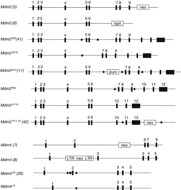
The p53 protein is undetectable in most embryonic and adult tissues.12 However, p53 transcription can be measured by RT-PCR (Marine J-C, unpublished data) suggesting that the p53 protein undergoes constitutive degradation in vivo. Treatment of cells with 26S proteasome inhibitors leads to accumulation of ubiquitin-p53 conjugates indicating that p53 degradation occurs in a ubiquitin-proteasome-dependent manner.13 The Mdm2 protein is the first p53 E3-ubiquitin ligase described, and induces p53 polyubiquitination and degradation when overexpressed.14, 15, 16, 17 More recently, the idea that Mdm2 mediates monomeric p53 ubiquitination on multiple lysine residues rather than poly-ubiquitination has been proposed.18 As chains of multiple ubiquitin molecules are necessary for efficient protein degradation, the data suggested that Mdm2 might not be sufficient for optimal degradation of p53, and that other proteins must aid in polyubiquitination and degradation of p53 in vivo. p300 was proposed to promote polyubiquitination of p53-monoubiquitinated molecules acting as an E4 ligase.19 More recent data indicated that the ability of Mdm2 to monoubiquitinate or polyubiquitinate p53 depends on its expression level.20 Low levels of Mdm2 induce monoubiquitination and nuclear export of p53, whereas high levels promote its polyubiquitination and nuclear degradation. Thus, these distinct mechanisms may be exploited in different physiological settings. For example, Mdm2-mediated polyubiquitination and nuclear degradation of p53 could play a critical role in suppressing p53 function during the later stages of a DNA-damage response or when Mdm2 is malignantly overexpressed. In contrast, Mdm2-mediated monoubiquitination and subsequent cytoplasmic translocation of p53 would represent an important means of p53 regulation in unstressed cells.21 Importantly, decreased expression of Mdm2 in mice harboring a hypomorphic and a null allele of mdm2 led to activation of p53 function without concomitant increase in p53 protein levels.11 Thus, the reduced levels of Mdm2 in these mice (about 30% of the level in wild-type tissues) are sufficiently high to allow efficient degradation of p53. Alternatively, other p53 ubiquitin ligases might compensate for the decrease in Mdm2 expression in vivo. A number of other p53 ubiquitin ligases, such as Pirh2, Cop-1, Yin Yang1 and ARF/BP1, have indeed been discovered and shown to function in a Mdm2-independent manner.22, 23, 24, 25 Together these data challenge the conventional view that Mdm2 is essential for p53 turnover in vivo. A clear answer to this key issue could have come from studies of the _mdm2_-deletion mutants. Unfortunately, because of the very early embryonic lethality, it has been technically difficult to evaluate the physiological role of Mdm2 in the regulation of p53 stability in this setting. However, a series of recent studies using conditional alleles clearly demonstrate that p53 degradation in vivo occurs in a strict Mdm2-dependent manner. Conditional inactivation of mdm2 in cardiomyocytes, in neuronal progenitor cells and terminally differentiated smooth muscle cells (SMCs) of the gastro-intestinal (GI) tract was achieved using an mdm2 floxed allele and various specific Cre transgenic lines (Figure 1; Boesten et al., unpublished data).26, 27 In all these experimental settings, loss of mdm2 leads to a dramatic accumulation of the p53 protein in vivo. In addition, similar results were obtained using an alternative genetic approach. Mice carrying a transcriptional stop element flanked by _lox_P recombination sites (_lox_P-stop-_lox_P, LSL) upstream of the coding region of the p53 gene were used (p53 LSL-LSL A Ventura, D Tuveson and T Jacks, unpublished data). Since this p53 knock-in allele is silenced by the stop element, this mutation could be transferred into the _mdm2_-null, _mdm4_-null and _mdm2/mdm4_-double null backgrounds. Conditional expression of p53 was achieved in tissues in vivo by crossing these compound mice with various specific Cre transgenic lines. Consistent with the recent data described above, specific expression of p53 in neuronal progenitor cells and postmitotic neurons of mice lacking mdm2 lead to accumulation of p53.28
Figure 1
A comparison of the different mdm2 and mdm4 alleles that have been generated. Exons are numbered and represented as black boxes. Frt sites are shown as filled circles, loxP sites are represented as diamonds. Neo, neomycin resistance gene; HPRT, hypoxanthine/aminopterin/thymidine resistance gene; puro, puromycin resistance gene ; LTR, long terminal repeat
Finally, the generation by gene targeting of various mouse mutants encoding p53 variants provide additional support, even if less direct, for a key role of Mdm2 in the regulation of p53 levels in vivo. Mutation of two amino acids (L25Q and W26S), essential for the Mdm2–p53 interaction, generates a stable p53 protein.29, 30 Similarly, a p53 mutant lacking the proline-rich domain (p53ΔP), a region that appears to modulate Mdm2 binding and Mdm2-mediated degradation in vitro,31, 32 indeed exhibits a significantly shorter half-life than wild-type p53 in vivo (Toledo et al., unpublished data).
All in all, Mdm2 is required to maintain p53 at low levels both in proliferating progenitor cells and in, terminally differentiated, postmitotic cells. The data suggest that, even if other E3 or E4 ligases might contribute to degradation of p53, this process occurs in a strict Mdm2-dependent manner. Further germline and/or conditional knockout studies are required to determine the physiological contribution of additional ubiquitin ligases in the regulation of p53 turnover.
Mdm2 and its Role in the Regulation of p53 Transcriptional Activity
In addition to promoting p53 degradation, Mdm2 binds p53 in its transactivation domain and it has been proposed that this interaction interferes with the recruitment of the basal transcription machinery and/or essential coactivator(s).33 Moreover, Mdm2 was also reported to promote NEDD8 conjugation of p53, a modification that inhibits its transcriptional activity.34 Finally, Mdm2 induces monoubiquitination of histones surrounding the p53-response elements resulting in transcriptional repression.35
Surprisingly, recent genetic studies fail to support a role for Mdm2 in the regulation of p53 transcriptional activity per se. Conditional inactivation of mdm2 leads to a dramatic increase in p53 levels accompanied by an increase in transcriptional activity. Indeed loss of mdm2 in neuronal progenitor cells and in mouse embryonic fibroblasts (MEFs) leads to enhanced transcription of p53 target genes as measured by quantitative PCR analysis.27, 28 However, when the transcriptional activity of p53 was normalized to the amount of protein present, no significant difference could be observed in cells with or without mdm2.28 In other words, stabilized p53 in cells lacking mdm2 is not transcriptionally more potent than in cells expressing Mdm2.
The analysis of mice encoding a mutant p53 allele generated by homologous recombination that lacks the proline-rich domain (p53ΔP) led to a similar observation (Toledo et al., unpublished data). The p53ΔP protein exhibited increased sensitivity to Mdm2-dependent degradation and decreased transactivation capacity, correlating with deficient cell cycle arrest and reduced apoptotic responses. Strikingly, the compromised function of p53ΔP failed to rescue mdm2 deficiency, but allowed full rescue of mdm4 deficiency (see below). Importantly, deletion of one single mdm2 gene copy significantly increased p53ΔP levels, leading to increased transactivation of p53 target genes. However, p53ΔP did not appear more active on a per molecule basis in p53 ΔP/ΔP mdm2+/− cells than in p53 ΔP/ΔP cells. Thus, two very distinct in vivo approaches suggest that Mdm2 does not control p53 transcriptional activity per se.
However, this conclusion should be taken with caution. For instance, since association with Mdm2 leads to rapid degradation of p53, it cannot be excluded that, the majority of the p53 molecules detected in Mdm2 competent cells are not physically associated with Mdm2. If this is indeed the case, then it is not surprising that the transcriptional activity of p53, when normalized to the total amount of p53 present, is identical in cells with or without Mdm2. It should also be noted that these experiments were essentially performed in cultured MEFs under nonphysiological oxygen levels. In these conditions, MEFs accumulate significantly more DNA damage,36 a situation known to modulate the levels and behavior of the Mdm2, Mdm4 and p53 proteins (see accompanying review by G Wahl). Thus, until a careful quantification of the respective protein levels of all relevant players and a precise evaluation of the various complexes formed in physiological settings, this proposal has to be considered as tentative.
Regardless, these data strongly argue that the primary physiological function of Mdm2 is to promote p53 degradation, instead of controlling its transcriptional activity.
Mdm2 is a Critical Survival Factor both in Proliferating and Terminally Differentiated Cells
Interestingly, increased p53 levels and activity upon mdm2 loss led to p53-mediated cell death in neuronal progenitors cells, and in cardiomyocytes as well as in postmitotic neurons or SMCs of the GI tract (Boesten et al., unpublished data).26, 27, 28 Thus, Mdm2 is required to maintain p53 at low levels and to suppress the ability of p53 to induce cell death both in proliferating as well as in quiescent cells in vivo. Of note, activation of p53 in all these cell types resulted in a classical caspase-dependent apoptotic cell death, except in the SMCs of the GI tract. Surprisingly, activation of p53 in these cells resulted in caspase-independent cell death with a necrotic morphotype (Boesten et al., unpublished data).
These data highlight the ability of p53 to function at its full potential in vivo in cells lacking mdm2, and thus in absence of detectable genotoxic lesions and/or activating post-translational modifications. Indeed, increased basal level of DNA damage and ATM-mediated activating phosphorylation could not be detected in Cre-expressing cells in vivo. These data are consistent with the observation that a small molecule Mdm2 antagonist leads to activation of p53 target genes and biological responses in vivo in the absence of phosphorylation induced by stress-activated kinases.37
Mdm2 and Mdm4 are Required to Control p53 in Neuronal Cells
The generation of _mdm2_-null mice clearly revealed the in vivo significance of Mdm2 in p53 inhibition.5, 6 Mdm4 was later discovered and identified as a novel p53-binding partner.38 In mice, mdm4 loss also resulted in a p53-dependent embryo lethal phenotype.7, 8, 9 Thus, it was a surprise to find that two p53 negative regulators were critical in regulating p53 activity in vivo. One possible scenario, based on the observation that Mdm2 and Mdm4 heterodimerize, is that the two proteins work together to inhibit p53.39, 40 However, the molecular mechanisms underlining the abilities of both Mdm proteins to regulate p53 activity have yet to be clarified in vivo.10 Conditional alleles have now been developed that yield further insight into how and in what cell types Mdm2 and Mdm4 regulate p53 (Figure 1).11, 26, 41, 42
To test whether Mdm2 and Mdm4 are required to restrain p53 activity in a single cell type, both mdm2 and mdm4 were conditionally inactivated in neuronal progenitors.27 In addition, conditional expression of p53 was restored specifically in neuronal progenitor cells or in postmitotic cells of mice lacking mdm2 and/or mdm4.28 Both studies led to the same conclusion: Mdm2 and Mdm4 are essential for keeping p53 activity in check, in a nonredundant manner, in the developing nervous system. Loss of mdm4 led to milder phenotypes than loss of mdm2, but loss of both genes synergized to produce an even more dramatic cell loss phenotype. In fact, the severity of defects in the neural epithelium varies with different combinations of null alleles such that the phenotype is mildest in _mdm4_-null mice, greater in _mdm4_−/− mdm2+/−, and even greater in _mdm2_-null mice. The _mdm2_−/− mdm4+/− mice show a worse phenotype than _mdm2_-null and the severest phenotype is seen in mdm2 mdm4 double null mice.27, 28 Importantly, all phenotypes disappear in the absence of p53. Thus, this gradation of severity that depends on the combination of alleles further supports the synergistic function of Mdm2 and Mdm4 in regulating p53. These observations demonstrate that both Mdm2 and Mdm4 are required to inhibit p53 activity in the same cell type and confirm the notion that Mdm2 cannot compensate for mdm4 loss in vivo, at least in this particular cell type. Together, these data are consistent with specific, non-overlapping roles for Mdm4 in the regulation of the Mdm2–p53 interplay. Importantly, this conclusion is not only true in proliferating progenitor cells but also in postmitotic neuronal cells.
Interestingly, Cre-mediated conditional expression of p53 in mice lacking mdm4 lead to cell cycle progression delay in progenitor cells and to apoptosis in postmitotic neurons.28 These observations confirm the ability of Mdm4 to regulate both p53-mediated cell cycle arrest and apoptosis.8 This view is further supported by the classical conditional approach since specific loss of mdm4 in the central nervous system (CNS) induced both apoptosis and cell cycle arrest.27
In one of the reported mdm4 germline mutations, widespread p53-dependent apoptosis was observed in the neuroepithelium as early as E10.5.8 In contrast, conditional expression of one p53 allele in the neuronal progenitor cells of mice harboring the same mdm4 mutation delays cell cycle progression.28 One possible explanation is that activation of apoptosis requires expression from both p53 alleles. However, a strong argument against this hypothesis is our demonstration that loss of one p53 allele does not rescue apoptosis in the _mdm4_-null mutants.8 Together, these findings suggest that, while proper cell cycle regulation of the NPCs is dependent on the presence of functional Mdm4 in a cell-autonomous manner, the requirement for Mdm4 in neuronal progenitor cell survival may be non-cell-autonomous.
Minor Changes in Mdm2 and Mdm4 Levels Reveal Important Phenotypes
The genetic experiments outlined above further support the idea that minor changes in Mdm2 or Mdm4 levels also contribute to specific phenotypes. Specific deletion of mdm2 in the CNS using Cre recombination indicated a severe depletion of neural cells in embryogenesis. These experiments were actually performed in two different ways, one involving the traditional mechanism of using a conditional mdm2 allele that was lost upon Cre expression, the other with a p53 conditional loss-of-function allele that could be reactivated in neural cells lacking the other p53 allele and mdm2. In these experiments, there was no difference in phenotype even though mice had one or two copies of functional p53. The finding that p53 heterozygosity does not rescue the _mdm2_-null phenotype, even partially, further supports the dominant nature of the _mdm2_-null phenotype.5 A hypomorphic p53 allele with deletion of the proline-rich domain (described above) also could not rescue this phenotype (Toledo et al., unpublished data). These data imply that the presence of any functional p53 is detrimental for viability in the absence of Mdm2.
However, in the generation of an mdm2 conditional allele, Mendrysa et al.,11 generated a hypomorphic mdm2 allele. This allele, mdm2 puro in combination with a _mdm2_-null allele results in approximately 30% of the total levels of Mdm2 and the mice exhibited fascinating phenotypes. The mice were born and appeared normal at first, but later showed a reduction in body weight, and mild anemia which was the result of depletion of red blood cells (80% of normal), white blood cells (30% of normal) and lymphocytes (37% of normal). Since these phenotypes were eliminated in a _p53_-null background, these data indicate that the hematopoetic system in the mouse is the most sensitive to small decreases in Mdm2 levels and thus to small increases in p53 activity.
Minor changes in Mdm2 levels may have more subtle phenotypes later in life. Mice with 30% of total Mdm2 levels had decreased incidence of tumors in the small intestine in an _APC_min/+ background.43 Additionally, a single nucleotide polymorphism identified in the human mdm2 promoter that increases slightly the levels of Mdm2 is associated with an increased age of tumor onset in Li-Fraumeni syndrome patients already compromised for p53 levels.44 These data indicate that small changes in Mdm2 may have some long-term consequences and function as a modifier of a tumor phenotype.
The results get more interesting when mdm4 loss was examined in the CNS. The traditional way of deleting mdm4 specifically in the CNS yielded mice that died in utero or right after birth. These mice have flat heads caused by the absence of a large mass of cells in the brain and the presence of a large cavity.27 In contrast, mice generated in an _mdm4_-null background with one p53 allele that was reactivated with the same Cre recombinase and one _p53-_null allele yielded mice that were viable, but exhibit microcephaly, cerebellar defects and ataxia.28 Thus, the phenotype associated with mdm4 loss in the neural progenitor cells appears more severe than the phenotype induced by conditional expression of one p53 allele in the same cells lacking mdm4. This difference in survival is likely due to a gene dosage effect and is a consequence of the difference in the number of transcriptionally active copies of the p53 gene. These data point to the observation that small differences in p53 levels can mean the difference between life and death and further highlight the importance of maintaining a delicate balance between Mdm2, Mdm4 and p53 proteins to allow appropriate control of p53 activity in vivo.
Interestingly, the microcephaly, cerebellar defects and ataxia seen in mice lacking mdm4 with one functional p53 allele28 is reminescent of neurological defects observed in individuals with Nijmegen breakage syndrome (NBS, caused by a hypomorphism in NBS1), ataxia telangiectasia (AT), and AT-like disorder. Specific inactivation of the mouse ortholog of NBS1 (Nbn or Nbs1) in neural tissues results in a very similar combination of neurological abnormalities.45 Loss of Nbn caused proliferation arrest in neuronal progenitor cells and apoptosis in postmitotic neurons. Consistent with a role for Nbn in double-strand break repair, high basal chromosomal aberrations were observed in _NBS_-deficient neural cells. p53 is activated by ATM, a DNA damage-induced kinase known to directly phosphorylate p53 and disrupt interactions of p53 with Mdm2 and possibly Mdm4. Thus, inactivation of mdm4 or activation of the ATM-mediated DNA damage response leads to identical p53 response in neuronal cells and similar neurological abnormalities. Notably, as in the p53 LSL/- _mdm4_−/− Nestin-Cre+ mice, the cerebellum in the _NBS_-CNS-del mice also seems to be the major target. The reasons for the extreme sensitivity of the cerebellum to increased p53 activity is not known, but provides a rational explanation for the high selective pressure for mutational inactivation of p53 during medulloblastoma development.46
Tissue-Specific Differences of p53 Inhibition by Mdm2 and Mdm4
Conditional inactivation of mdm4 was also conducted in cardiomyocytes and SMCs of the GI tract (Boesten et al., unpublished data).26 In contrast to loss of mdm2, loss of mdm4 in these various cell types, leads to phenotypes ranging from not obvious to only minor. Mice lacking mdm4 in the heart were born at the correct ratio and appeared normal. However, the mice died prematurely for yet unknown reasons. Further investigations are required to determine whether the cause of death is a consequence of heart failure. Similarly, whereas mdm2 inactivation in the SMCs of the GI tract leads to rapid cell loss and a lethal phenotype, inactivation of mdm4 in the SMCs did not lead to any obvious phenotype. The data suggest that inhibition of p53 by Mdm4 is only required in a restricted number of cell types. However, interpretation of these results is complicated. Indeed, even in cells in which Mdm4 function was shown to be critical, such as the neuronal progenitor and postmitotic cells, in contrast to mdm2, loss of mdm4 consistently led to only a moderate increase in p53 activity in vivo. This difference can be explained, at least in part, by the fact that p53 activates the transcription of mdm2 but not mdm4.47 Thus, in absence of mdm4, p53 transcriptional activity is enhanced leading to the stimulation of the p53–Mdm2-negative feedback loop. In agreement, mdm4 loss leads to a moderate increase in Mdm2 protein levels in vitro and an increase in _mdm2_-transcription in vivo (Toledo et al., unpublished data).27, 28 The stimulation of mdm2 transcription therefore complicates the interpretation of the results from mdm4 deficiency. In one particular example, overexpression of an mdm2 transgene rescues the embryonic lethality associated with mdm4 deficiency,48 indicating that high levels of Mdm2 compensate for mdm4 loss. Thus, as an alternative to the simplistic view of tissue-specific function for Mdm4, increased Mdm2 levels might better compensate for mdm4 loss in specific cell types.
Nevertheless, at the molecular level, the difference in the severity of the phenotypes observed following mdm2 or mdm4 loss is most likely due to the fact that loss of Mdm2 leads to dramatic accumulation of the p53 protein, whereas loss of mdm4 does not cause significant increase in p53 levels in vivo (see below).
Mdm4 does not Modulate p53 Levels Independently of Mdm2
The role of Mdm4 in the control of p53 and Mdm2 stability is unclear. Mdm4 was reported to act as a ubiquitin ligase in vitro49 but Mdm4 overexpression in cells does not lead to p53 ubiquitination and degradation.50, 51 However, Mdm4 might regulate p53 stability indirectly, by stabilizing Mdm2. Indeed, transfection studies suggest that Mdm4 stabilizes Mdm2, perhaps by interfering with its autoubiquitination.40, 51 Another report, however, suggested that Mdm4 stimulates not only Mdm2-mediated ubiquitination of p53, but also Mdm2 self-ubiquitination.52
In vivo, p53 levels stay below the limit of detection in both Western blotting and immunohistochemistry assays, when its expression was restored in progenitor and in postmitotic neuronal cells lacking mdm4.28 Similarly, p53 staining was not detected in the neural progenitor cells conditionally inactivated for Mdm4 at E10.5. In contrast, clear p53 staining was observed in _mdm2_-inactivated cells at the same stage of development.27 However, interpretation of these data are again complicated by the existance of the p53–Mdm2-negative feedback loop. To assess the role of Mdm4 in the regulation of p53 stability and activity in vivo, one therefore needs to compare the consequences of inactivation of both mdm2 and mdm4 with loss of mdm2 alone. Using such a strategy, loss of both mdm2 and mdm4 does not lead to any further increase in p53 levels compared to loss of mdm2 alone, suggesting that Mdm4 does not participate in the regulation of p53 stability independently of Mdm2.28 However, whether it does so in a Mdm2-dependent manner remains unclear. Indeed, two possibilities remain: (i) Mdm4 does not actively participate in the regulation of p53 levels. mdm4 loss would then lead to increased p53 turn-over as a result of the stimulation of the p53–Mdm2-negative feedback loop. Moreover, since Mdm4 might even compete with Mdm2 for p53 binding, loss of mdm4 could further increase the availability of p53 for Mdm2 binding and therefore Mdm2-mediated degradation. In these, nonmutually exclusive, scenari, loss of mdm4 would result in decreased p53 levels. (ii) Mdm4 stimulates Mdm2-mediated degradation of p53, as proposed earlier,52 and therefore contributes to the regulation of p53 degradation in an Mdm2-dependent manner. In this case, overall p53 levels would be the result of a decrease in Mdm2 activity but, at the same time, an increase in Mdm2 expression. Recent data favor the second possibility because overall p53 levels do not decrease in p53 LSL/- _mdm4_−/− MEFs after Cre expression, despite the stimulation of the p53–Mdm2-negative feedback loop, and because a slight increase in p53 immunostaining was observed in tissues in vivo from p53 LSL/- mdm2+/− _mdm4_−/− NES-Cre embryos compared to p53 LSL/- mdm2+/− mdm4+/− NES-Cre embryos.28 However, in order to formally resolve this issue, it will be important to carefully measure p53 half-life in the various genetic backgrounds and, if possible, in an in vivo setting.
Additionally, the p53ΔP mouse model also enabled evaluation of Mdm4 function (Toledo et al., unpublished data). This hypomorphic p53 mutant presents the unique property of being able to fully rescue mdm4 deficiency. In this case, the consequences of mdm4 loss were observed in a compromised p53 context, but did not require Cre expression. The results show that in the absence of Mdm4, the transactivation of mdm2 is stimulated, leading to increased Mdm2 protein levels. In contrast to previous transfection studies,40 these data suggest that Mdm4 does not affect Mdm2 protein stability significantly. This was indeed directly tested and the half-life of Mdm2 was found to be similar in p53 ΔP/ΔP cells and p53 ΔP/ΔP _mdm4_−/− cells, as well as in p53 LSL/- and p53 LSL/- _mdm4_−/− MEFs (Toledo et al., unpublished data).28 Moreover, also consistent with the data described above, loss of mdm4 alone did not lead to any significant increase in p53ΔP levels. However, the accumulation of p53ΔP after DNA damage was significantly reduced in _mdm4_-null fibroblasts. Thus, increased Mdm2 levels, as a result of the stimulation of the feedback loop, degrades p53ΔP more efficiently, implying that Mdm4 has little or no effect on Mdm2-mediated p53 degradation in this particular cellular context. Although this last conclusion is not entirely consistent with the data described above, both sets of data are completely consistent in showing that Mdm2 can induce efficient degradation of p53 in vivo in the absence of mdm4.
Mdm4 Inhibits p53 Transcriptional Activity and Contributes to the Overall Inhibition of p53 Independent of Mdm2
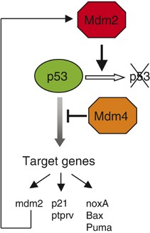
The contribution of Mdm4 to the regulation of p53 transcriptional activity has also remained unclear. High levels of Mdm4 inhibit p53 transcriptional activity;38 however, whether Mdm4 can interfere with this activity in an Mdm2-independent manner in vivo is unknown. The approaches described above provide genetic evidence that Mdm4 inhibits p53 transcriptional activity independent of Mdm2.28 Loss of mdm4, in cells lacking mdm2, indeed causes an increase in p53 activity in cultured MEFs. Importantly, this finding was confirmed in various in vivo settings. Mice null for both mdm2 and mdm4 in the CNS were generated. These mice exhibited a phenotype that was more severe and appeared earlier than the phenotype seen with loss of mdm2.27 Similarly, the extent of p53-mediated apoptosis, upon Cre-mediated p53 expression, was significantly greater in the neuroepithelium and in postmitotic cells of mice lacking both mdm2 and mdm4 than in mice lacking mdm2 alone.28 Together, the data support a model in which Mdm2 and Mdm4 cooperate in vivo to limit p53 activity, irrespectively of the proliferation/differentiation status of the cells. They also indicate that the primary function of Mdm2 is to prevent accumulation of the p53 protein, whereas Mdm4 regulates p53 transcriptional activity. A similar model was deduced from the analysis of p53ΔP regulation by Mdm2 and Mdm4 (Toledo et al., unpublished data). As mentioned above, the deletion of one single mdm2 gene copy significantly increased p53ΔP levels, leading to increased transactivation of p53 target genes. However, p53ΔP did not appear more active, on a per molecule basis, in p53 ΔP/ΔP mdm2+/− cells than in p53 ΔP/ΔP cells. In contrast, total loss of mdm4 resulted in less abundant, but more active p53ΔP Together, the data suggest a functional complementarity of Mdm2 and Mdm4, in which Mdm4 functions as a major inhibitor of p53 transcriptional activity, whereas Mdm2 serves to mainly regulate p53 stability (Figure 2).
Figure 2
A model for cooperative controls of the p53 protein levels and transcriptional activity by Mdm4 and Mdm2. See text for details. Bold arrows indicate that genetic evidence for the associated activities are provided by the papers reviewed herein
Lack of Genetic Evidence for a p53-Independent Role of Mdm2 and Mdm4 Under Physiological Expression Levels
Mdm2, and to a lesser extent Mdm4, has been implicated in the regulation of the stability and/or the activity of several proteins playing a key role in the control of cell proliferation such as the retinoblastoma protein pRb, the heterodimer E2F1/DP1, Numb and Smads.53, 10 However, the relevance of these interactions has not been firmly established genetically. Several lines of evidence do not support p53-independent functions for regulation of Mdm2 and Mdm4 under physiological conditions. p53/mdm2 and p53/mdm4 double null mice and cells were shown to be undistinguishable from their _p53_−/− counterparts,54, 55 suggesting that the predominant function of these two proteins is to regulate p53. In addition, all phenotypes from the recent mdm2 and mdm4 conditional knockout studies are entirely p53 dependent (Boesten et al., unpublished data).26, 27, 28 However, it still has to be excluded that Mdm2 and Mdm4 regulate the activity of other proteins in a redundant manner. In order to test this possibility, we examined a number of in vitro growth properties of MEFs lacking functional p53 and both Mdm2 and Mdm4. There was, for instance, no significant difference in the proliferative rates, saturation densities or in the G1/S ratio of _p53_−/− fibroblasts, _p53_−/−_Mdm2_−/− or _p53_−/− _Mdm2_−/− _Mdm4_−/− fibroblasts. The ability of these cells to survive and proliferate when plated at very low densities (colony formation at clonal densities) was comparable. Finally, these cells also show comparable ability to enter S phase following DNA damage through a p53-independent pathway. The data provide genetic evidence that Mdm2 and Mdm4 do not regulate essential cell proliferation or cell cycle control events independently of p53 (Froment P and Marine J-C, unpublished data). In other words, the data argue for a unique physiological function of both Mdm2 and Mdm4 in the regulation of p53 activity in vivo. Importantly, however, these data do not exclude the possibility that these two proteins when overexpressed affect the activity of other proteins and of p53-independent pathways. This possibility is of great interest, since both proteins are aberrantly expressed in a number of human primary tumors.56, 57 Along these lines, it has been proposed recently that Mdm2 overexpression could contribute to cancer development by destabilizing the retinoblastoma protein Rb.58
The Mdm4–Mdm2–p53 Interplay as a Target for Therapeutic Intervention
There is evidence that transformed cells are more sensitive to p53-induced apoptosis than their normal counterparts, and that thus activation of p53 might cause tumor-specific cell killing. Activation of the p53 reponse becomes, therefore, an attractive therapeutic goal. As such, Mdm2 ubiquitination activity and the physical interaction between p53 and Mdm2 have recently become the targets for the development of new cancer therapeutic strategies.59, 60, 61 These approaches rely on effective activation of the p53 tumor suppressor function in the cancer cells, and should be cytostatic to normal proliferating or resting host cells. At first, some of the recent studies reviewed herein raise concerns about putative overall p53-dependent toxicity of such approaches. Mdm2 is critical for controlling p53 levels in both normal proliferating and terminally differentiated cells. One would predict that interfering with Mdm2 ligase activity or with the p53/Mdm2 interaction in vivo will be detrimental not only for cancer cells, but also for most normal host cells. However, this interpretation is essentially based on complete loss of function studies, which is very unlikely to occur with the use of small antagonist molecules in vivo. In support of the use of Mdm2 antagonists, decreased Mdm2 expression in mice affects primarily homeostatic tissues11 and considerably reduced tumor formation in vivo.43 Recent data suggest that Mdm4 may also serve as a clinically relevant therapeutic target. Reducing the gene dosage of either Mdm2 and Mdm4 only marginally affected the growth of oncogene-induced tumors in p53 ΔP/ΔP mice. However, far more significant effects were observed by decreasing the expression of both Mdm2 and Mdm4, or by complete ablation of Mdm4 activity (Toledo et al., unpublished data). In addition, amplification and ectopic expression of Mdm4 occur in a substantial fraction of lung, breast and colon tumors, the three most common human cancers.57 As the p53-binding domains of Mdm2 and Mdm4 are similar and require the same amino acids in p53 for interaction, antagonists of the p53–Mdm2 interaction might also antagonize Mdm4 binding to p53. However, some differences between the p53–Mdm2 and p53–Mdm4 interactions have been reported,62 indicating the need to search for optimal Mdm4 antagonists. In addition, the in vivo studies reviewed here, which revealed that Mdm2 and Mdm4 regulate p53 in distinct and synergistic ways, provide both the rationale and experimental evidence that optimal antagonists to Mdm2 and Mdm4 could synergize to ensure strong p53 activation in tumors.

