A new redox-cofactor vitamin for mammals (original) (raw)
In animals, lysine is an essential amino acid (that is, one that cannot be made in the body) that is degraded after use to 2-aminoadipic 6-semialdehyde (AAS), which is subsequently oxidized to 2-aminoadipic acid (AAA; Fig. 1a, right arrows)4. The molecular features of the AAS-dehydrogenase (AASDH; EC 1.2.1.31) enzyme that catalyses this oxidation of AAS are unknown.
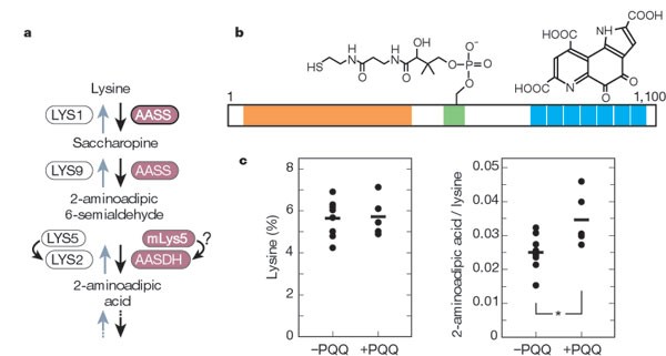
Figure 1: The role of pyrroloquinoline quinone (PQQ) in mammalian lysine degradation.
a, Metabolic pathways of lysine: right arrows, lysine degradation in mammals; left arrows, lysine biosynthesis in yeast. In yeast, LYS2 is post-translationally 4′-phosphopantetheinylated in a reaction catalysed by LYS5. AASS, AAS-synthetase; AASDH, AAS-dehydrogenase; mLys5, mammalian homologue of LYS5. b, Arrangement of putative binding domains in murine U26 protein. The adenylation domain (Pfam 00501), thiolation domain (00550), and PQQ-binding motifs (01011) are shown in orange, green and blue, respectively. Serine 592 in the thiolation domain is 4′-phosphopantetheinylated and PQQ (right) is non-covalently bonded by the seven repeats of the PQQ-binding motif near the carboxy terminus. c, Peripheral blood concentrations of lysine and 2-aminoadipic acid in PQQ-supplemented and PQQ-deprived mice. Lysine is quantified as a percentage of the total amino acids (left; determined using an amino-acid analyser) and 2-aminoadipic acid is normalized with respect to lysine (right). Horizontal bars, average for each group of mice. Asterisk denotes P = 0.010.
In yeast, lysine is synthesized from AAA by the reverse pathway (Fig. 1a, left arrows)5. The reduction of AAA to AAS requires two enzymes that are encoded by the Lys2 and Lys5 genes: the LYS2 protein has NADPH-dependent AAA-reductase activity, whereas LYS5 catalyses the post-translational 4′-phosphopantetheinylation of LYS2 (ref. 6). A human homologue of Lys5 has been cloned7, raising the possibility that there is a structural analogue of LYS2 in animals.
To find the analogue of LYS2, we searched the Drosophila genome databases for gene product(s) containing functional domains, adenylation and thiolation domains, which serve to capture the substrate in LYS2 (ref. 6). A BLAST search detected two gene products, EBONY (DDBJ/EMBL/GenBank accession number, CAA11962) and U26 (accession number, AAF52679); EBONY is β-alanyl-dopamine synthetase, but the function of U26 was unknown. In contrast to yeast LYS2, which contains a binding domain for the reducing cofactor NADPH, Drosophila U26 contains several repeats of the binding motif for PQQ (an oxidizing cofactor); this motif is conserved among bacterial PQQ-dependent dehydrogenases8.
To investigate whether U26 could be AAS-dehydrogenase, we cloned mouse U26 complementary DNA (accession number, AB095954), which encodes a protein of 1,100 amino acids that is 23% identical and 43% similar to Drosophila U26. Mouse U26 contains one set of adenylation/thiolation domains and seven PQQ-binding motifs (Fig. 1b). The six to eight tandem repeats are also present in bacterial PQQ-dependent dehydrogenases8, indicating that mouse U26 could be a PQQ-dependent dehydrogenase.
Northern-blot analysis showed that mouse U26 messenger RNA is expressed in all of the tissues that we tested, with expression being notably high in the heart, liver and kidney (results not shown). In mice fed on a lysine-rich diet (containing 2,000-fold more free lysine than normal), blood concentrations of lysine and AAA were markedly increased (2.4- and 1.5-fold, respectively). The level of U26 mRNA (normalized to that of glyceraldehyde-3-phosphate dehydrogenase) in the liver and heart of lysine-loaded mice was significantly higher (1.6- and 1.4-fold, respectively; P = 0.008 for each) than in control mice. Mouse AAS-synthetase (AASS in Fig. 1a) and Lys5 transcripts were also increased in the lysine-loaded mice (results not shown). These results indicate that U26, as well as AAS-synthetase and LYS5, is involved in the lysine-degradation pathway in mice.
To test the importance of PQQ in AAS-dehydrogenase activity, we examined the effects of a PQQ-deficient diet on mice. After being fed on a chemically defined diet2,3 that was either devoid of PQQ or supplemented with PQQ (1,000 ng PQQ·2Na per g of food), we measured the blood levels of lysine and AAA in the two groups of mice. AAA concentrations were significantly decreased in PQQ-deprived mice, whereas lysine levels remained the same (Fig. 1c); the intermediates saccharopine (Fig. 1a) and AAS were not detected. This decrease in AAA may be indicative of impaired AAS-dehydrogenase (U26) activity resulting from a PQQ deficiency in deprived mice, pointing to a requirement for PQQ as a redox cofactor for the proper functioning of AAS-dehydrogenase.
PQQ is found in various foods, including vegetables and meat9,10. When mice are fed a PQQ-deficient diet, they grow slowly, have fragile skin and a reduced immune response, and do not reproduce well2,3. On the basis of our demonstration of its molecular function, we propose that PQQ should be classified as a new B vitamin, joining niacin/nicotinic acid (vitamin B3) and riboflavin (vitamin B2), the redox-cofactor derivatives of which are NAD+/NADP+ and FAD/FMN, respectively.
