Bee venom phospholipase A2 ameliorates motor dysfunction and modulates microglia activation in Parkinson’s disease alpha-synuclein transgenic mice (original) (raw)
Introduction
Parkinson’s disease (PD) is the most extensively perceived synucleinopathy because it is the most general movement disorder, with a prevalence of ~1% at 65 years of age, increasing to 4–5% by 85 years of age.1 Increasing evidence shows that neuroinflammation has a significant role in the pathogenesis of PD.2 Activated microglia and recruited peripheral macrophages release neurotoxic species, such as proinflammatory cytokines, and promote neurodegeneration.3, 4, 5, 6 Activated microglia/macrophages are classified into M1 or M2 phenotypes. When they are polarized to release neurotoxic factors, such as inflammatory cytokines, reactive oxygen species (ROS), nitric oxide synthase (NOS) and glutamate, they are defined by the M1 phenotype. However, when they release diverse neuroprotective molecules, such as anti-inflammatory cytokines, neurotrophins and membrane receptors, they are defined by the M2 phenotype.7 An increased ratio of M1 over M2 microglia/macrophages has been reported in various neurodegenerative pathologies, such as Alzheimer's disease, and in the elderly brain; modulation of microglia polarization has been suggested as a neuroprotective therapy.8, 9 In the PD brain, microglia proliferate early in the disease process, remaining comparatively static and unrelated to the extent of striatal degeneration or clinical severity.10 However, microglia polarization to proinflammatory M1 or anti-inflammatory M2 phenotypes has been poorly studied in PD; the cytokine levels from autopsy studies have suggested that both M1 and M2 microglia may coexist in a brain with Parkinson’s disease.11 In addition, studies conducted in a transgenic α-synuclein (Syn) A53T model have provided crucial insight into the dichotomous response, which microglia can activate.
Bee venom (BV) has been used in traditional medicine to cure some immune-related diseases, such as rheumatoid arthritis, in humans and animals.12, 13 Recently, several clinical trials have shown that BV might be beneficial in the treatment of neurodegenerative diseases, including PD.14, 15 Our recent study demonstrated that the modulation of peripheral immune tolerance by regulatory T cells (Tregs) may contribute to the neuroprotective effect of BV in the 1-methyl-4-phenyl-1,2,3,6-tetrahydropyridine (MPTP) model of PD.16 Bee venom phospholipase A2 (bvPLA2) is an enzyme that breaks down membrane phospholipids to produce free fatty acids and lysophospholipids.17, 18 PLA2 can be classified into three broad classes based on its cellular distribution: secreted or extracellular type (sPLA2), cytosolic or intracellular type (cPLA2) and Ca2+-independent PLA2. bvPLA2 is of the sPLA2 type; sPLA2 is generally present in the venom of snakes, bees, cnidarians and scorpions. sPLA2 has a central role in various cellular responses, including phospholipid metabolism, signal transduction and the initiation and regulation of inflammatory and immune responses.19, 20, 21, 22 bvPLA2 is a typical Group III sPLA2 member and comprises ~12% of the dry weight of venom in the European honeybee, Apis mellifera.17, 23 bvPLA2 has a wide variety of pharmacological properties, including anti-HIV activity, myotoxicity and neurite outgrowth induction.24, 25 However, the efficacy and the mechanism of the action of bvPLA2 that is involved in PD remains largely unknown.
In this study, we evaluated for the first time the motor dysfunction of A53T human α-Syn Tg mice and age-matched non-transgenic (nTg) littermates using a modified pole test. In addition, α-Syn aggregation and M1/M2 polarization were analyzed in the spinal cord.
Materials and methods
Animals and treatment
Animal procedures were approved by the University of Kyung Hee Institutional Animal Care and Usage Committee and were in accordance with the Guide for the Care and Use of Laboratory Animals of the National Institutes of Health. Human A53T α-Syn transgenic (A53T Tg) mice (B6;C3-Tg(Prnp-SNCA*A53T)83Vle/J) were purchased from The Jackson Laboratory (Bar Harbor, ME, USA). All animals were kept in a pathogen-free environment on a 12-h light/dark cycle and had access to food and water ad libitum. The A53T Tg mice were randomized into five groups. The mice received repeated intraperitoneal treatment of bvPLA2 (0.2 or 1 mg kg−1) or phosphate-buffered saline (PBS). Starting at 3 months of age, the mice received PBS and bvPLA2 once a week for 3 months until they were 6 months old.
Cell culture and treatment
The murine BV-2 microglial cell line was maintained in RPMI 1640 medium (Welgene, Gyeongsan, Korea) supplemented with 10% fetal bovine serum (Welgene, Gyeongsan, Korea), 100 U ml−1penicillin and 100 μg ml−1 streptomycin (Invitrogen Life Technologies, Rockville, MD, USA). The cells were cultured every 2–3 days until the cells became 80% confluent. The cells were incubated at 37 °C with 95% humidity and 5% CO2 for all experiments. For polarization, the cells were seeded in six-well plates at a density of 5 × 10⁵ cells per ml and treated the following day. Immediately before treatment, the cells were washed twice with serum-free RPMI medium and supplemented with 2 ml of warm serum-free RPMI medium containing experimental treatments. The cells were pretreated with 0.1, 1 or 10 μg ml−1 bvPLA2 for 30 min, then 1 μg ml−1 lipopolysaccharides (LPS) (Sigma-Aldrich, St Louis, MO, USA) or 20 ng ml−1 murine recombinant interleukin (IL)-4 (R&D Systems, Minneapolis, MN, USA) were added to each well. After 24 h of treatment, the cells were rinsed twice with PBS and collected for RNA extraction.
RNA extraction and quantitative real-time PCR
Total RNA was extracted from BV-2 cells using an Easy-BLUE RNA extraction kit (iNtRON Biotechnology, Inc., Seongnam, Korea). The RNA quality and concentration were determined using a NanoDrop spectrophotometer (NanoDrop Technologies, Inc., ND-1000; Wilmington, DE, USA) and standardized to the lowest concentration with RNase free water. The RNA was reverse transcribed to cDNA using CycleScript Reverse Transcriptase and Random Oligonucleotide Primers (Bioneer, Deajeon, Korea) following the manufacturer’s instructions. Quantitative real-time PCR was performed using the SensiFAST SYBR No-ROX Kit (Bioline, Taunton, MA, USA) and analyzed with the LightCycler 480 system (Roche Ltd, Basel, Switzerland). The PCR reactions were subjected to 55 cycles of denaturation at 95 °C for 10 s, annealing at 72 °C for 10 s and extension at 60 °C for 10 s, with fluorescence measured at the end of each cycle. The data are expressed as 2−ΔΔCT for the experimental gene normalized to GAPDH, and presented as the fold change relative to the saline-treated control. The following primers were used: TNF-α for: 5′-TTCTGTCTACTGAACTTCGGGGTGATCGGTCC-3′; TNF-α rev: 5′-GTATGAGATAGCAAATCGGCTGACGGTGTGGG-3′; iNOS for: 5′-GGCAGCCTGTGAGACCTTTG-3′; iNOS rev: 5′-CATTGGAAGTGAAGCGTTTCG-3′; Arg1 for: 5′-AGACAGCAGAGGAGGTGAAGAG-3′; Arg1 rev: 5′-CGAAGCAAGCCAAGGTTAAAGC-3′; MMR for: 5′-AGTGGCAGGTGGCTTATG-3′; MMR rev: 5′-GGTTCAGGAGTTGTTGTG-3′; GAPDH for: 5′-ACCCAGAAGACTGTGGATGG-3′; GAPDH rev: 5′-CACATTGGGGGTAGGAACAC-3′
Measurement of motor activity
The pole test has been used to measure motor coordination and balance in mouse models of PD.26 The pole test was performed 12 weeks after starting the experiment. In the test, the mice were placed on top of a rough-surfaced iron pole (50 cm in length and 0.8 cm in diameter) and could climb down to the base of the pole. The number of times that it took for the mouse to turn completely downward (time to turn; _T_-turn) and then to reach the floor (locomotion activity time; _T_-LA) were measured, with a cutoff limit of 30 s. The average of the three measurements was used as the result.
Tissue preparation
Transcardial perfusion was performed with a saline solution containing 0.5% sodium nitrate and heparin (10 U ml−1); then, fixation was performed with 4% paraformaldehyde in 0.1 M phosphate buffer. Each brain was dissected, post-fixed overnight in buffered 4% paraformaldehyde at 4 °C and stored in a 30% sucrose solution at 4 °C until it sank. Freeze sectioning was performed on a sliding microtome to generate 30-μm-thick coronal sections.
Immunofluorescence staining and quantification
For immunofluorescence staining, A53T Tg and non-transgenic littermate wildtype (WT) mice were transcardially perfused with PBS followed by 4% paraformaldehyde. The brain and spinal cord were removed and post-fixed in buffered 4% paraformaldehyde for 6 h. After immersion in a 30% sucrose solution, the frozen spinal cord was cut into 30-μm-thick coronal sections. The slices were washed with PBS, incubated for 24 h at room temperature with human anti-α-synuclein (Santa Cruz Biotechnology, Dallas, TX, USA), and diluted 1:200 with 0.5% bovine serum albumin in PBS. The sections were then incubated with AlexaFluor 594 goat anti-rabbit (1:200, Vector Laboratories, Burlingame, CA, USA) for 2 h in the dark. The sections were washed thoroughly and mounted with fluorescent mounting medium (Vector Laboratories). The number of cells labeled in the dorsal column was counted using a confocal microscope equipped with argon and HeNe1 lasers (LSM-5; Zeiss, Jena, Germany). Fluorescent images were sequentially acquired for the red and green channels to prevent the crossover of signals from the green to red or from the red to green channels. The images were viewed as z-stacks, both as a series of optical sections and as a projection of merged images.
Statistical analysis
All of the values are expressed as the mean±s.e.m. The statistical significance (P<0.05 for all analyses) was assessed by one-way analysis of variance followed by Tukey’s post hoc test for multiple comparisons using Prism 5.01 software (GraphPad Software Inc., San Diego, CA, USA).
Results
Inhibitory effects of bvPLA2 on motor impairment in A53T Tg mice
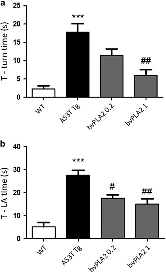
The pole test is a useful method for evaluating the motor dysfunction caused by PD. Prolonged _T_-turn and _T_-LA are known characteristic motor dysfunctions of PD in mice. To confirm the effect of bvPLA2 on motor dysfunction in an in vivo PD model, we treated mice with bvPLA2 or PBS and carried out the pole test. The _T_-turn and _T_-LA of the A53T Tg group were markedly prolonged, at 17.8±2.3 s and 27.5±2.15 s, respectively, compared with the WT group (_T_-turn: 2.3 ± 0.84 s; _T_-LA: 5.1±1.87 s; Figure 1). However, the _T_-turn time for the bvPLA2 1 mg kg−1 per day treated group was shortened to 6 ±1.54 s, and the _T_-LA time of the bvPLA2 0.2 and 1 mg kg−1 per day treated groups were decreased to 17.5±1.46 s and 15±2.21 s, respectively (Figure 1).
Figure 1
Inhibtory effects of bvPLA2 on motor impairment (_T_-turn and _T_-LA) in A53T Tg mice. A53T Tg mice were pretreated with 0.2 or 1 mg kg−1 bvPLA2 for 3 months. (a) The amount of time for the mice to turn completely downward (_T_-turn) was recorded. (b) The amount of time for the mice to reach the floor (_T_-LA) was recorded. The values indicate the mean±s.e.m. ***P <0.001, compared with the WT groups; #P<0.05, ##P<0.01, compared with the A53T Tg group. Tg, transgenic; WT, wild type
Inhibitory effects of bvPLA2 on α-syn pathology in A53T Tg mice
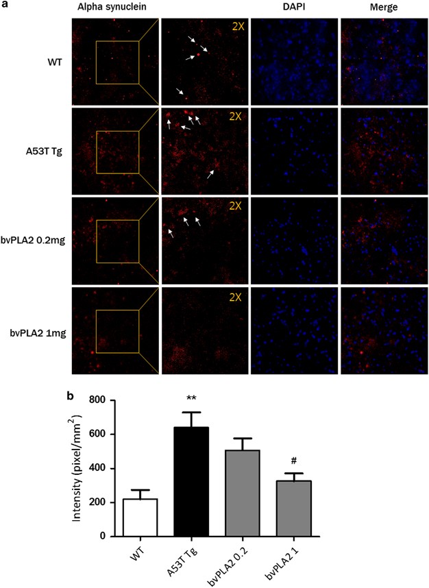
Because α-Syn is known to contribute to PD pathogenesis in various ways,27 it is the gold standard in the evaluation of PD. To confirm the effect of bvPLA2 on the α-Syn pathology, α-Syn immunostaining was performed using spinal cord tissue sections from 6-month-old A53T Tg mice and age-matched WT mice. The intensity of α-Syn in the dorsal column region of the spinal cord was significantly increased in A53T Tg mice (640±89) compared with the WT group (220±54, P<0.01; Figure 2). bvPLA2 treatment effectively inhibited α-Syn pathology (1 mg kg−1, 326±45, P<0.05; Figure 2) compared with A53T Tg mice.
Figure 2
bvPLA2 inhibits α-Syn in the spinal cord in A53Tg mice. (a) Confocal microscopic imaging of α-Syn fluorescence (red) in the spinal cord. The nuclei were counterstained with DAPI (blue). The insets are a × 2 magnification of the boxed area. The representative immunostained α-Syn inclusions are indicated as arrows. (b) The confocal image shown in a was analyzed. The values indicate the mean±s.e.m. **P<0.01 compared with the WT group, #P<0.05 compared with the A53T Tg group. α-Syn, α-Synuclein; WT, wild type.
Treatment with bvPLA2 induces microglial deactivation in the spinal cord
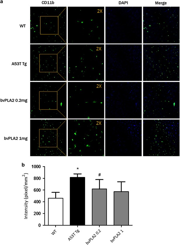
The activation of microglial cells is also one of the characteristics of PD. To determine whether the neuroprotective effect of bvPLA2 resulted from the inhibition of microglial activation in the spinal cord, we performed immunohistochemistry with anti-CD11b to detect microglia/macrophages in the spinal cord sections (Figure 3). In WT mice, few CD11b+ microglia/macrophages, with resting morphologies characterized by small cell bodies and thin processes, were observed in the spinal cords. In contrast, numerous CD11b+ microglia/macrophages, with activated morphologies characterized by larger cell bodies and thick processes, were obvious in the spinal cords of A53T Tg mice. In A53T Tg mice treated with bvPLA2, the number of activated microglia/macrophages decreased in the spinal cord.
Figure 3
Treatment with bvPLA2 induces microglial deactivation in the spinal cord. (a) A section was selected and processed for CD11b immunofluorescence. Confocal microscopic observation revealed microglia fluorescence (green) in the spinal cord. The nuclei were counterstained with DAPI (blue). The insets are × 2 magnification of the boxed area. The representative immunostained α-Syn inclusions are indicated as arrows. (b) The confocal image shown in (a) was analyzed. The values indicate the mean±s.e.m. *P<0.05 compared with the WT group, #P<0.05 compared with the A53T Tg group. α-Syn, α-Synuclein; Tg, transgenic; WT, wild type.
Treatment with bvPLA2 normalizes the ratio of M1/M2 microglial phenotypes
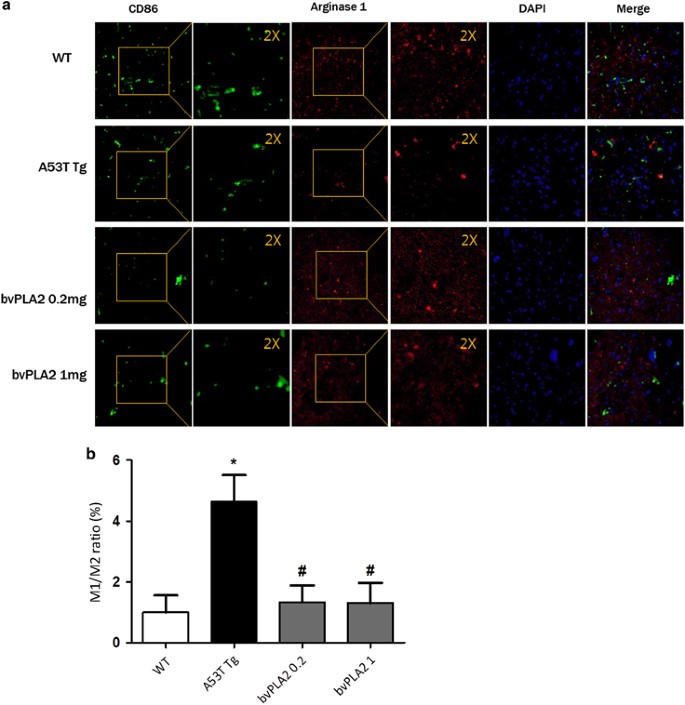
While M1 microglia produce toxic substances to neurons, such as proinflammatory cytokines and reactive oxygen species, M2 microglia produce anti-inflammatory and tissue repair factors to promote survival and repair. We tested the effects of bvPLA on the M1/M2 microglial phenotypes using immunofluorescence double-labeling and confocal microscopy. Specifically, we documented the distribution and magnitude of microglia/macrophages expressing M1 (CD86) and M2 (Arginase 1) phenotypic markers (Figure 4). The M1:M2 ratio decreased markedly upon bvPLA2 treatment, mostly because of the increase of labeling for M2 phenotypic markers (Figure 4).
Figure 4
bvPLA2 changes the M1/M2 ratio in the spinal cords of A53T Tg mice. (a) Confocal microscopic imaging of CD86+M1 macrophage fluorescence (red) and Arginase I fluorescence (green) M2 macrophages in the spinal cord. The nuclei were counterstained with DAPI (blue). The insets are a × 2 magnification of the boxed area. The representative immunostained α-Syn inclusions are indicated as arrows. (b) The confocal image shown in a was analyzed. The values indicate the mean±s.e.m. *P<0.05 compared with the WT group, #P<0.05 compared with the A53T Tg group. Tg, transgenic; WT, wild type.
bvPLA2 influences the polarization of BV-2 microglial cells to the M2 phenotype
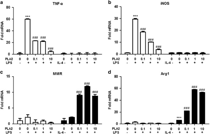
We tested the effects of bvPLA2 on the M1/M2 polarization of microglial cells by quantifying the gene expression upon exposure to M1-inducing conditions (LPS) or M2-inducing conditions (IL-4). Tumor necrosis factor alpha (TNF-α) and inducible nitric oxide synthase (iNOS) served as M1 phenotypic markers, whereas macrophage mannose receptor (MMR; CD206) and arginase-1 (Arg1) served as M2 markers. The messenger RNA (mRNA) levels of TNF-α and iNOS were significantly increased compared with the vehicle control when the cells were exposed to LPS (Figure 5). bvPLA2 effectively blocked the polarization of BV-2 cells to the M1 phenotype in a dose-dependent manner during LPS exposure. No changes were observed in M1 markers during IL-4 exposure. MMR was slightly upregulated and Arg1 was significantly increased by IL-4 compared with the vehicle control. The mRNA levels of both were significantly increased upon bvPLA2 treatment when the cells were exposed to IL-4. There were no significant differences in M2 markers during LPS exposure. Taken together, these results demonstrate that bvPLA2 promotes M2 microglia polarization
Figure 5
bvPLA2 directly induces M2-like polarization of BV-2 microglial cells. Cells were pretreated with bvPLA2 for 30 min and stimulated with LPS or IL-4 for 24 h. The gene expression profile of BV-2 microglial cells was determined by quantitative real-time PCR. (a, b): M1-phenotypic marker (A, TNF-α; B, iNOS) and (c, d): M2-phenotypic marker (C, MMR; D, Arg1) mRNA expression in BV-2 microglial cells. The fold increase in each gene was normalized against the level of the unstimulated (vehicle-treated) control. The values indicate the mean±s.e.m., with each experiment performed in triplicate. ***P<0.0001 compared with the control group, ###P<0.0001 compared with the LPS or IL-4 only treated groups. BV, bee venom; bvPLA2, BV-derived phospholipase A2; IL, interleukin; iNOS, inducible nitric oxide synthase; LPS, lipopolysaccharides; MMR, macrophage mannose receptor; mRNA, messenger RNA; TNF, tumor necrosis factor.
Discussion
We report that progressive degeneration in A53T Tg mice and the improvements resulting from bvPLA2 were associated with α-Syn accumulation in the spinal cord. At the behavioral level, these alterations were accompanied by significantly reduced _T_-turn and T-locomotor times in a modified pole test. The behavior effects of bvPLA2 were accompanied by differential changes in the expression of inflammatory mediators in the spinal cords of A53T Tg mice. Specifically, treatment with bvPLA2 reduced the expression of the microglial activation maker CD11b and the M1 marker CD86 in the spinal cords of A53T Tg mice. In contrast, bvPLA2 treatment increased the expression of the M2 marker arginase 1 in the spinal cords of A53T Tg mice. These in vivo results were confirmed by in vitro experiments using BV-2 microglia. Although bvPLA2 treatment promoted M2 differentiation, M1 differentiation was inhibited by bvPLA2 treatment.
In recent years, several strains of genetically modified mice have been generated as models for studying PD, including the BAC-Tg3 mice reported by Hilton et al. and the A30P+A53T transgenic mice reported by Ikeda et al.28 In the present study, we used A53T Tg mice, a model expressing mutant α-Syn under the control of the mouse prion protein promoter. The A53T mutant human α-Syn homozygous mice showed severe motor disturbance, with accumulation of α-Syn in the brain, similar to patients with the A53T mutation.1 A53T Tg mice have many similarities with human neuronal α-synucleinopathies (especially familial PD) because of the A53T α-Syn mutation. Specifically, the PrP-driven expression of human A53T α-Syn results in a mid- to late-onset neurodegenerative disorder that coincides with the accumulation of filamentous α-Syn cytoplasmic inclusions throughout the neuraxis, similar to patients with the A53T mutation.29 No α-Syn pathology was detected in young animals expressing A53T human α-Syn, and these animals did not present an overt neurological impairment.
These results indicate that the pathological features that mimic PD in A53T mice remain stable. Previous studies have reported the pharmacological efficacy of bvPLA2 in various PD models. bvPLA2 was shown to have an important role in MPTP-induced neurotoxicity and oxidative stress in the nigrostriatal system.30 Our study demonstrated that treatment with bvPLA2 resulted in a significant restoration of motor dysfunction in PD mice. These results suggest that bvPLA2 treatment may be effective at ameliorating locomotive impairment caused by PD. α-Syn is a presynaptic neuronal protein that is linked genetically and neuropathologically to PD31, 32, 33 and multiple system atrophy.34 α-Syn is not only a biomarker for diagnosing PD but is also an important target for PD therapy. We demonstrated that treatment with bvPLA2 is a feasible and effective way to decrease α-Syn in A53T Tg mice. However, the detailed mechanism by which bvPLA2 reduces α-Syn remains unclear and requires further exploration.
Microglia are the resident macrophages of the brain; they have crucial roles in the development and preservation of the neural environment.35 However, in the presence of adverse stimuli, microglia can induce chronic, damaging inflammation that ultimately leads to neuronal cell death.36 Microglia are readily activated during most neuropathological conditions, such as PD and Alzheimer’s disease.37 Our results show that bvPLA2 significantly decreases microglia activation in the spinal cord, suggesting that a decrease in microglia activation may contribute to the therapeutic effect of bvPLA2. Multiple reports have suggested that the phenotype of activated microglia (M1/M2) controls the repair and regeneration response following nerve injury.38, 39 When we tested the effects of bvPLA2 on the M1/M2 polarization of microglial cells, bvPLA2 treatment promoted M2 polarization and suppressed M1 polarization in BV-2 microglia. These observations strongly support a dual role for activated microglia in human brains with ongoing synucleinopathies. M1 microglia are thought to be proinflammatory (IL-1β, TNF-α) and result in deleterious neuroinflammation, whereas M2 microglia (IL-10, neurotrophins) are considered to promote neuroprotection.39, 40, 41, 42 Therefore, some researchers suggest that future immunomodulatory therapies for PD should focus on promoting the M2 profile over the M1 profile to promote regeneration and neuroprotection.9 Our results show that bvPLA2 significantly promotes the M2 profile over the M1 profile in A53T Tg mice and BV-2 microglia. We cannot rule out the possibility of the involvement of other immune cells in addition to macrophages, such as Tregs, in the effects of bvPLA2.
Previously, we reported that bee venom ameliorated MPTP-induced PD via Treg induction.16 We also revealed that bvPLA2 induced a Treg population via CD206 on DCs.43 Recently, we revealed that bvPLA2 protects dopaminergic neurons by inhibiting neuroinflammatory responses in an MPTP-induced PD mouse model.44 In the study, bvPLA2 had no effect on mice depleted of Tregs by anti-CD25 Abs, and we also demonstrated that bvPLA2-CD206-PGE2-EP2 signaling promotes Treg differentiation. Thus, it is reasonable to predict that the increase in Tregs mediated by bvPLA2 treatment could reduce neuroinflammation in A53T Tg mice.
These findings suggest that treatment with bvPLA2 ameliorates motor deficits and reduces α-Syn in A53T Tg mice. These effects are associated with a decrease in the M1/M2 ratio and the activation of microglia in A53T Tg mice. These results provide strong evidence for the use of bvPLA2 as a novel strategy for PD treatment.
References
- Giasson BI, Duda JE, Quinn SM, Zhang B, Trojanowski JQ, Lee VM . Neuronal alpha-synucleinopathy with severe movement disorder in mice expressing A53T human alpha-synuclein. Neuron 2002; 34: 521–533.
Article CAS Google Scholar - Hirsch EC, Breidert T, Rousselet E, Hunot S, Hartmann A, Michel PP . The role of glial reaction and inflammation in Parkinson's disease. Ann N Y Acad Sci 2003; 991: 214–228.
Article CAS PubMed Google Scholar - Barcia C, Sanchez Bahillo A, Fernandez-Villalba E, Bautista V, Poza YPM, Fernandez-Barreiro A et al. Evidence of active microglia in substantia nigra pars compacta of parkinsonian monkeys 1 year after MPTP exposure. Glia 2004; 46: 402–409.
Article PubMed Google Scholar - McGeer PL, Schwab C, Parent A, Doudet D . Presence of reactive microglia in monkey substantia nigra years after 1-methyl-4-phenyl-1,2,3,6-tetrahydropyridine administration. Ann Neurol 2003; 54: 599–604.
Article CAS PubMed Google Scholar - Orr CF, Rowe DB, Halliday GM . An inflammatory review of Parkinson's disease. Prog Neurobiol 2002; 68: 325–340.
Article CAS PubMed Google Scholar - Sawada M, Imamura K, Nagatsu T . Role of cytokines in inflammatory process in Parkinson's disease. J Neural Transm Suppl 2006; 70: 373–381.
Article CAS Google Scholar - Varnum MM, Ikezu T . The classification of microglial activation phenotypes on neurodegeneration and regeneration in Alzheimer's disease brain. Arch Immunol Ther Exp (Warsz) 2012; 60: 251–266.
Article CAS Google Scholar - Hoozemans JJ, Veerhuis R, Rozemuller JM, Eikelenboom P . Neuroinflammation and regeneration in the early stages of Alzheimer's disease pathology. Int J Dev Neurosci 2006; 24: 157–165.
Article CAS PubMed Google Scholar - Sanchez-Guajardo V, Barnum CJ, Tansey MG, Romero-Ramos M . Neuroimmunological processes in Parkinson's disease and their relation to alpha-synuclein: microglia as the referee between neuronal processes and peripheral immunity. ASN Neuro 2013; 5: 113–139.
Article CAS PubMed Google Scholar - Gerhard A, Pavese N, Hotton G, Turkheimer F, Es M, Hammers A et al. In vivo imaging of microglial activation with [11C](R)-PK11195 PET in idiopathic Parkinson's disease. Neurobiol Dis 2006; 21: 404–412.
Article CAS PubMed Google Scholar - Rojo AI, Innamorato NG, Martin-Moreno AM, De Ceballos ML, Yamamoto M, Cuadrado A . Nrf2 regulates microglial dynamics and neuroinflammation in experimental Parkinson's disease. Glia 2010; 58: 588–598.
Article PubMed Google Scholar - Kwon YB, Lee HJ, Han HJ, Mar WC, Kang SK, Yoon OB et al. The water-soluble fraction of bee venom produces antinociceptive and anti-inflammatory effects on rheumatoid arthritis in rats. Life Sci 2002; 71: 191–204.
Article CAS PubMed Google Scholar - Son DJ, Lee JW, Lee YH, Song HS, Lee CK, Hong JT . Therapeutic application of anti-arthritis, pain-releasing, and anti-cancer effects of bee venom and its constituent compounds. Pharmacol Ther 2007; 115: 246–270.
Article CAS PubMed Google Scholar - Cho SY, Shim SR, Rhee HY, Park HJ, Jung WS, Moon SK et al. Effectiveness of acupuncture and bee venom acupuncture in idiopathic Parkinson's disease. Parkinsonism Relat Disord 2012; 18: 948–952.
Article PubMed Google Scholar - Mirshafiey A . Venom therapy in multiple sclerosis. Neuropharmacology 2007; 53: 353–361.
Article CAS PubMed Google Scholar - Chung ES, Kim H, Lee G, Park S, Bae H . Neuro-protective effects of bee venom by suppression of neuroinflammatory responses in a mouse model of Parkinson's disease: role of regulatory T cells. Brain Behav Immun 2012; 26: 1322–1330.
Article CAS PubMed Google Scholar - Habermann E . Bee and wasp venoms. Science 1972; 177: 314–322.
Article CAS PubMed Google Scholar - Linderoth L, Peters GH, Jorgensen K, Madsen R, Andresen TL . Synthesis of sn-1 functionalized phospholipids as substrates for secretory phospholipase A2. Chem Phys Lipids 2007; 146: 54–66.
Article CAS PubMed Google Scholar - Dennis EA, Rhee SG, Billah MM, Hannun YA . Role of phospholipase in generating lipid second messengers in signal transduction. FASEB J 1991; 5: 2068–2077.
Article CAS PubMed Google Scholar - Dennis EA . Diversity of group types, regulation, and function of phospholipase A2. J Biol Chem 1994; 269: 13057–13060.
CAS PubMed Google Scholar - Mukherjee AB, Miele L, Pattabiraman N . Phospholipase A2 enzymes: regulation and physiological role. Biochem Pharmacol 1994; 48: 1–10.
Article CAS PubMed Google Scholar - Granata F, Frattini A, Loffredo S, Del Prete A, Sozzani S, Marone G et al. Signaling events involved in cytokine and chemokine production induced by secretory phospholipase A2 in human lung macrophages. Eur J Immunol 2006; 36: 1938–1950.
Article CAS PubMed Google Scholar - Dennis EA . The growing phospholipase A2 superfamily of signal transduction enzymes. Trends Biochem Sci 1997; 22: 1–2.
Article CAS PubMed Google Scholar - Fenard D, Lambeau G, Maurin T, Lefebvre JC, Doglio A . A peptide derived from bee venom-secreted phospholipase A2 inhibits replication of T-cell tropic HIV-1 strains via interaction with the CXCR4 chemokine receptor. Mol Pharmacol 2001; 60: 341–347.
Article CAS PubMed Google Scholar - Nakashima S, Kitamoto K, Arioka M . The catalytic activity, but not receptor binding, of sPLA2s plays a critical role for neurite outgrowth induction in PC12 cells. Brain Res 2004; 1015: 207–211.
Article CAS PubMed Google Scholar - Ogawa N . Dopamine neurotransmission and treatments for Parkinson's disease in the molecular biology era. Eur Neurol 1997; 38 (Suppl 1): 2–5.
Article CAS PubMed Google Scholar - Lu J, Sun F, Ma H, Qing H, Deng Y . Comparison between alpha-synuclein wild-type and A53T mutation in a progressive Parkinson's disease model. Biochem Biophys Res Commun 2015; 464: 988–993.
Article CAS PubMed Google Scholar - Ikeda M, Kawarabayashi T, Harigaya Y, Sasaki A, Yamada S, Matsubara E et al. Motor impairment and aberrant production of neurochemicals in human alpha-synuclein A30P+A53T transgenic mice with alpha-synuclein pathology. Brain Res 2009; 1250: 232–241.
Article CAS PubMed Google Scholar - Roostaee A, Beaudoin S, Staskevicius A, Roucou X . Aggregation and neurotoxicity of recombinant alpha-synuclein aggregates initiated by dimerization. Mol Neurodegener 2013; 8: 5.
Article CAS PubMed PubMed Central Google Scholar - Chalimoniuk M, Stolecka A, Zieminska E, Stepien A, Langfort J, Strosznajder JB . Involvement of multiple protein kinases in cPLA2 phosphorylation, arachidonic acid release, and cell death in in vivo and in vitro models of 1-methyl-4-phenylpyridinium-induced parkinsonism—the possible key role of PKG. J Neurochem 2009; 110: 307–317.
Article CAS PubMed Google Scholar - Stefanis L . alpha-Synuclein in Parkinson's disease. Cold Spring Harb Perspect Med 2012; 2: a009399.
Article PubMed PubMed Central Google Scholar - Oliveras-Salva M, Van der Perren A, Casadei N, Stroobants S, Nuber S, D'Hooge R et al. rAAV2/7 vector-mediated overexpression of alpha-synuclein in mouse substantia nigra induces protein aggregation and progressive dose-dependent neurodegeneration. Mol Neurodegener 2013; 8: 44.
Article PubMed PubMed Central Google Scholar - Sacino AN, Thomas MA, Ceballos-Diaz C, Cruz PE, Rosario AM, Lewis J et al. Conformational templating of alpha-synuclein aggregates in neuronal-glial cultures. Mol Neurodegener 2013; 8: 17.
Article CAS PubMed PubMed Central Google Scholar - Prusiner SB, Woerman AL, Mordes DA, Watts JC, Rampersaud R, Berry DB et al. Evidence for alpha-synuclein prions causing multiple system atrophy in humans with parkinsonism. Proc Natl Acad Sci USA 2015; 112: E5308–E5317.
Article CAS PubMed Google Scholar - Kraft AD, Harry GJ . Features of microglia and neuroinflammation relevant to environmental exposure and neurotoxicity. Int J Environ Res Public Health 2011; 8: 2980–3018.
Article PubMed PubMed Central Google Scholar - Block ML, Hong JS . Microglia and inflammation-mediated neurodegeneration: multiple triggers with a common mechanism. Prog Neurobiol 2005; 76: 77–98.
Article CAS PubMed Google Scholar - McGeer PL, Itagaki S, Boyes BE, McGeer EG . Reactive microglia are positive for HLA-DR in the substantia nigra of Parkinson's and Alzheimer's disease brains. Neurology 1988; 38: 1285–1291.
Article CAS Google Scholar - Kigerl KA, Gensel JC, Ankeny DP, Alexander JK, Donnelly DJ, Popovich PG . Identification of two distinct macrophage subsets with divergent effects causing either neurotoxicity or regeneration in the injured mouse spinal cord. J Neurosci 2009; 29: 13435–13444.
Article CAS PubMed PubMed Central Google Scholar - David S, Kroner A . Repertoire of microglial and macrophage responses after spinal cord injury. Nat Rev Neurosci 2011; 12: 388–399.
Article CAS PubMed Google Scholar - Cherry JD, Olschowka JA, O'Banion MK . Neuroinflammation and M2 microglia: the good, the bad, and the inflamed. J Neuroinflammation 2014; 11: 98.
Article PubMed PubMed Central Google Scholar - Olah M, Biber K, Vinet J, Boddeke HW . Microglia phenotype diversity. CNS Neurol Disord Drug Targets 2011; 10: 108–118.
Article CAS PubMed Google Scholar - Jimenez S, Baglietto-Vargas D, Caballero C, Moreno-Gonzalez I, Torres M, Sanchez-Varo R et al. Inflammatory response in the hippocampus of PS1M146L/APP751SL mouse model of Alzheimer's disease: age-dependent switch in the microglial phenotype from alternative to classic. J Neurosci 2008; 28: 11650–11661.
Article CAS PubMed Google Scholar - Kim H, Lee H, Lee G, Jang H, Kim SS, Yoon H et al. Phospholipase A2 inhibits cisplatin-induced acute kidney injury by modulating regulatory T cells by the CD206 mannose receptor. Kidney Int 2015; 88: 550–559.
Article CAS PubMed Google Scholar - Chung ES, Lee G, Lee C, Ye M, Chung HS, Kim H et al. Bee venom phospholipase A2, a novel Foxp3+ regulatory T cell inducer, protects dopaminergic neurons by modulating neuroinflammatory responses in a mouse model of Parkinson's Disease. J Immunol 2015; 195: 4853–4860.
Article CAS PubMed Google Scholar
Acknowledgements
This work was supported by a National Research Foundation of Korea (NRF) grant funded by the Korean government (Ministry of Education, Science, and Technology, MEST) (no. 2013-068954).
Author information
Authors and Affiliations
- Department of Physiology, College of Korean Medicine, Kyung Hee University, Seoul, Republic of Korea
Minsook Ye, Chanju Lee, Joo Hyun Song & Hyunsu Bae - Korean Medicine (KM)-Application Center, Korea Institute of Oriental Medicine (KIOM), Daegu, Republic of Korea
Hwan-Suck Chung - Acupuncture and Meridian Science Research Center, College of Korean Medical Science Graduate School, Kyung Hee University, Seoul, Republic of Korea
Insop Shim - Department of Anatomy-Pointlogy, College of Korean Medicine, Gachon University, Seongnam, Republic of Korea
Youn-Sub Kim
Authors
- Minsook Ye
- Hwan-Suck Chung
- Chanju Lee
- Joo Hyun Song
- Insop Shim
- Youn-Sub Kim
- Hyunsu Bae
Corresponding author
Correspondence toHyunsu Bae.
Ethics declarations
Competing interests
The authors declare no conflict of interest.
Rights and permissions
This work is licensed under a Creative Commons Attribution-NonCommercial-ShareAlike 4.0 International License. The images or other third party material in this article are included in the article’s Creative Commons license, unless indicated otherwise in the credit line; if the material is not included under the Creative Commons license, users will need to obtain permission from the license holder to reproduce the material. To view a copy of this license, visit http://creativecommons.org/licenses/by-nc-sa/4.0/
About this article
Cite this article
Ye, M., Chung, HS., Lee, C. et al. Bee venom phospholipase A2 ameliorates motor dysfunction and modulates microglia activation in Parkinson’s disease alpha-synuclein transgenic mice.Exp Mol Med 48, e244 (2016). https://doi.org/10.1038/emm.2016.49
- Received: 28 August 2015
- Revised: 25 January 2016
- Accepted: 22 February 2016
- Published: 08 July 2016
- Issue Date: July 2016
- DOI: https://doi.org/10.1038/emm.2016.49
