Hedgehog regulates smoothened activity by inducing a conformational switch (original) (raw)
Main
The HH morphogen controls many key development processes, with different thresholds specifying distinct outcomes1,2,3,4. In Drosophila wing discs, HH proteins secreted by posterior (P) compartment cells move into the anterior (A) compartment to form a local concentration gradient5,6. Low levels of HH suffice to induce the expression of decapentaplegic (dpp), whereas high levels are required to induce patched (ptc) and engrailed (en) (Supplementary Fig. 1)7,8,9.
The reception system for HH consists of a twelve-transmembrane protein, PTC, as the HH receptor and a seven-transmembrane protein smoothened (SMO) as the signal transducer10,11,12,13. In Drosophila, HH binding to PTC abrogates its inhibition on SMO and induces extensive phosphorylation of the SMO cytoplasmic tail by protein kinase A (PKA) and casein kinase I (CKI), leading to SMO cell surface accumulation and activation14,15,16,17. How phosphorylation promotes SMO cell surface accumulation is not understood. In addition, phosphorylation may regulate SMO activity through mechanism(s) other than controlling its cell surface abundance.
Regulation of SMO by multiple Arg clusters
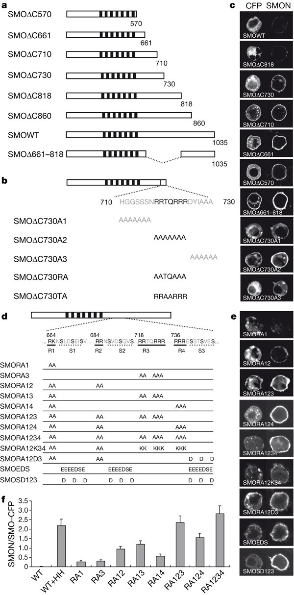
Our previous study indicates that phosphorylation may regulate SMO cell surface abundance by either preventing its endocytosis and/or promoting its recycling15. To investigate further how SMO cell surface expression is regulated, we generated a set of C-terminally truncated SMO variants and examined their subcellular localization using a cell-based assay (Fig. 1). Deletion up to amino acid 818 did not significantly change SMO subcellular distribution; however, further deletions resulted in progressively increased cell surface expression (Fig. 1a, c), implying that multiple negative regulatory elements exist between amino acids 661–818.
Figure 1: Regulation of SMO cell surface expression by multiple Arg clusters.
a, SMO deletion mutants with CFP (not shown) fused to their C termini. Filled boxes indicate the transmembrane domains. b, Ala-scan mutagenesis of SMOΔC730 with the last 20 amino acids and corresponding substitutions shown underneath. c, e, Cell surface expression of the indicated SMO mutants. S2 cells were transfected with the indicated CFP-tagged SMO constructs, followed by immunostaining with anti-SMON antibody before membrane permeabilization15. The SMON column indicates cell surface staining, whereas the CFP column indicates the total protein distribution. SMOΔ730RA and SMOΔ730TA behaved like SMOΔ710 and SMOΔ730, respectively (data not shown). d, A schematic drawing of a full-length SMO with the sequences of the four Arg clusters (R1–R4) and three phosphorylation clusters (S1–S3) shown underneath. SMO variants with the indicated substitutions are listed. f, Ratio of cell surface level (SMON signal) to total level (CFP signal) of protein for wild-type and indicated mutant forms of SMO. For SMO variants, n = 20; error bars, 1 s.d.
SMOΔC710 exhibits consistently higher cell surface expression than SMOΔC730 (Fig. 1c), indicating that amino acids 710–730 may harbour a negative element(s). Ala-scan mutagenesis, which substituted multiple residues to Ala, identified the Arg residues in RRTQRRR as critical for preventing SMO cell surface accumulation (Fig. 1b, c; data not shown). Interestingly, multiple Arg clusters, arbitrarily named R1 to R4, are located between amino acids 661–818, a region critical for blocking SMO cell surface accumulation (Fig. 1d). We therefore introduced into the full-length SMO Arg to Ala (RA) mutations in individual, or combinations of, Arg clusters. SMO variants with one Arg cluster mutated did not exhibit significant change in their cell surface expression; however, mutating two or more Arg clusters caused a gradual increase in SMO cell surface expression (Fig. 1d–f; data not shown), suggesting that multiple Arg clusters cooperate to restrict SMO cell surface accumulation.
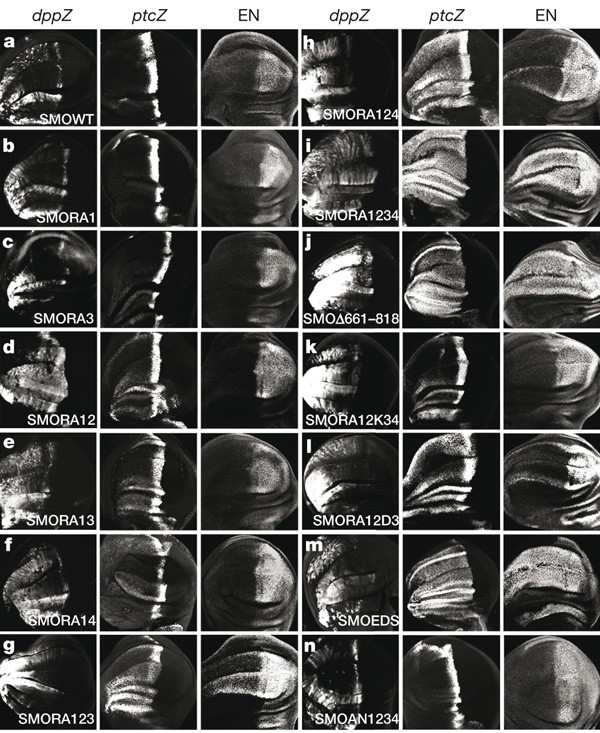
To determine whether the Arg clusters negatively regulate SMO activity, SMO variants with one or more mutated Arg clusters were expressed in wing discs using the MS1096 Gal4 driver. SMO variants with one mutated Arg cluster exhibited low levels of basal activity similar to that of wild-type SMO, as is evident from the ectopic expression of dpp but not ptc and en (Fig. 2a–c). However, SMO variants with two or more mutated Arg clusters exhibited a progressive increase in their constitutive signalling activities (Fig. 2d–i). Thus, SMO activity is inversely correlated with the number of functional Arg clusters. We also mutated several Arg clusters in the membrane-proximal region of the SMO cytoplasmic tail and observed no effect on SMO cell surface expression and activity (Supplementary Fig. 2). Hence, the Arg clusters between amino acids 661–818 are specifically involved in SMO autoinhibition.
Figure 2: In vivo activities of SMO variants.
a–n, Wing discs expressing the indicated SMO variants from MS1096 were immunostained to show the expression of dpp-lacZ (dppZ), ptc-lacZ (ptcZ) and EN. dppZ, ptcZ and en are induced by low, intermediate and high levels of HH, respectively. The levels of SMO activity inversely correlate with the number of intact Arg clusters (b–i). An internal deletion removing amino acids 661–818 resulted in high levels of constitutive SMO activity (j). SMORA12K34 (k) has similar activity to SMORA12 (compare to d). SMORA12D3 (l) exhibited constitutive activity similar to that of SMORA124 (h). m, Substitution of the three PKA/CKI phosphorylation clusters with acidic clusters led to high constitutive activity similar to that of SMOSD123 (ref. 15). n, SMOAN1234 (see Fig. 4a) did not exhibit higher basal activity than SMOWT (compare to a).
Phosphorylation counteracts the Arg motifs
Increasing the number of phosphorylation-mimetic mutations in PKA/CKI sites resulted in a graded increase in SMO cell surface level and activity15, which phenocopies the effect of increasing the number of RA mutations, indicating that phosphorylation may activate SMO by antagonizing the Arg motifs. Consistently, an internal deletion that removes both the phosphorylation and Arg clusters (SMOΔ661–818) results in high levels of SMO cell surface expression and activity (Figs 1a, c and 2j).
It is intriguing that the Arg clusters are situated adjacent to the PKA/CKI phosphorylation clusters (Fig. 1d). In fact, R1, R2 and R4 are part of the PKA phosphorylation consensus site, R/KRXS. The juxtaposition of the Arg and phosphorylation clusters may allow precise control of SMO activity because phosphorylation at individual clusters may only neutralize the negative influence of adjacent Arg clusters. To test this, we constructed SMORA12D3 and found it behaved like SMORA124 (Fig. 1d, e; compare Fig. 2l with 2h), suggesting that phosphorylation at S3 (Fig. 1d) neutralizes the negative effect of R4.
Because Arg carries positive charge whereas phosphorylation brings in negative charge, phosphorylation may antagonize the Arg clusters by neutralizing their positive charges. In support of this model, we found that R3 and R4 can be functionally substituted by Lys, because SMORA12K34 behaved like SMORA12 rather than SMORA1234 (Fig. 1d, e, 2k). Furthermore, SMOEDS, which has three PKA/CKI phosphorylation clusters replaced by a stretch of acidic amino acids (Fig. 1d), exhibited high levels of cell surface expression and signalling activity similar to the phosphorylation-mimetic SMO variant, SMOSD123 (Fig. 1d, e, 2m; ref. 15), suggesting that the exact sequence composition of the phosphorylation clusters is not critical, but rather the negative charges they carry are important.
HH induces increased proximity of SMO cytoplasmic tails
Although SMO activity correlates with its cell surface levels, HH may induce SMO activation through additional mechanism(s) such as dimerization and/or conformational change18,19. To test these possibilities, we employed fluorescence resonance energy transfer (FRET) analysis, which measures the transfer of energy between yellow fluorescent protein (YFP) and cyan fluorescent protein (CFP) as a function of distance20. We initially constructed two pairs of tagged SMO with CFP/YFP either fused to the C terminus (SMO–CFPC/SMO–YFPC) or inserted at an amino-terminal position (SMO–CFPN/SMO–YFPN) of SMO (Fig. 3a, b; Supplementary Fig. 3). As controls, we constructed CFP/YFP-tagged forms of frizzled 2 (FZ2) and RAB5.
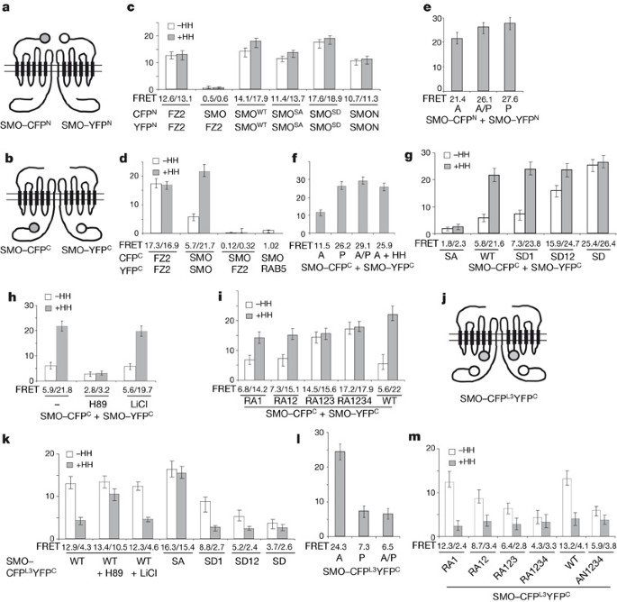
Figure 3: Regulation of both conformation and proximity of SMO cytoplasmic tails.
a, b, j, Cartoons of SMO–CFPN/SMO–YFPN (a), SMO–CFPC/SMO–YFPC (b), and SMO–CFPL3YFPC dimers (j). The filled and open circles indicate CFP and YFP, respectively. c, d, g–i, k, m, FRET efficiency (y axis and numbers below bars) from the indicated CFP/YFP-tagged constructs in S2 cells treated with or without HH-conditioned medium, and with or without PKA inhibitor H89 or GSK3 inhibitor LiCl (mean ± s.d., n ≥ 10). SMOSA has three PKA sites mutated to Ala, whereas SMOSD1, SMOSD12 and SMOSD have PKA and CKI sites in one, two and three phosphorylation clusters converted to Asp, respectively (c, g, k; ref. 15). SMON lacks a cytoplasmic tail (c). RA1, RA12, RA123 and RA1234 have one, two, three and four Arg clusters mutated to Ala, respectively (i, m; see Fig. 1d). AN1234 has four C-terminal acidic clusters mutated to Ala (m; see Fig. 4a). HH increased FRETC between SMO–CFPC/SMO–YFPC (d) and decreased FRETL3C from SMO–CFPL3YFPC (k), both of which were blocked by the SA mutations (g) or H89 but not by LiCl (h, k). Phospho-mimetic or RA mutations progressively increased basal FRETC (g, i) whereas they gradually decreased FRETL3C (k, m). e, f, l, FRET efficiency between SMO–CFPN/SMO–YFPN (e), SMO–CFPC/SMO–YFPC (f), or from SMO–CFPL3YFPC (l) expressed in wing discs (mean ± s.d., n ≥ 5). A, A-compartment cells away from the A/P boundary; P, P-compartment cells; A/P, A-compartment cells adjacent to the A/P boundary; A + HH, A-compartment cells expressing UAS-HH.
Consistent with a previous finding that FZ family members form constitutive dimers/oligomers21, we observed high FRET between FZ2–CFPC/FZ2–YFPC (17.3 ± 1.9%) or FZ2–CFPN/FZ2–YFPN (12.6 ± 1.1%) in S2 cells (Fig. 3c, d). Under similar conditions, FRET between SMO–CFPN/SMO–YFPN (referred to as FRETN) was 14.1 ± 1.4% (Fig. 3c), whereas FRET between SMO–CFPC/SMO–YFPC (FRETC) was 5.7 ± 1.3% (Fig. 3d). HH stimulation significantly increased FRETC to 21.7 ± 1.5% (Fig. 3d), but only modestly increased FRETN (Fig. 3c). FRET between control pairs (SMO/FZ2 or SMO/RAB5) was ≤1.0% (Fig. 3c, d). In addition, CFP- and YFP-tagged SMO colocalized whereas SMO–CFP barely overlapped with FZ2–YFP (Supplementary Fig. 4). Even in S2 cells stimulated with HH, in which SMO accumulated on the cell surface and overlapped with FZ2, FRET between SMO/FZ2 remained low (Fig. 3c, d, and Supplementary Fig. 4). Furthermore, over fourfold changes in SMO signal intensity did not significantly affect FRETC (Supplementary Fig. 5).
In wing discs, FRETN was high in both A and P compartments regardless of HH (Fig. 3e), whereas FRETC in A-compartment cells distant from the A/P boundary was relatively low but increased significantly in P-compartment cells and in A-compartment cells exposed to HH or lacking PTC (Fig. 3f; Supplementary Figs 6–7a). The high basal FRETN suggests that SMO forms a constitutive dimer/oligomer (dimer is used hereafter for simplicity), as is the case for the FZ family. Constitutive SMO dimerization was confirmed by immunoprecipitation assays (Supplementary Figs 8 and 9). SMO dimerization is likely to be mediated by SMON, which includes the N-terminal extracellular domain and transmembrane helices, because SMON–CFPN and SMON–YFPN colocalized and produced high basal FRET (Fig. 3c).
The low basal but high HH-induced FRETC suggests that the two SMO cytoplasmic tails within a dimer are separated from each other but HH signalling increases their proximity. To investigate whether increased proximity is accompanied by a conformational change, we generated a doubly tagged SMO (SMO–CFPL3YFPC) with CFP inserted into the third intracellular loop (L3) and YFP fused to the C terminus (Fig. 3j; Supplementary Fig. 3). SMO–CFPL3YFPC responded to HH and possessed signalling activity (Supplementary Figs 10, 11). In S2 cells, basal FRET from SMO–CFPL3YFPC (referred to as FRETL3C) was 12.9 ± 1.2% but dropped to 4.3 ± 0.8% after HH treatment (Fig. 3k; Supplementary Fig. 11). In wing discs, FRETL3C was 24.3 ± 2.1% in A-compartment cells distant from the A/P boundary, but dropped to 6.5 ± 1.5% in A-compartment cells near the A/P boundary or to 7.3 ± 1.6% in P-compartment cells (Fig. 3l; Supplementary Fig. 12). FRETL3C also reduced to 5.9 ± 1.2% in A-compartment ptc mutant clones (Supplementary Fig. 7b). The high basal FRETL3C is probably due to close proximity between the C terminus and L3 of the same SMO molecule (Supplementary Fig. 13). These results suggest that SMO adopts a closed inactive conformation with its C terminus in close proximity to L3, in quiescent cells. HH promotes SMO to adopt an open active conformation in which its C terminus moves away from L3 but closer to the C terminus of its binding partner.
Phosphorylation regulates SMO conformation
To determine if conformational change and increased proximity of SMO cytoplasmic tails is regulated by phosphorylation, we mutated three PKA sites (Ser 667, Ser 687 and Ser 740) to Ala (SA) or substituted them and adjacent CKI sites with Asp (SD123 or SD for simplicity)15. The HH-induced increase in FRETC or decrease in FRETL3C was blocked by the SA mutation as well as a PKA inhibitor H89 (Fig. 3g, h, k), whereas the SD123 substitution resulted in high basal FRETC but low basal FRETL3C (Fig. 3g, k). In contrast, neither the basal nor the HH-induced FRETN was significantly affected by the SA or SD123 mutation (Fig. 3c), suggesting that constitutive SMO dimerization is not regulated by phosphorylation, but conformational change and increased proximity of SMO cytoplasmic tails are triggered by phosphorylation. Mutating multiple Arg clusters also resulted in high basal FRETC but low basal FRETL3C (Fig. 3i, m), suggesting that the Arg motifs keep SMO cytoplasmic tails in a closed inactive conformation.
To assess direct physical interaction between SMO cytoplasmic tails and its regulation by phosphorylation, we applied the CytoTrap yeast two-hybrid assay (Methods Summary). Wild-type SMO cytoplasmic tail (SMOCWT) failed to self-associate, whereas phospho-mimetic SMO cytoplasmic tail (SMOCSD) could self-associate and also interact weakly with SMOWT (Supplementary Fig. 14), indicating that phosphorylation of the SMO cytoplasmic tail may promote self-association.
Our previous study suggests that graded SMO activities are governed by SMO phosphorylation levels15. To determine if increasing SMO phosphorylation could induce gradual changes in SMO conformation, we compared FRETC and FRETL3C for several phosphorylation-mimetic forms of SMO. SMOSD1, SMOSD12 and SMOSD contain Ser to Asp substitution in one, two and three phosphorylation clusters, respectively, and exhibit progressively higher levels of basal activity15. Interestingly, they also exhibited a progressive increase in basal FRETC (Fig. 3g) and gradual decrease in basal FRETL3C (Fig. 3k). Furthermore, FRETC progressively increased, whereas FRETL3C gradually decreased, when increasingly more Arg clusters were mutated (Fig. 3i, m). Thus, increasing SMO phosphorylation seems to induce progressive changes in SMO conformation by antagonizing the Arg clusters. SMO may adopt a series of conformational states determined by its phosphorylation levels. Alternatively, SMO may switch in equilibrium between two distinct conformational states: a closed inactive conformation and an open active conformation; phosphorylation increases the probability for individual SMO to adopt the open active conformation.
Arg clusters mediate intramolecular interaction
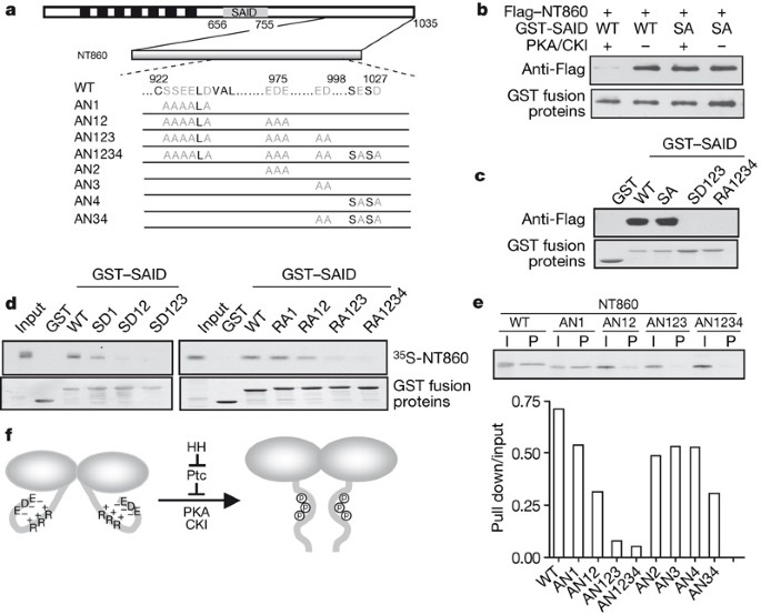
To determine how Arg clusters keep SMO in a closed inactive conformation, we tested the possibility that they might be involved in intramolecular interactions. A glutathione _S_-transferase (GST) fusion protein (GST–SMO656–755) that contains the SMO region between amino acids 656–755 (referred to hereafter as SAID for SMO auto-inhibitory domain) was tested for interaction with a set of C-terminal fragments, and a minimal SAID interacting fragment (NT860) was identified that contains the C-terminal region between amino acids 860–1035 (Supplementary Fig. 15). SAID–NT860 interaction was diminished by PKA/CKI phosphorylation as well as RA or SD123 mutations (Fig. 4b, c), and the binding affinity gradually decreased with more phosphorylation or Arg clusters mutated (Fig. 4d). The importance of Arg clusters in the SAID–NT860 interaction indicates that the association may be mediated by electrostatic interactions. Indeed, mutating several acidic clusters in the C-terminal half of NT860 gradually diminished the SAID–NT860 interaction (Fig. 4a, e).
Figure 4: The Arg clusters mediate intramolecular electrostatic interaction.
a, Diagram of SMO with the SAID indicated by a grey box, and NT860 with indicated substitutions. b, GST pull-down assay using GST–SAID and S2 cell extracts expressing the indicated Flag-tagged SMO C-terminal fragments. c, d, GST pull-down experiments using wild-type or the indicated mutant GST–SAID and S2 cell extracts expressing Flag-tagged NT860 (c) or in vitro translated 35S-labelled NT860 (d). e, Autoradiography (upper panel) and quantification (lower panel) of a GST pull-down assay using GST–SAID and in vitro translated 35S-labelled wild-type (WT) or mutant (AN) NT860. I, input; P, pulled-down protein. The binding affinity was indicated by the ratio of pulled-down protein (pull down) to input. f, A model for regulating SMO conformation by multiple Arg clusters and HH-induced phosphorylation; see text for detail.
The electrostatic interaction between NT860 and SAID may result in a folding back of the SMO cytoplasmic tail to form a closed conformation (Fig. 4f). Consistently, mutating the acidic clusters (SMOAN1234) resulted in decreased basal FRETL3C (Fig. 3m). However, unlike RA mutations, which not only caused conformational change but also promoted SMO cell surface accumulation, SMOAN1234 exhibited little if any cell surface expression and did not exhibit high levels of constitutive activity (Fig. 2n), indicating that both cell surface accumulation and conformational change may be critical for SMO activity.
Clustering of the SMO cytoplasmic tail activates the HH pathway
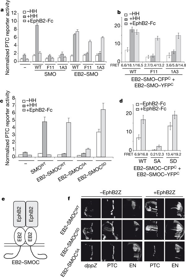
To assess the biological significance of SMO dimerization, we analysed two SMO mutants with point mutations in the N-terminal extracellular domain: SMO1A3 is encoded by a hypomorphic allele such that Cys 90 is substituted to Ser; and SMOF11 is encoded by a strong allele that changes Cys 155 to Tyr (ref. 22). Both mutations reduced basal as well as HH-induced FRETN and FRETC, with SMOF11 exhibiting more severe defects (Supplementary Fig. 16). Immunoprecipitation assays indicated that SMO1A3 and SMOF11 failed to dimerize with SMOWT (Supplementary Fig. 17a). Unlike SMOWT, neither SMO1A3 nor SMOF11 was phosphorylated in response to HH (Supplementary Fig. 17b). In addition, both SMO1A3 and SMOF11 lost HH-induced activity (Fig. 5a).
Figure 5: Clustering of SMO cytoplasmic tails triggers HH pathway activation.
a, c, The ptc-luc reporter assay in cl-8 cells transfected with the indicated SMO expression constructs and treated with or without HH-conditioned medium or EphB2-Fc. Error bars, 1 s.d. (triplicate wells). b, d, FRET between wild-type or mutant pairs of EB2–SMO–CFPC/EB2–SMO–YFPC (b) or EB2–SMOC–CFPC/EB2–SMOC–YFPC (d) expressed in S2 cells treated with or without HH-conditioned medium or EphB2-Fc (mean ± s.d., n ≥ 10). e, Cartoon of the EB2–SMOC–EphB2 complex. f, Wing discs expressing the indicated SMO constructs with or without EphB2Z under the control of the MS1096 Gal4 driver were immunostained to show the expression of dpp-lacZ (dppZ), PTC and EN. Of note, EB2–SMOCSD exhibited higher basal activity than EB2–SMOCWT because it induced higher levels of ectopic dppZ and also induced ectopic albeit low levels of ptc. When coexpressed with EphB2Z, both EB2–SMOCWT and EB2–SMOCSD ectopically activated high levels of ptc and low levels of en. In contrast, EB2–SMOCSA failed to activate any HH target genes.
If loss of SMO activity was due to compromised dimerization, restoring dimerization to these mutants should rescue their activities. To test this, we developed an inducible dimerization system by taking advantage of the observation that the mammalian receptor tyrosine kinase EphB2 forms a hetero-tetramer with its ligand ephrin B2 (EB2; also known as Efnb2) to trigger bidirectional signalling23. Accordingly, we constructed EB2–SMO chimaeric proteins in which the extracellular domain of EB2 was inserted into the SMO N-terminal extracellular domain (Supplementary Fig. 3). When expressed in cl-8 cells, EB2–SMO1A3 and EB2–SMOF11 failed to be activated by HH-conditioned medium; however, they were activated when cells were exposed to the soluble pre-clustered EphB2 extracellular domain, EphB2-Fc (Fig. 5a). In addition, FRETC between mutant pairs of EB2–SMO–CFPC/EB2–SMO–YFPC increased significantly in response to EphB2-Fc but not HH (Fig. 5b).
To determine if dimerization of the SMO cytoplasmic tail suffices to activate the HH pathway, we constructed EB2–SMO cytoplasmic-tail chimaeric proteins in which the intracellular domain of EB2 was replaced by the wild-type (EB2–SMOCWT), phosphorylation-deficient (EB2–SMOCSA), or phosphorylation-mimetic (EB2–SMOCSD) SMO cytoplasmic tail (Fig. 5e, and Supplementary Fig. 3). In both cl-8 cells and wing discs, EB2–SMOCWT exhibited low basal activity but was markedly stimulated by EphB2 (Fig. 5c, f). Furthermore, EB2–SMOCWT activated the HH pathway independent of endogenous SMO (Supplementary Fig. 18). EB2–SMOCWT also induced FU phosphorylation in response to EphB2-Fc (Supplementary Fig. 19a). In addition, FRET between EB2–SMOCWT–CFPC/EB2–SMOCWT–YFPC increased significantly in response to EphB2-Fc (Fig. 5d).
EB2–SMOCSA did not significantly activate any HH target genes even after clustering by EphB2 (Fig. 5c, f). PKA-site mutation may lock the cytoplasmic tails in a closed inactive conformation that prevents their association. Consistent with this model, FRET between EB2–SMOCSA–CFPC/EB2–SMOCSA–YFPC remained low after EphB2-Fc treatment (Fig. 5d). EphB2-Fc treatment induced phosphorylation of EB2–SMOC, which was abolished by the SA mutation and H89 (Supplementary Fig. 19b), suggesting that EphB2/EB2-induced clustering of SMO cytoplasmic tails promoted their phosphorylation and close proximity, leading to HH pathway activation.
EB2–SMOCSD exhibited high basal activity, yet its activity was further enhanced by EphB2 (Fig. 5c, f). In addition, FRET between EB2–SMOCSD–CFPC/EB2–SMOCSD–YFPC increased after EphB2-Fc treatment (Fig. 5d). Thus, even though ‘phosphorylated’ SMO cytoplasmic tails may adopt an open conformation that allows them to interact more avidly, as suggested by their high basal FRETC (Fig. 5d), EphB2/EB2-induced clustering further increased their proximity, leading to enhanced pathway activation. These results further underscore the importance of close proximity between SMO cytoplasmic tails for pathway activation.
Regulation of mammalian SMO
In response to SHH and PTC inactivation, mammalian SMO (Smo) translocates to primary cilia, which is thought to trigger pathway activation24,25,26. To determine if SHH may also regulate Smo conformation, we constructed C- or N-terminally CFP/YFP-tagged Smo or a doubly tagged Smo with CFP inserted into the second intracellular loop (L2) and YFP fused to the C terminus (Supplementary Fig. 20a). All tagged forms exhibited activities similar to that of the untagged wild-type form (Supplementary Fig. 20b). Like Drosophila SMO, Smo also exhibited high basal FRETN and low basal FRETC; however, SHH as well as an oncogenic mutation (A1)27 induced significant increases in FRETC (Supplementary Fig. 20c, d). In addition, both SHH and the A1 mutation reduced FRET from Smo–CFPL2YFPC (FRETL2C; Supplementary Fig. 20e), indicating that Smo may also exist as a constitutive dimer and that SHH induces a conformational change, leading to increased proximity of Smo cytoplasmic tails. Interestingly, induced clustering of full-length Smo through the ephrin B2/EphB2 system also triggered pathway activation; however, unlike Drosophila SMO, clustering of Smo cytoplasmic tails failed to activate the pathway (Supplementary Fig. 21). It is possible that other intracellular domains such as L3 may be essential for inducing the active conformation of Smo and/or recruiting the intracellular signalling complex because point mutations in L3 inactivate Smo28.
Vertebrate SMO proteins contain multiple conserved clusters of basic residues in their cytoplasmic tails, including a long stretch of Arg/Lys residues in the central region (Supplementary Fig. 22a). Interestingly, mutating this long stretch of Arg/Lys residues to Ala resulted in constitutive activity of Smo, increased FRETC and decreased FRETL2C (Supplementary Fig. 22b–e), indicating that Smo may employ an Arg/Lys cluster to regulate its conformation and activity.
Discussion
The prevalent view regarding SMO regulation is that SMO is activated as a result of subcellular compartmentation14,15,24,25,26,29. Here we provide substantial evidence that SMO activity is also regulated by a conformational switch. In particular, we identified an autoinhibitory domain (SAID) in the Drosophila SMO cytoplasmic tail, containing multiple Arg clusters that keep SMO in a closed inactive conformation through intracellular electrostatic interaction (Fig. 4f). HH-induced phosphorylation disrupts such interaction and triggers a conformational switch and increased proximity of SMO cytoplasmic tails, which may further promote recruitment and interaction of intracellular signalling complexes30,31,32,33. Our results also indicate that the Arg clusters may promote endocytosis and degradation of SMO, whereas multiple phosphorylation events neutralize the negative effect of the Arg clusters either by inhibiting endocytosis and/or promoting recycling of SMO.
A striking feature of the SAID domain is that it contains multiple regulatory modules each of which consists of an Arg cluster linked to a phosphorylation cluster. The pairing of positive and negative regulatory elements may offer precise regulation, because phosphorylation at a given cluster may only neutralize adjacent negative element(s), leading to an incremental change in SMO activity. We propose that increasing phosphorylation gradually neutralizes the negative effect of multiple Arg clusters, leading to a progressive increase in SMO cell surface expression and activity (Supplementary Fig. 23). Thus, by employing multiple Arg clusters as inhibitory elements that are counteracted by differential phosphorylation, SMO acts as a rheostat to translate graded HH signals into distinct responses.
Methods Summary
smo 3 and ptc IIW are strong alleles of smo and ptc, respectively (http://flybase.bio.indiana.edu/). MS1096, ptc-Gal4, dpp-lacZ, _UAS_-smo_–_CFP C /UAS-smo_–_YFP C and their mutant derivatives have been described15. Drosophila smo and mouse Smo constructs were generated using the pUAST and pGE vectors, respectively. Amino acid substitutions were generated using PCR-based mutagenesis. Fly transformants were generated by standard P-element mediated transformation. Multiple independent transgenic lines were tested for activity. Immunostaining was carried out as described34. S2 and cl-8 cells were cultured as described35,36. Treatment of transfected cells with HHN-conditioned medium and ptc-luc reporter assays were carried out as described32. Cell surface staining was carried out as described15. NIH-3T3 cells were cultured in DMEM medium. Mammalian reporter assays were performed essentially as described27. For GST pull-down assays, S2 cell lysates or reticulocytes with in vitro translated 35S-labelled proteins were incubated with GST fusion proteins absorbed on glutathione beads. Proteins bound to the beads were separated on SDS–PAGE, followed by western blot or autoradiography. Immunoprecipitation and western blot analysis were carried out using standard protocols. Yeast two-hybrid assays were carried out using Stratagene’s CytoTrap system according to the manufacturer’s instructions. For FRET analysis, a Zeiss LSM510 confocal microscope was used. CFP was excited at 458 nm wavelength and the emission was collected through a BP 480–520 nm filter. YFP was excited at 514 nm wavelength and the emission was collected through a BP 535–590 nm filter. CFP signal was obtained once before (BP) and once after (AP) photobleaching YFP using the full power of the 514 nm laser line for 1–2 min at the top half of each cell or selected disc area, leaving the bottom as an internal control. The intensity change of CFP was analysed using the Metamorph software (Universal Imaging). The efficiency of FRET was calculated using the formula: FRET% = [(CFPAP - CFPBP)/CFPAP] × 100.
Online Methods
Constructs and transgenes
SMO C-terminal deletion constructs were generated by PCR amplification of the corresponding coding sequences, followed by subcloning into a pUAST vector containing CFP coding sequence so that the CFP was fused to the C terminus of each SMO deletion mutant. For Ala-scan mutagenesis and F11, 1A3, RA, SA and SD mutations, substitutions were generated by PCR-based site-directed mutagenesis. For UAS-Smo_–_CFP L3 YFP C , the CFP coding sequence was inserted between SMO amino acids 451–452, and YFP was fused to the C terminus. To construct mutant forms of SMO–CFPC/YFPC or SMO–CFPL3YFPC, the corresponding mutant sequences were swapped by using a unique _Spe_I site in the seventh transmembrane domain. To construct wild-type or mutant forms of SMO–CFPN/YFPN, CFP/YFP was inserted in frame into a unique _Sfi_I site near the N-terminal region. To construct EB2–SMOC chimaerical proteins, the intracellular domain of EB2 was replaced by wild-type or mutant forms of the SMO cytoplasmic tail (amino acids 556–1035). To construct EB2–SMOWT, EB2–SMOF11 and EB2–SMOA11, the extracellular domain of EB2 was fused to SMO sequence encoding amino acids 33–1035. EphB2Z contains a full-length EphB2 fused to β-galactosidase to facilitate oligomerization37. To generate GST–SMO fusion constructs, smo complementary DNA fragments encoding amino acids 656–755 with wild-type sequence or point mutations were amplified by PCR and inserted between _Not_I and _Eco_RI sites in the pGEX4T-2 vector. To generate Flag-tagged SMO C-terminal fragments such as SMO-NT860, the corresponding cDNA fragments were amplified by PCR and subcloned to a pUAST-Flag vector. To construct Smo–CFPC/YFPC, CFP/YFP was fused in frame to the Smo C terminus. To construct Smo–CFPN/YFPN, CFP/YFP was inserted in frame after amino acid 31. To construct Smo–CFPL2YFPC, the CFP coding sequence was inserted between Smo residues 355 and 356, and YFP was fused in frame to the C terminus. For EB2–Smo, the extracellular domain of EB2 was fused N-terminally to the full-length Smo. For EB2–SmoC, the intracellular domain of EB2 was replaced by the Smo cytoplasmic tail (amino acids 544–793). Multiple independent transgenic lines were tested for each construct. MS1096, ptc-gal4, dpp-lacZ, UAS-smo_–_CFP C /YFP C and their mutant derivative have been described15.
Cell culture, immunoprecipitation, GST pull-down, western blot, immunostaining and luciferase reporter assay
S2 and cl-8 cells were cultured as described35,36. Transfection was carried out using the Calcium Phosphate Transfection Kit (Speciality Media). HH-condition medium treatment was carried out as described32. Immunoprecipitation and western blot analysis were carried out using standard protocols. For cell surface staining, transfected cells were fixed with 4% paraformaldehyde and incubated with primary antibody in PBS for 30 min at room temperature, followed by incubation with secondary antibody in PBT. For GST pull-down assays, GST fusion proteins absorbed on glutathione beads were washed three times with ice-cold PBS containing 1% NP40. Cell lysates from S2 cells expressing tagged SMO C-terminal fragments or reticulocytes with in vitro translated 35S-labelled SMO C-terminal fragments were then added and the mixtures were incubated at 4 °C for 1 h with occasional mixing. Proteins bound to the beads were washed five times with PBS plus 1% NP40 before separation on SDS–PAGE, followed by western blot or autoradiography. For EphB2-Fc treatment, EphB2-Fc chimaera (R&D Systems) and goat anti-human IgG Fc (Jackson Immunoresearch Labs) were mixed for 4 h at 4 °C before being added into cultured cells. NIH-3T3 cells were cultured in DMEM containing 10% bovine calf serum and antibiotics penicillin/streptomycin at 5% CO2 in a humidified incubator. Transfection of NIH-3T3 cells was carried out using FuGENE6 (Roche). Briefly, after transfection for 2 days, cell culture medium was changed to DMEM with 0.5% bovine calf serum with or without recombinant mouse SHHN (R&D Systems). Mammalian reporter assays were performed essentially as described27. Immunostaining of imaginal discs was carried out as described34. Antibodies used in this study were: rabbit anti-βGal (Cappel), mouse anti-PTC (from I. Guerrero), mouse anti-EN (DSHB), rabbit and mouse anti-Flag (Sigma), mouse anti-SMON (DSHB) and mouse anti-Myc (Santa Cruz).
FRET analysis using confocal microscopy
For FRET analysis of cultured cells, CFP- and YFP-tagged constructs were transfected into S2 cells together with an ub-Gal4 expression vector15. Transfected cells were treated with or without HH-conditioned medium. For maximal HH signalling strength, a UAS-HH expression construct was also included in the transfection32. Cells were washed with PBS, fixed with 4% formaldehyde for 20 min, and mounted on slides in 80% glycerol. For FRET analysis of wing discs, smo transgenes were expressed with MS1096 (for analysis of A- or P-compartment cells) or _ptc_-Gal4 (for analysis of A-compartment cells near the A/P boundary). Late third instar wing discs were fixed with 4% formaldehyde and mounted on slides in 80% glycerol. Fluorescence signals were acquired with the ×100 objective of a Zeiss LSM510 confocal microscope. Each data set was based on 10–15 individual cells. In each cell, four to five regions of interest in photobleached area were selected for analysis.
Yeast two-hybrid assay
The prey and bait plasmids were constructed using the C-terminal fragment of SMO (amino acids 641–1035).
References
- Ingham, P. W. & McMahon, A. P. Hedgehog signaling in animal development: paradigms and principles. Genes Dev. 15, 3059–3087 (2001)
Article CAS PubMed Google Scholar - Heemskerk, J. & DiNardo, S. Drosophila hedgehog acts as a morphogen in cellular patterning. Cell 76, 449–460 (1994)
Article CAS PubMed Google Scholar - Roelink, H. et al. Floor plate and motor neuron induction by different concentrations of the amino-terminal cleavage product of sonic hedgehog autoproteolysis. Cell 81, 445–455 (1995)
Article CAS PubMed Google Scholar - Riddle, R. D., Johnson, R. L., Laufer, E. & Tabin, C. Sonic hedgehog mediates the polarizing activity of the ZPA. Cell 75, 1401–1416 (1993)
Article CAS PubMed Google Scholar - Hooper, J. E. & Scott, M. P. Communicating with Hedgehogs. Nature Rev. Mol. Cell Biol. 6, 306–317 (2005)
Article CAS Google Scholar - Jia, J. & Jiang, J. Decoding the Hedgehog signal in animal development. Cell. Mol. Life Sci. 63, 1249–1265 (2006)
Article CAS PubMed Google Scholar - Basler, K. & Struhl, G. Compartment boundaries and the control of Drosophila limb pattern by hedgehog protein. Nature 368, 208–214 (1994)
Article ADS CAS PubMed Google Scholar - Tabata, T. & Kornberg, T. B. Hedgehog is a signaling protein with a key role in patterning Drosophila imaginal discs. Cell 76, 89–102 (1994)
Article CAS PubMed Google Scholar - Strigini, M. & Cohen, S. M. A Hedgehog activity gradient contributes to AP axial patterning of the Drosophila wing. Development 124, 4697–4705 (1997)
CAS PubMed Google Scholar - van-den-Heuval, M. & Ingham, P. W. smoothened encodes a receptor-like serpentine protein required for hedgehog signalling. Nature 382, 547–551 (1996)
Article ADS Google Scholar - Alcedo, J., Ayzenzon, M., Von Ohlen, T., Noll, M. & Hooper, J. E. The Drosophila smoothened gene encodes a seven-pass membrane protein, a putative receptor for the Hedgehog signal. Cell 86, 221–232 (1996)
Article CAS PubMed Google Scholar - Chen, Y. & Struhl, G. Dual roles for Patched in sequestering and transducing Hedgehog. Cell 87, 553–563 (1996)
Article CAS PubMed Google Scholar - Stone, D. M. et al. The tumour-suppressor gene patched encodes a candidate receptor for Sonic hedgehog. Nature 384, 129–134 (1996)
Article ADS CAS PubMed Google Scholar - Denef, N., Neubuser, D., Perez, L. & Cohen, S. M. Hedgehog induces opposite changes in turnover and subcellular localization of patched and smoothened. Cell 102, 521–531 (2000)
Article CAS PubMed Google Scholar - Jia, J., Tong, C., Wang, B., Luo, L. & Jiang, J. Hedgehog signalling activity of smoothened requires phosphorylation by protein kinase A and casein kinase I. Nature 432, 1045–1050 (2004)
Article ADS CAS PubMed Google Scholar - Zhang, C., Williams, E. H., Guo, Y., Lum, L. & Beachy, P. A. Extensive phosphorylation of Smoothened in Hedgehog pathway activation. Proc. Natl Acad. Sci. USA 101, 17900–17907 (2004)
Article ADS CAS PubMed PubMed Central Google Scholar - Apionishev, S., Katanayeva, N. M., Marks, S. A., Kalderon, D. & Tomlinson, A. Drosophila Smoothened phosphorylation sites essential for Hedgehog signal transduction. Nature Cell Biol. 7, 86–92 (2005)
Article CAS PubMed Google Scholar - Schlessinger, J. Cell signaling by receptor tyrosine kinases. Cell 103, 211–225 (2000)
Article CAS PubMed Google Scholar - Bourne, H. R. How receptors talk to trimeric G proteins. Curr. Opin. Cell Biol. 9, 134–142 (1997)
Article CAS PubMed Google Scholar - Centonze, V. E., Sun, M., Masuda, A., Gerritsen, H. & Herman, B. Fluorescence resonance energy transfer imaging microscopy. Methods Enzymol. 360, 542–560 (2003)
Article CAS PubMed Google Scholar - Kaykas, A. et al. Mutant Frizzled 4 associated with vitreoretinopathy traps wild-type Frizzled in the endoplasmic reticulum by oligomerization. Nature Cell Biol. 6, 52–58 (2004)
Article CAS PubMed Google Scholar - Nakano, Y. et al. Functional domains and sub-cellular distribution of the Hedgehog transducing protein Smoothened in Drosophila . Mech. Dev. 121, 507–518 (2004)
Article CAS PubMed Google Scholar - Himanen, J. P. & Nikolov, D. B. Eph signaling: a structural view. Trends Neurosci. 26, 46–51 (2003)
Article CAS PubMed Google Scholar - Huangfu, D. & Anderson, K. V. Signaling from Smo to Ci/Gli: conservation and divergence of Hedgehog pathways from Drosophila to vertebrates. Development 133, 3–14 (2006)
Article CAS PubMed Google Scholar - Corbit, K. C. et al. Vertebrate Smoothened functions at the primary cilium. Nature 437, 1018–1021 (2005)
Article ADS CAS PubMed Google Scholar - Rohatgi, R., Milenkovic, L. & Scott, M. P. Patched1 regulates hedgehog signaling at the primary cilium. Science 317, 372–376 (2007)
Article ADS CAS PubMed Google Scholar - Taipale, J. et al. Effects of oncogenic mutations in Smoothened and Patched can be reversed by cyclopamine. Nature 406, 1005–1009 (2000)
Article ADS CAS PubMed Google Scholar - Varjosalo, M., Li, S. P. & Taipale, J. Divergence of hedgehog signal transduction mechanism between Drosophila and mammals. Dev. Cell 10, 177–186 (2006)
Article CAS PubMed Google Scholar - Zhu, A. J., Zheng, L., Suyama, K. & Scott, M. P. Altered localization of Drosophila Smoothened protein activates Hedgehog signal transduction. Genes Dev. 17, 1240–1252 (2003)
Article CAS PubMed PubMed Central Google Scholar - Jia, J., Tong, C. & Jiang, J. Smoothened transduces Hedgehog signal by physically interacting with Costal2/Fused complex through its C-terminal tail. Genes Dev. 17, 2709–2720 (2003)
Article CAS PubMed PubMed Central Google Scholar - Ruel, L., Rodriguez, R., Gallet, A., Lavenant-Staccini, L. & Therond, P. P. Stability and association of Smoothened, Costal2 and Fused with Cubitus interruptus are regulated by Hedgehog. Nature Cell Biol. 5, 907–913 (2003)
Article CAS PubMed Google Scholar - Lum, L. et al. Hedgehog signal transduction via Smoothened association with a cytoplasmic complex scaffolded by the atypical kinesin, Costal-2. Mol. Cell 12, 1261–1274 (2003)
Article CAS PubMed Google Scholar - Ogden, S. K. et al. Identification of a functional interaction between the transmembrane protein Smoothened and the kinesin-related protein Costal2. Curr. Biol. 13, 1998–2003 (2003)
Article CAS PubMed PubMed Central Google Scholar - Jiang, J. & Struhl, G. Protein kinase A and Hedgehog signalling in Drosophila limb development. Cell 80, 563–572 (1995)
Article CAS PubMed Google Scholar - Zhang, W. et al. Hedgehog-regulated costal2-kinase complexes control phosphorylation and proteolytic processing of cubitus interruptus. Dev. Cell 8, 267–278 (2005)
Article CAS PubMed Google Scholar - van Leeuwen, F., Harryman Samos, C. & Nusse, R. Biological activity of soluble wingless protein in cultured Drosophila imaginal disc cells. Nature 368, 342–344 (1994)
Article ADS CAS PubMed Google Scholar - Dravis, C. et al. Bidirectional signaling mediated by ephrin-B2 and EphB2 controls urorectal development. Dev. Biol. 271, 272–290 (2004)
Article CAS PubMed Google Scholar
Acknowledgements
We thank B. Wang and S. Thet for technical assistance, L. Zhang for assistance with yeast two-hybrid experiments, Q. Shi for help with Smo constructs, M. Chumley, L. Lum, P. Beachy, I. Guerrero and DSHB for reagents, and K. Wharton and H. Kramer for comments. Y.Z. was supported by a National Scientist Development Grant from the American Heart Association. J.J. was supported by grants from the National Institutes of Health, the Leukemia & Lymphoma Society Scholar Program, and the Robert A. Welch Foundation. J.J. is a Eugene McDermott Endowed Scholar of Biomedical Science at UTSW.
Author Contributions Y.Z. and C.T. participated in the design, execution and analysis of experiments, and the preparation of the manuscript. Y.Z. conducted and analysed all the FRET experiments. C.T. identified the Arg clusters. J.J. participated in the design and analysis of experiments and writing the manuscript.
Author information
Author notes
- Chao Tong
Present address: Present address: Department of Molecular and Human Genetics, Baylor College of Medicine, Houston, Texas 77030, USA., - Yun Zhao and Chao Tong: These authors contributed equally to this work.
Authors and Affiliations
- Department of Developmental Biology, and,
Yun Zhao, Chao Tong & Jin Jiang - Department of Pharmacology, University of Texas Southwestern Medical Center, Dallas, Texas 75390, USA,
Jin Jiang
Authors
- Yun Zhao
You can also search for this author inPubMed Google Scholar - Chao Tong
You can also search for this author inPubMed Google Scholar - Jin Jiang
You can also search for this author inPubMed Google Scholar
Corresponding author
Correspondence toJin Jiang.
Ethics declarations
Competing interests
The authors declare no competing financial interests.
Supplementary information
Rights and permissions
About this article
Cite this article
Zhao, Y., Tong, C. & Jiang, J. Hedgehog regulates smoothened activity by inducing a conformational switch.Nature 450, 252–258 (2007). https://doi.org/10.1038/nature06225
- Received: 04 June 2007
- Accepted: 07 September 2007
- Published: 24 October 2007
- Issue Date: 08 November 2007
- DOI: https://doi.org/10.1038/nature06225
