Hepatic stellate cells hold the key to liver fibrosis (original) (raw)
- Research Highlight
- Published: 10 December 2013
Liver
Nature Reviews Gastroenterology & Hepatology volume 11, page 74 (2014)Cite this article
Subjects
New research published in Nature Communications has confirmed hepatic stellate cells (HSCs) as key contributors towards liver fibrosis in vivo irrespective of the underlying disease. These cells gave rise to the majority of myofibroblasts present in the liver in mouse models of toxic, cholestatic and fatty liver disease. The findings highlight HSCs as a primary target for the development of novel antifibrotic therapies, an important step forward given the lack of available therapies for liver fibrosis.
“Data from the literature from the past 30 years has suggested HSCs to be important contributors [in liver fibrosis], however, all these studies have relied on experiments in cultured or isolated HSCs, and their role in promoting fibrosis in injured livers has never been directly shown,” explains author Robert Schwabe. Moreover, the underlying liver disease was thought to determine which cell type contributed to myofibroblast development. As such, the researchers developed a new transgenic mouse model (LratCre mice co-expressing a fluorescent marker on HSCs) to track the fate of HSCs during liver fibrosis in a variety of settings.

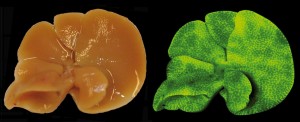
Mouse liver with biliary fibrosis expressing LratCre and a green fluorescent reporter for HSCs (right). HSCs form typical macroscopic septa in this model. Courtesy of R. F. Schwabe.
Schwabe and colleagues found that the majority (82–96%) of myofibroblasts had come from a HSC lineage, irrespective of the underlying aetiology of liver disease. Genetically labelled HSCs and myofibroblast markers co-localized on fluorescence microscopic images of samples taken from injured livers, with the characteristic septal pattern of fibrosis observed. According to the findings, HSCs were the crucial contributor to fibrogenesis in the liver in many settings: chemical-induced liver injury (induction with either carbon tetrachloride or thioacetamide); cholestatic liver disease (in bile-duct ligation, diet and genetic models); and NASH (methionine–choline-deficient diet model).
Notably, HSCs were not derived from bone marrow and were confirmed as a liver-resident cell population. Moreover, the researchers determined that HSCs do not function as epithelial progenitors and have no role in the generation of new hepatocytes in the context of liver injury, a matter of much controversy previously.
The investigators hope to use their new mouse model to investigate further functions of HSCs. In particular, they plan to track HSCs in models of liver cancer to confirm whether these cells are a component of the tumour microenvironment and interact with tumour cells.
References
- Mederacke, I. et al. Fate tracing reveals hepatic stellate cells as dominant contributors to liver fibrosis independent of its aetiology. Nat. Commun. 4, 2823 (2013)
Article Google Scholar
Authors
- Katrina Ray
You can also search for this author inPubMed Google Scholar
Rights and permissions
About this article
Cite this article
Ray, K. Hepatic stellate cells hold the key to liver fibrosis.Nat Rev Gastroenterol Hepatol 11, 74 (2014). https://doi.org/10.1038/nrgastro.2013.244
- Published: 10 December 2013
- Issue Date: February 2014
- DOI: https://doi.org/10.1038/nrgastro.2013.244