Structural basis of receptor recognition by SARS-CoV-2 (original) (raw)
The sudden emergence and rapid spread of SARS-CoV-2 is endangering global health and economy1,2. SARS-CoV-2 has caused many more infections, deaths and economic disruptions than SARS-CoV in 2002–20035,6. The origin of SARS-CoV-2 remains unclear. Bats are considered the original source of SARS-CoV-2 because a closely related coronavirus, RaTG13, has been isolated from bats7. However, the molecular events that led to the possible bat-to-human transmission of SARS-CoV-2 are unknown. Clinically approved vaccines or drugs that specifically target SARS-CoV-2 are also lacking. Receptor recognition by coronaviruses is an important determinant of viral infectivity, pathogenesis and host range8,9. It presents a major target for vaccination and antiviral strategies10. Here we elucidate the structural and biochemical mechanisms of receptor recognition by SARS-CoV-2.
Receptor recognition by SARS-CoV has been extensively studied. A virus-surface spike protein mediates the entry of coronavirus into host cells. The spike protein of SARS-CoV contains a RBD that specifically recognizes ACE2 as its receptor3,4. A series of crystal structures of the SARS-CoV RBD from different strains in complex with ACE2 from different hosts has previously been determined3,11,12. These structures showed that SARS-CoV RBD contains a core and a receptor-binding motif (RBM); the RBM mediates contacts with ACE2. The surface of ACE2 contains two virus-binding hotspots that are essential for SARS-CoV binding. Several naturally selected mutations in the SARS-CoV RBM surround these hotspots and regulate the infectivity, pathogenesis, and cross-species and human-to-human transmissions of SARS-CoV3,11,12.
Because of the sequence similarity between the spike proteins of SARS-CoV and SARS-CoV-2, it was recently predicted that SARS-CoV-2 also uses ACE2 as its receptor13, which has been validated by other studies7,14,[15](#ref-CR15 "Hoffmann, M. et al. SARS-CoV-2 cell entry depends on ACE2 and TMPRSS2 and Is blocked by a clinically proven protease inhibitor. Cell https://doi.org/10.1016/j.cell.2020.02.052
(2020)."),[16](/articles/s41586-020-2179-y#ref-CR16 "Walls, A. C. et al. Structure, function, and antigenicity of the SARS-CoV-2 spike glycoprotein. Cell https://doi.org/10.1016/j.cell.2020.02.058
(2020)."). Here we determined the structural basis of receptor recognition by SARS-CoV-2 and compared the ACE2-binding affinity among SARS-CoV-2, SARS-CoV and RaTG13. Our findings identify the molecular and structural features of the SARS-CoV-2 RBM that result in tight ACE2 binding. They provide insights into the animal origin of SARS-CoV-2, and can help to guide intervention strategies that target SARS-CoV-2–ACE2 interactions.
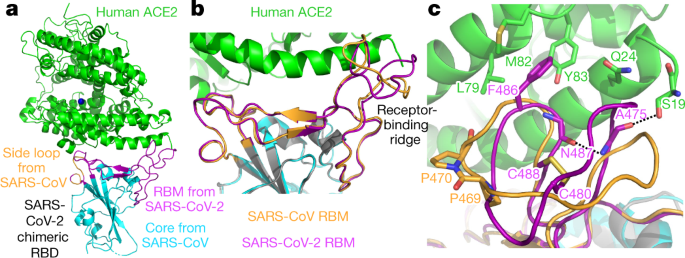
To understand the structural basis of ACE2 recognition by SARS-CoV-2, we aimed to crystallize the SARS-CoV-2 RBD–ACE2 complex. Our strategy was informed by previous crystallization of the SARS-CoV RBD–ACE2 complex3. In this crystal form, the core of the SARS-CoV RBD (along with the ACE2 surface) was mainly involved in crystal lattice contact; the essential ACE2-binding residues in the SARS-CoV RBM were buried at the RBD–ACE2 interface and did not affect crystallization. To facilitate crystallization, we designed a chimeric RBD that uses the core from the SARS-CoV RBD as the crystallization scaffold and the RBM from SARS-CoV-2 as the functionally relevant unit (Fig. 1a and Extended Data Fig. 1). To further enhance crystallization, we improved the ACE2-binding affinity of the chimeric RBD by keeping a short loop from the SARS-CoV RBM, which maintains a strong salt bridge between Arg426 of the RBD and Glu329 of ACE2 (Extended Data Fig. 2a). This loop sits on the side of the binding interface, away from the main binding interface. We expressed and purified the chimeric RBD and ACE2, and crystallized the complex under the same conditions and in the same crystal form as those used for the SARS-CoV RBD–ACE2 complex. On the basis of X-ray diffraction data, we determined the structure of the chimeric RBD–ACE2 complex by molecular replacement using the structure of the SARS-CoV RBD–ACE2 complex as the search template. We refined the structure to 2.68 Å (Extended Data Table 1 and Extended Data Fig. 3). The structure of this chimeric RBD–ACE2 complex, particularly in the RBM region, is highly similar to another recently determined structure of the SARS-CoV-2 wild-type RBD–ACE2 complex[17](/articles/s41586-020-2179-y#ref-CR17 "Lan, J. et al. Structure of the SARS-CoV-2 spike receptor-binding domain bound to the ACE2 receptor. Nature https://doi.org/10.1038/s41586-020-2180-5
(2020)."), confirming that the chimeric RBD is a successful design.
Fig. 1: Structure of the SARS-CoV-2 chimeric RBD complexed with ACE2.
a, Crystal structure of the SARS-CoV-2 chimeric RBD complexed with ACE2. ACE2 is shown in green. The RBD core is shown in cyan. The RBM is shown in magenta. A side loop in RBM is shown in orange. A zinc ion in ACE2 is shown in blue. b, Comparison of the conformations of the ridge in SARS-CoV-2 RBM (magenta) and SARS-CoV RBM (orange). c, Comparison of the conformations of the ridge from another viewing angle. In the SARS-CoV RBM, a proline-proline-alanine motif is shown. In the SARS-CoV-2 RBM, a newly formed hydrogen bond, Phe486, and some of the interactions of the ridge with the N-terminal helix of ACE2 are shown.
The overall structure of the chimeric RBD–ACE2 complex is similar to that of the SARS-CoV RBD–ACE2 complex (Fig. 1a). Similar to the SARS-CoV RBM, SARS-CoV-2 RBM forms a gently concave surface with a ridge on one side; it binds to the exposed outer surface of the claw-like structure of ACE2 (Fig. 1a). The strong salt bridge between SARS-CoV RBD and ACE2 became a weaker (as judged by the longer distance of the interaction), but still energetically favourable, N–O bridge between Arg439 of the chimeric RBD and Glu329 of ACE218 (Extended Data Fig. 2b). In comparison to the SARS-CoV RBM, the SARS-CoV-2 RBM forms a larger binding interface and more contacts with ACE2 (Extended Data Fig. 4a, b). Our structural model also contained glycans attached to four ACE2 sites and one RBD site (Extended Data Fig. 5a). The glycan attached to Asn90 of ACE2 forms a hydrogen bond with Arg408 of the RBD core (Extended Data Fig. 5b); this glycan-interacting arginine is conserved between SARS-CoV-2 and SARS-CoV (Extended Data Fig. 1). The overall structural similarity in ACE2 binding by SARS-CoV-2 and SARS-CoV supports a close evolutionary relationship between the two viruses.
We measured the binding affinities between each of the three RBDs (SARS-CoV-2, chimeric and SARS-CoV) and ACE2 using surface plasmon resonance (SPR) (Extended Data Figs. 4c, 6). We found that the chimeric RBD has a higher ACE2-binding affinity than the SARS-CoV-2 RBD, consistent with the introduced N–O bridge between the chimeric RBD and ACE2. Both the chimeric and SARS-CoV-2 RBDs have significantly higher ACE2-binding affinities than the SARS-CoV RBD. These dissociation constant _K_d values are consistent with other SPR studies12,19, although the exact _K_d values vary depending on the specific approaches of each SPR experiment (Extended Data Table 2). Here we investigate the structural differences between the RBMs of SARS-CoV-2 and SARS-CoV that account for their different ACE2-binding affinities.
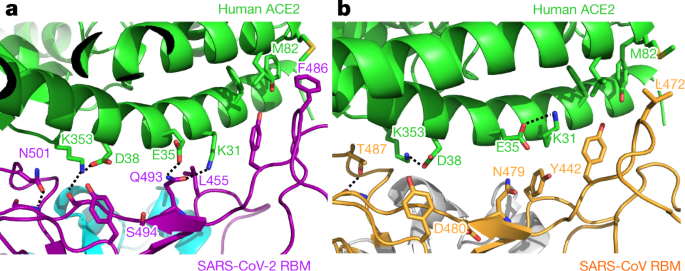
A marked structural difference between the RBMs of SARS-CoV-2 and SARS-CoV is the conformation of the loops in the ACE2-binding ridge (Fig. 1b, c). In both RBMs, one of the ridge loops contains an essential disulfide bond and the region between the disulfide-bond-forming cysteines is variable (Fig. 1c and Extended Data Fig. 1). Specifically, human and civet SARS-CoV strains and bat coronavirus Rs3367 all contain a three-residue motif proline-proline-alanine in this loop; the tandem prolines allow the loop to take a sharp turn. By contrast, SARS-CoV-2 and bat coronavirus RaTG13 both contain a four-residue motif glycine-valine/glutamine-glutamate/threonine-glycine; the two relatively bulky residues and two flexible glycines enable the loop to take a different conformation (Fig. 1c and Extended Data Fig. 1). Because of these structural differences, an additional main-chain hydrogen bond forms between Asn487 and Ala475 in the SARS-CoV-2 RBM, causing the ridge to take a more compact conformation and the loop containing Ala475 to move closer to ACE2 (Fig. 1c). As a consequence, the ridge in the SARS-CoV-2 RBM forms more contacts with the N-terminal helix of ACE2 (Extended Data Fig. 4b). For example, the N-terminal residue Ser19 of ACE2 forms a new hydrogen bond with the main chain of Ala475 of the SARS-CoV-2 RBM, and Gln24 in the N-terminal helix of ACE2 also forms a new contact with the SARS-CoV-2 RBM (Fig. 1c and Extended Data Fig. 4b). Moreover, compared with the corresponding Leu472 of the SARS-CoV RBM, Phe486 of the SARS-CoV-2 RBM points in a different direction and inserts into a hydrophobic pocket involving Met82, Leu79 and Tyr83 of ACE2 (Figs. 1c, 2a, b). In comparison to the SARS-CoV RBM, these structural changes in the SARS-CoV-2 RBM are more favourable for ACE2 binding.
Fig. 2: Structural details at the interface between the SARS-CoV-2 RBM and ACE2.
a, The interface between the SARS-CoV-2 RBM and ACE2. b, The interface between SARS-CoV RBM and ACE2.
In comparison to the SARS-CoV RBM–ACE2 interface, subtle yet functionally important structural changes take place near the two virus-binding hotspots at the SARS-CoV-2 RBM–ACE2 interface (Fig. 2a, b). At the SARS-CoV–ACE2 interface, two virus-binding hotspots were previously identified11,12: hotspot Lys31 (that is, hotspot 31) consists of a salt bridge between Lys31 and Glu35, and hotspot Lys353 (that is, hotspot 353) consists of a salt bridge between Lys353 and Asp38. Both salt bridges are weak, as judged by the relatively long distance of these interactions. Burial of these weak salt bridges in hydrophobic environments on virus binding would enhance their energy, owing to a reduction in the dielectric constant. This process is facilitated by interactions between the hotspots and nearby RBD residues. First, at the SARS-CoV RBM–ACE2 interface, hotspot 31 requires support from Tyr442 of the SARS-CoV RBM (Fig. 2b). In comparison, at the SARS-CoV-2 RBM–ACE2 interface, Leu455 of the SARS-CoV-2 RBM (corresponding to Tyr442 of the SARS-CoV RBM) has a less bulky side chain, providing less support to Lys31 of ACE2. As a result, the structure of hotspot 31 has rearranged: the salt bridge between Lys31 and Glu35 breaks apart, and each of the residues forms a hydrogen bond with Gln493 of the SARS-CoV-2 RBM (Fig. 2a). Second, at the SARS-CoV RBM–ACE2 interface, hotspot 353 requires support from the side-chain methyl group of Thr487 of the SARS-CoV RBM, whereas the side-chain hydroxyl group of Thr487 forms a hydrogen bond with the RBM main chain (which fixes the conformation of the Thr487 side chain) (Fig. 2b). In comparison, at the SARS-CoV-2 RBM–ACE2 interface, Asn501 of the SARS-CoV-2 RBM also has its conformation fixed through a hydrogen bond between its side chain and the RBM main chain; correspondingly, its side chain provides less support to hotspot 353 than the corresponding Thr487 of the SARS-CoV RBM does (Fig. 2a). Consequently, Lys353 of ACE2 takes a slightly different conformation, forming a hydrogen bond with the main chain of the SARS-CoV-2 RBM while maintaining the salt bridge with Asp38 of ACE2 (Fig. 2a). Thus, both hotspots have adjusted to the reduced support from nearby RBD residues, yet still become well-stabilized at the SARS-CoV-2 RBM–ACE2 interface.
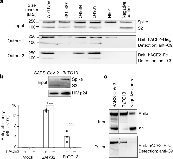
To corroborate the structural observations, we characterized ACE2-binding affinities of the SARS-CoV-2 spike that contains mutations in critical ACE2-interacting residues. To this end, protein pull-down assays were performed, with purified recombinant ACE2 as the bait and cell-associated SARS-CoV-2 spike as the target (Fig. 3a). For cross-validation, we used ACE2 with two different tags, His6 and Fc. The SARS-CoV-2 spike contained one of the following RBM changes: 481–487 (481-NGVEGFN-487 in SARS-CoV-2 were mutated to TPPALN as in SARS-CoV), Q493N (Gln493 in SARS-CoV-2 was mutated to an asparagine as in human SARS-CoV), Q493Y (Gln493 in SARS-CoV-2 was mutated to a tyrosine as in bat RaTG13) and N501T (Asn501 in SARS-CoV-2 was mutated to a threonine as in human SARS-CoV). The results showed that all of these introduced mutations reduced the ACE2-binding affinity of the SARS-CoV-2 spike. They confirm that the structural features of the SARS-CoV-2 RBM, including the ACE2-binding ridge and the hotspots-stabilizing residues, all contribute to the high ACE2-binding affinity of SARS-CoV-2.
Fig. 3: Biochemical data showing the interactions between SARS-CoV-2 or bat RaTG13 spike and ACE2.
a, Protein pull-down assay using ACE2 as the bait and cell-associated SARS-CoV-2 spike molecules (wild type and mutants) as the targets. Top, cell-expressed SARS-CoV-2 spike. Middle, pull-down results using His6-tagged ACE2. Bottom, pull-down results using Fc-tagged ACE2. MERS-CoV spike was used as a negative control. b, Entry of SARS-CoV-2 and bat RaTG13 pseudoviruses into ACE2-expressing cells. Top, packaged SARS-CoV-2 and bat RaTG13 pseudoviruses. HIV p24 was detected as an internal control. Bottom, pseudovirus entry efficiency. Mock, no pseudoviruses. Data are mean + s.d. A comparison (two-tailed Student’s _t_-test) between SARS-CoV-2 with ACE2 (n = 3 independent samples) and SARS-CoV-2 without ACE2 (n = 4 independent samples) showed a significant difference (P < 1.16 × 10⁻⁸). A comparison (two-tailed Student’s _t_-test) between RaTG13 with ACE2 (n = 3 independent samples) and RaTG13 without ACE2 (n = 4 independent samples) showed a significant difference, P = 0.0097. Individual data points are shown as black dots. ***P < 0.001; **P < 0.01. c, Protein pull-down assay using ACE2 as the bait and cell-associated RaTG13 spike as the target. All experiments were repeated independently three times with similar results.
Having compared ACE2 recognition by SARS-CoV-2 and SARS-CoV, we further investigated human ACE2 binding by bat RaTG13. To this end, we performed a pseudovirus entry assay in which retroviruses pseudotyped with RaTG13 spike (that is, RaTG13 pseudoviruses) were used to enter ACE2-expressing human cells (Fig. 3b). The results showed that RaTG13 pseudovirus entry into the cells depends on ACE2. Additionally, RaTG13 spike was not cleaved on the pseudovirus surface. SARS-CoV-2 pseudovirus entry also depends on ACE2, but its spike was cleaved to S2 on the pseudovirus surface (probably because of a furin site insertion[16](/articles/s41586-020-2179-y#ref-CR16 "Walls, A. C. et al. Structure, function, and antigenicity of the SARS-CoV-2 spike glycoprotein. Cell https://doi.org/10.1016/j.cell.2020.02.058
(2020).")) (Fig. 3b). Moreover, we performed a protein pull-down assay using ACE2 as the bait and cell-associated RaTG13 spike as the target (Fig. 3c). We found that the RaTG13 spike was pulled down by ACE2. Therefore, similar to SARS-CoV-2, bat RaTG13 binds to human ACE2 and can use human ACE2 as its entry receptor.
The current SARS-CoV-2 outbreak has become a global pandemic. Previous structural studies on SARS-CoV have established receptor recognition as an important determinant of SARS-CoV infectivity, pathogenesis and host range9. On the basis of the structural information presented here, along with biochemical data, we discuss the receptor recognition and evolution of SARS-CoV-2.
We will first discuss how well SARS-CoV-2 recognizes ACE2 in comparison to SARS-CoV. We show that, compared with SARS-CoV, the SARS-CoV-2 RBM contains structural changes in the ACE2-binding ridge, largely caused by a four-residue motif (residues 482–485: Gly-Val-Glu-Gly). This structural change allows the ridge to become more compact and form better contacts with the N-terminal helix of ACE2 (Fig. 1b, c). In addition, Phe486 of the SARS-CoV-2 RBM inserts into a hydrophobic pocket (Fig. 1c). The corresponding residue in the SARS-CoV RBM is a leucine, which probably forms a weaker contact with ACE2 owing to its smaller side chain. Finally, both virus-binding hotspots are more stabilized at the RBM–ACE2 interface through interactions with the SARS-CoV-2 RBM. As previous studies have shown11,12, these hotspots on ACE2 are important for coronavirus binding, because they involve two lysine residues that need to be accommodated properly in hydrophobic environments. Neutralizing the charges of the lysines is key to the binding of coronavirus RBDs to ACE2. The SARS-CoV-2 RBM has evolved strategies to stabilize the two hotspots: Gln493 and Leu455 stabilize hotspot 31, whereas Asn501 stabilizes hotspot 353 (Fig. 2a). Our biochemical data confirm that the SARS-CoV-2 RBD has a significantly higher ACE2-binding affinity than the SARS-CoV RBD and that the above structural features of the SARS-CoV-2 RBM contribute to the high ACE2-binding affinity of SARS-CoV-2 RBD (Fig. 3a). Thus, both structural and biochemical data reveal that the SARS-CoV-2 RBD recognizes ACE2 better than SARS-CoV RBD does.
Next, we investigated how SARS-CoV-2 may have been transmitted from bats to humans. First, we found that bat RaTG13 uses human ACE2 as its receptor (Fig. 3b, c), suggesting that RaTG13 may infect humans. Second, as with SARS-CoV-2, bat RaTG13 RBM contains a similar four-residue motif in the ACE2-binding ridge, supporting the notion that SARS-CoV-2 may have evolved from RaTG13 or a RaTG13-related bat coronavirus (Extended Data Table 3 and Extended Data Fig. 7). Third, the L486F, Y493Q and D501N residue changes from RaTG13 to SARS-CoV-2 enhance ACE2 recognition and may have facilitated the bat-to-human transmission of SARS-CoV-2 (Extended Data Table 3 and Extended Data Fig. 7). A lysine-to-asparagine mutation at the 479 position in the SARS-CoV RBD (corresponding to the 493 position in the SARS-CoV-2 RBD) enabled SARS-CoV to infect humans3. Fourth, Leu455 contributes favourably to ACE2 recognition, and it is conserved between RaTG13 and SARS-CoV-2; its presence in the SARS-CoV-2 RBM may be important for the bat-to-human transmission of SARS-CoV-2 (Extended Data Table 3 and Extended Data Fig. 7). Host and viral factors other than receptor recognition also have important roles in the cross-species transmission of coronaviruses20,21. Nevertheless, the identified receptor-binding features of the SARS-CoV-2 RBM may have facilitated SARS-CoV-2 to transmit from bats to humans (Extended Data Fig. 7).
We then examined whether intermediate hosts were involved in the potential bat-to-human transmission of SARS-CoV-2. Because bat coronavirus RaTG13 binds to human ACE2, one possibility is that there is no intermediate host. Alternatively, pangolins have been proposed to be an intermediate host[22](/articles/s41586-020-2179-y#ref-CR22 "Lam, T. T. et al. Identifying SARS-CoV-2 related coronaviruses in Malayan pangolins. Nature https://doi.org/10.1038/s41586-020-2169-0
(2020)."). The structural information provided in this study enables us to inspect and understand the important RBM residues in coronaviruses isolated from pangolins. Two coronaviruses, CoV-pangolin/GD and CoV-pangolin/GX, have been isolated from pangolins from two different locations in China: Guangdong (GD) and Guangxi (GX), respectively. The RBM of the CoV-pangolin/GD contains Leu455, the 482–485 loop, Phe486, Gln493 and Asn501 (Extended Data Table 3), all of which are favourable for ACE2 recognition. The RBM of CoV-pangolin/GX contains Leu455 and the 482–485 loop, both of which are favourable for ACE2 recognition, and it also contains Leu486, Glu493 and Thr501 (Extended Data Table 3), all of which are less favourable for ACE2 recognition. Therefore, CoV-pangolin/GD potentially recognizes human ACE2 well, whereas CoV-pangolin/GX does not. Hence, pangolins from Guangdong, but not pangolins from Guangxi, could potentially pass coronaviruses to humans. However, many other factors determine the cross-species transmission of coronaviruses20,21, and the above analysis will need to be verified experimentally.
Finally, this study helps to inform intervention strategies. First, neutralizing monoclonal antibodies that target the SARS-CoV-2 RBM can prevent the virus from binding to ACE2, and are therefore promising antiviral drugs. Our structure has laid out all of the functionally important epitopes in the SARS-CoV-2 RBM that can potentially be targeted by neutralizing antibody drugs. Thus, this study can help to guide the development and optimization of these antibody drugs. Second, the RBD itself can function as a subunit vaccine10,23. The functionally important epitopes in the SARS-CoV-2 RBM that were identified in this study can guide structure-based design of highly efficacious RBD vaccines. Such a structure-based strategy for subunit vaccine design has previously been developed24. This strategy may be helpful in designing SARS-CoV-2 RBD vaccines. Overall, this study can help to inform structure-based intervention strategies that target receptor recognition by SARS-CoV-2.


