Complement mediated cell death is associated with DNA fragmentation (original) (raw)
Introduction
Since the publication of the seminal paper by Kerr et al,1 cell death has rigidly been classified as either necrotic or apoptotic. Necrotic death results from severe cell damage and is typified by osmotic dysregulation, cell swelling and, ultimately, cell lysis. Almost invariably inflammation occurs in the surrounding tissue.1,2,3 In contrast, apoptosis is typified by a controlled series of morphological and biochemical events which act in concert to prevent inflammation. Inflammation is clearly not a desirable outcome within a tissue and so it is no surprise that apoptosis is the predominant mode of death seen under physiological conditions in multicellular organisms.3,4,5,6
The changes which occur in an apoptotic cell include cytoplasmic shrinkage, membrane blebbing and chromatin condensation. Another event commonly observed in apoptotic cells, and one of the most readily detectable, is oligonucleosomal fragmentation of the nuclear DNA. This process is performed by endonuclease enzymes that cleave the DNA at nucleosomal distances, creating a so-called ‘DNA ladder’ which can be detected by agarose gel electrophoresis. DNA may also become degraded at late stages of necrosis, but fragmentation in this instance is predominantly due to the action of hydrolytic enzymes and does not result in internucleosomal cutting or a defined ladder.1,2,7 For this reason, detection of a DNA ladder is one of the most definitive diagnostic characteristics of apoptotic death.2,7,8
Recently it has become evident that the rigid characterisation of cell death as apoptotic or necrotic is too simplistic and that a broader range of death mechanisms than those typified by classic necrosis or apoptosis may well exist. Several reports demonstrate death mechanisms which are neither fully necrotic nor apoptotic, but which are a mixture of both.9,10,11 For this reason, it is now believed that a wide range of cell death mechanisms exists and that necrosis and apoptosis merely reflect the two physiological extremes. This proposal has led to the re-examination of a number of different modes of cell death and has recently led to the suggestion that hypoxia, classically considered to induce necrotic death, may involve typical features of apoptosis such as activation of endonuclease activity and signalling pathways.12
Like hypoxia, complement (C) induced death, resulting from C5b-9 lesions in target cell plasma membranes, has traditionally been cited as an inducer of necrotic death.2,3 However, a number of lines of evidence led us to believe that C induced death might also display features of apoptosis. Firstly, recent work by Papadimitriou et al13 has shown that chromatin condensation, typical of apoptosis, is present early during C attack of Ehrlich cells. Secondly, several authors have reported that C death, at least for nucleated cells, is not merely a function of osmotic swelling and lysis,14 implying a mechanism more complex than classical necrosis. Thirdly, under conditions where the culture media has a higher (more physiological) osmotic potential, C attacked target cells lose their intracellular contents less rapidly.15 Finally, and perhaps most intriguingly, in 1971 Shipley et al reported that C attack induced rapid fragmentation of target cell DNA.16 Unfortunately, the relevance of oligonucleosomal DNA fragmentation was not appreciated at this time and further observations were not reported. With these data in mind, we undertook an in vitro study utilising six different neoplastic B-cell lines, to assess the mechanism by which C attacked cells die.
Results
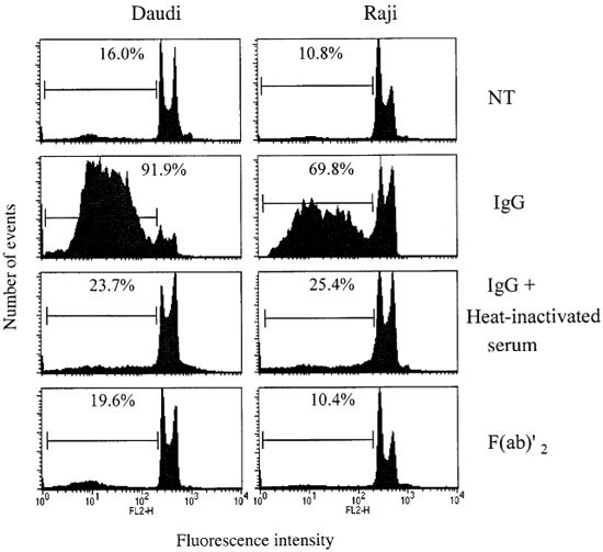
To determine whether DNA fragmentation, a typical feature of apoptosis, was occurring in target cells after C attack, we used a flow cytometric method.17 Apoptotic cells are seen in the sub-G1 peak of the DNA histograms (gated regions; see Figure 1), which represents degraded DNA. Figure 1 (top panels) shows that C attack in 30% human serum resulted in extensive DNA fragmentation of both Raji and Daudi B cells. To observe whether DNA fragmentation was dependent upon C activity, F(ab′)2 antibody (Ab) fragments were used instead of intact IgG, or C was rendered inactive through heating at 56°C for 30 min. Using either of these strategies, C damage was not induced (data not shown) and DNA fragmentation was not evident (Figure 1). Thus DNA fragmentation occurred only in the presence of intact Ab and C.
Figure 1
Requirement of C activity for the induction of DNA fragmentation. Daudi or Raji cells (5×10⁵ ml) were incubated with IgG or F(ab′)2 (7 μg/ml) Ab fragments for 30 min at room temperature, prior to the addition of 30% normal or heat-inactivated serum. A cell sample was then taken 1 h after the addition of serum to assess the degree of membrane permeability using the PI assay (data not shown). The remainder of the cells were incubated at 37°C without washing for a further 18–24 h. The extent of DNA fragmentation was then determined by the hypotonic PI assay. NT=no Ab added
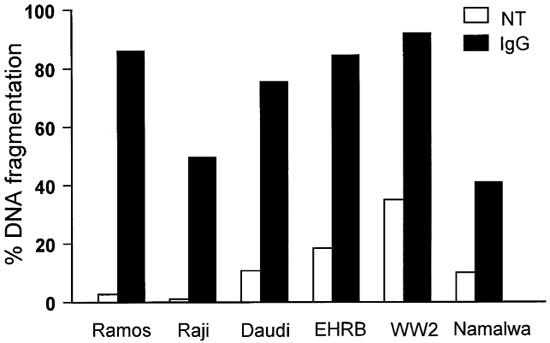
To determine whether this was a common phenomenon and not a peculiarity of the cell lines chosen, a further four cell lines were assessed. As shown in Figure 2, all lines tested exhibited abundant DNA fragmentation within 18 h of C attack. Three representative cell lines, Ramos-EHRB, Daudi and Raji, were selected for more detailed analysis. Similar results were obtained with serum from guinea pig, rat, sheep and rabbit, such that incubation with these sera generated DNA fragmentation in the target cells (data not shown).
Figure 2
DNA fragmentation induced following C attack on different cell lines. Each cell line was incubated with IgG (7 μg/ml) for 30 min at room temperature, prior to the addition of 30% human serum. Cells were then incubated and assessed as shown in Figure 1. NT=no Ab added
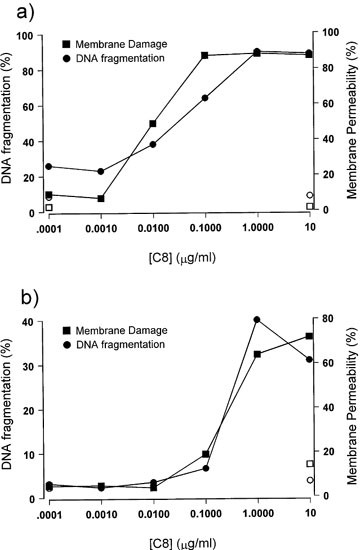
We next investigated the requirement for Membrane Attack Complex (MAC) formation in the induction of DNA fragmentation. In C8-depleted serum MAC cannot form and so addition of C8 protein becomes the rate-limiting factor for MAC formation. Therefore, the level of MAC that can form increases proportionally with the amount of C8 protein that is added. As shown for EHRB and Raji cells (Figure 3a,b) titration of C8 protein back into C8-depleted serum restored both membrane damaging (cell lysing) and DNA fragmentation activity to the serum in a dose-dependent fashion. In both cell lines, the amount of membrane permeability closely correlated with the extent of DNA fragmentation. These data demonstrate that C5b-9 MAC formation is both required for, and closely correlated with, the induction of DNA fragmentation.
Figure 3
Dependence of C lysis and DNA fragmentation on C8 protein. IgG was added to EHRB (a) or Raji (b) cells for 30 min at room temperature, prior to the addition of C8-deficient serum (30%) reconstituted with varying levels of C8 protein. The extent of membrane permeability was determined by the PI assay (squares) after 1 h. DNA fragmentation was then determined 18 h after addition of serum by the hypotonic PI assay, shown as circle symbols. Open symbols represent control samples and filled symbols represent results following addition of IgG
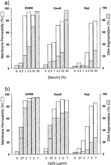
Figure 4 shows results from a series of experiments in which we further explored the link between MAC dependent membrane damage and DNA fragmentation, by measuring propidium iodide (PI) permeability and DNA fragmentation over a range of serum and Ab concentrations. Most informative were the experiments where the serum concentration was varied (Figure 4a). These experiments revealed that the extent of DNA fragmentation induced was clearly dependent on the concentration of serum used. Only at the highest concentrations of serum did we observe significant DNA fragmentation. The concentrations of serum required to induce significant levels of membrane permeability were lower than those necessary for DNA fragmentation: for example, in EHRB cells maximum membrane permeability was obtained at 1% serum, whereas maximum DNA fragmentation required 30% serum. In contrast, when Ab concentration was varied with a constant concentration of serum (Figure 4b), DNA fragmentation was maximal only in samples that displayed maximal membrane permeability. These data indicate that membrane permeability induced by C is a pre-requisite for subsequent DNA fragmentation, but that the membrane permeability alone is not sufficient and that once cells are damaged, it is the concentration of serum which determines the degree of DNA fragmentation. In addition, these data demonstrate that at levels of serum typically used by other workers (e.g. <20%) only low levels of DNA fragmentation were seen with Daudi and Raji cells, whilst at more physiological concentrations of serum (>20%), there was extensive DNA fragmentation. This perhaps explains why these observations have not been reported previously.
Figure 4
Dependence of DNA fragmentation on serum (a) and Ab (b) concentration. In (a), IgG (7 μg/ml) was added to Raji, Daudi, or EHRB cells, allowed to bind for 30 min at room temperature and varying concentrations of serum added. In (b), varying concentrations of IgG were added to a constant concentration of serum (20%). The degree of membrane permeability was determined by the PI assay after 30 min, represented as unshaded bars. The extent of DNA fragmentation was assessed via the hypotonic PI assay 18–24 h after the addition of serum, shown as shaded bars
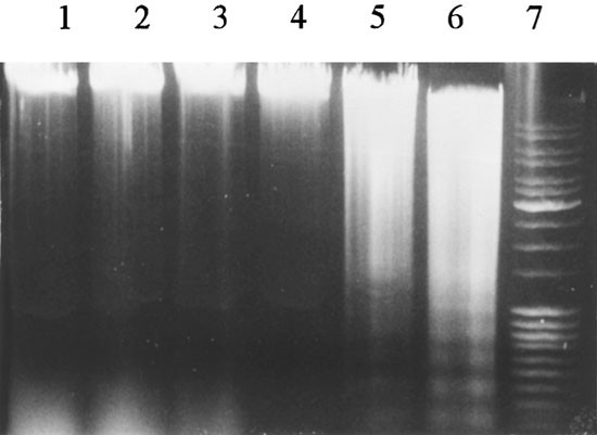
To assess whether the observed DNA fragmentation was apoptotic in nature, agarose gel analysis was performed. These data, shown in Figure 5 for Daudi cells, revealed that the DNA fragmentation induced was oligonucleosomal in nature and displayed discrete banded DNA ladder patterns typical of apoptosis.2,7,8 Bands on the gels were approximately 180–200 base-pairs apart, indicating their nucleosomal origins. In addition, the analysis supported the idea that fragmentation was a function of serum concentration, as increasing DNA fragmentation was observed with increasing levels of serum.
Figure 5
DNA fragmentation induced in Daudi cells after C attack in varying levels of serum. IgG (7 μg/ml) was added to 1–2×10⁶ Daudi cells for 30 min at room temperature and then varying levels of serum added and the cells incubated for 18 h at 37°C. Samples were then harvested, treated with Proteinase K, RNAse and subjected to agarose gel electrophoresis on a 1% gel for 2 h. Untreated control cells are shown in lane 1. In lanes 2–6 increasing levels of serum (1, 3, 10, 20, 30%) were added. A 100 bp molecular weight ladder is shown in lane 7
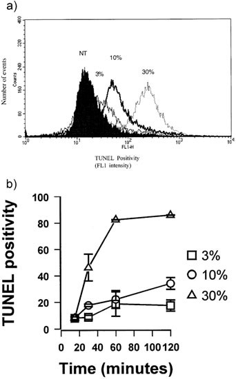
In the experiments detailed above, DNA fragmentation was detected after an overnight (18–24 h) incubation. In order to determine how soon after C attack the target cell DNA was degraded, the TUNEL technique was employed. This technique is a sensitive method for distinguishing apoptotic and necrotic cell death.18 TUNEL analysis (Figure 6) revealed that C-induced DNA fragmentation was due to the induction of apoptotic-like strand breaks. TUNEL positivity was observed as rapidly as 30 min after addition of serum (Figure 6b) and was evident at high levels after 1 h. The extent of TUNEL positivity was found to be a function of serum concentration. Presumably, these strand breaks subsequently lead to the formation of the apoptotic DNA ladders, as DNA ladders were observed as early as 2 h after the addition of serum (data not shown).
Figure 6
Assay of TUNEL positivity after C attack. EHRB cells were incubated with IgG (7 μg/ml) for 30 min at 20°C and then varying levels of serum (3, 10 or 30%) added and the cells incubated at 37°C. The extent of DNA strand breakage was assessed after 10, 30, 60 or 120 min via the TUNEL assay in (a). In (b) the means±S.D. of duplicate samples over the 2 h time course are shown. The histogram demonstrates the degree of TUNEL positivity after 1 h
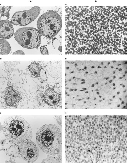
Given that apoptotic-like DNA fragmentation was apparent after C attack, we were intrigued to see whether any other features of apoptosis were evident. Electron microscopy (EM) revealed that following C attack, cells in near-physiological concentrations of serum retained far greater plasma and nuclear membrane integrity than cells subjected to a similar level of attack in the presence of low levels of serum, shown for EHRB cells in Figure 7. Thirty minutes after C attack, cells in high concentrations of serum (Figure 7 panel c) have generally intact cell and nuclear membranes, whereas those in low levels of serum demonstrate discontinuity of the plasma membrane, cell-lysis and nuclear leakage (Figure 7 panel b). Furthermore a proportion of the cells in high levels of serum retained nuclear integrity for up to 6 h (data not shown).
Figure 7
Light and electron microscopy of C attacked EHRB cells in the presence of high (30%) or low (3%) levels of serum. EHRB cells were incubated with IgG (7 μg/ml) for 30 min at 20°C and then either 3 or 30% serum was added. After 30 min at 37°C the cells were harvested, fixed by adding glutaraldehyde (1.5% final), washed and embedded in epon resin. For EM, thin (90 nm) sections were cut and stained with lead citrate. The views illustrated were gained at a magnification of 7500×and are shown in (A). In this figure, (a) represents normal untreated EHRB cells, (b) represents EHRB cells lysed in 3% serum and (c) represents cells attacked in 30% serum. For light microscopy, thick (0.5 μm) sections were cut and stained with toluene blue as shown in (B). Again, (a) represents normal untreated EHRB cells, (b) represents EHRB cells lysed in 3% serum and (c) represents cells attacked in 30% serum
Analysis of the EM data also demonstrated that chromatin was condensed following C attack in the presence of high levels of serum, in a manner similar to that reported by Papadimitriou for Ehrlich cells.13 This observation, in conjunction with the DNA fragmentation data, demonstrates that features of apoptosis were apparent during C-mediated death of these cells. However, although cells attacked in high levels of serum were more resistant to complete lysis, signs of osmotic swelling and intracellular leakage were evident and ultimately many of the cells lysed completely. This was reflected by the general increase in cell size and by organelle swelling; two features associated with necrotic death. Furthermore, membrane blebbing, a common feature of apoptosis, was not evident. Clearly then, although some of the hallmark features of apoptosis are apparent during C mediated death, not all were present and many of the membrane and cytoplasmic events more closely resembled a necrotic phenotype. Similar observations were made in all three cell lines examined.
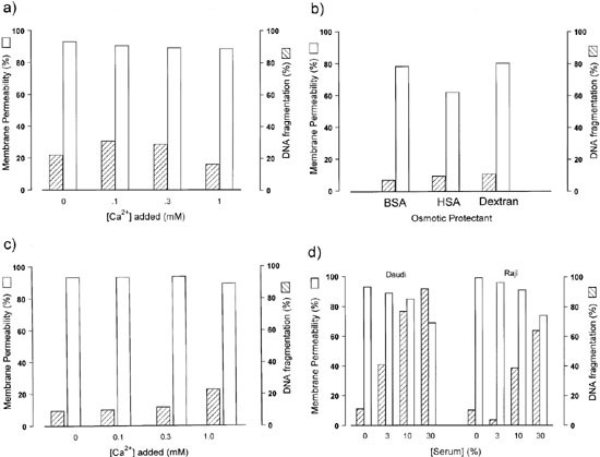
Once the nature of the DNA fragmentation had been verified in these cells, we wished to identify the mechanism by which it was induced. Ca2+ ions rapidly enter target cells after C attack19,20 and have been suggested as one of the effectors of C-induced cell death.21 Ca2+ is also a known co-factor for certain apoptotic endonucleases.2,8,22,23,24 As serum contains high levels of Ca2+ (1.1–1.23 mM)25 it was suggested that the serum concentration dependence for DNA fragmentation was due to differences in the concentration of Ca2+ present in the different levels of serum. To determine if this was the case, low levels of serum were supplemented with increasing levels of exogenous Ca2+ ions and assessed for their ability to induce DNA fragmentation (Figure 8a). Ramos-EHRB cells were used in these experiments, as in this cell line low levels of serum are sufficient to induce maximal levels of membrane permeability, without concurrently high levels of DNA fragmentation, enabling us to separate the phenomenon of cell lysis and DNA fragmentation. As shown in Figure 8a, low levels of serum supplemented with exogenous Ca2+ did not increase the DNA fragmentation observed.
Figure 8
Effect of additional Ca2+, osmotic protection or melittin on the extent of DNA fragmentation. In (a–c), EHRB cells were lysed in the presence of IgG (7 μg/ml) and 3% serum with (a) varying concentrations of exogenous Ca2+, (b) different osmotic protectants (dextran was added to a final concentration of 0.66 mM, and HSA and BSA were used at 30%), or (c) varying concentrations of exogenous Ca2+ and 0.33 mM dextran, added to the cultures. In (d), Raji or Daudi cells were incubated with melittin (5–20 μg/ml) in various doses of serum without IgG. Membrane permeability was determined 1 h after addition of serum; shown as open bars. DNA fragmentation was assessed 18 h after the addition of serum; shown as shaded bars
Alternatively, differences between the effects of high and low levels of serum might be explained on a basis of osmotic potential. Within this rationale, it was proposed that the osmotic protection afforded by high levels of serum would enable cells to survive long enough for intracellular endonucleases to become activated (possibly by the Ca2+ influx) and instigate DNA fragmentation. In contrast, low levels of serum would not inhibit osmotic swelling and cell lysis would occur prior to DNA fragmentation. To investigate this possibility, low levels of serum were supplemented with dextran, Human serum albumin (HSA) or Bovine serum albumin (BSA), to provide equivalent osmotic protection to that afforded by high levels of serum. These experiments, represented in Figure 8b, demonstrate that C attacked cells, in the presence of osmotic protectants, though protected from complete lysis, do not undergo significant DNA fragmentation in the absence of sufficiently high levels of serum. Next we investigated whether both osmotic protection and high levels of Ca2+ ions were sufficient to initiate DNA fragmentation (Figure 8c). These experiments, using dextran as the osmotic protectant, revealed that osmotically protected cells undergoing C attack in the presence of elevated levels of Ca2+ still did not undergo significant DNA fragmentation in the absence of high levels of serum.
To ascertain whether other forms of membrane permeability could induce DNA fragmentation, the bee venom derived pore-forming polypeptide melittin was used. DNA fragmentation was not observed following treatment with melittin in the absence of serum, although membrane permeability was induced (as measured by PI permeability). When serum was included (without Ab to activate C), DNA fragmentation was observed and in a dose-dependent manner. Furthermore, heat-inactivated serum was also able to induce DNA fragmentation in these experiments (data not shown) excluding the possibility that C activation is necessary. Together these data demonstrated that the DNA fragmentation induced was dependent upon a property of serum that was not related to osmotic potential, Ca2+ concentration or C activation.
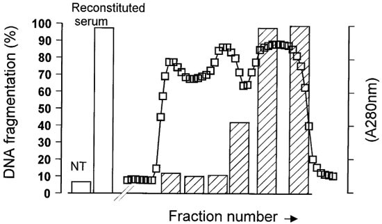
In view of these results, we decided to fractionate the components of serum and determine if the DNA fragmenting activity could be restricted to molecules of a given size. Fractionation was carried out using an ACA44 gel matrix. As illustrated in Figure 9, the major DNA fragmenting activity was found to be restricted to fractions corresponding to proteins of approximately 25–50 kDa in size. Fractionation of animal sera (guinea pig, sheep, rabbit) yielded similar results (data not shown), indicating that a similar factor is involved. Intriguingly, DNase I is 32–38 kDa26,27 and furthermore, is the only well known serum endonuclease.23,27,28,41
Figure 9
DNA fragmentation induced by various fractions of serum following separation on ACA44. Human serum was added to a pre-equilibrated ACA44 column and fractions collected. The approximate molecular weight of proteins in the different fractions was then determined by comparison with the elution profile of known molecular weight standards. 10% (v/v final) of fractionated serum was added to 3% native serum (to ensure membrane permeabilisation) and the DNA fragmentation activity of each fraction was determined using the hypotonic PI assay assessed 16 h after addition of serum. Also shown are the levels of DNA fragmentation induced with serum reconstituted from equivalent portions of the fractionated serum in the absence (NT) or presence of Ab
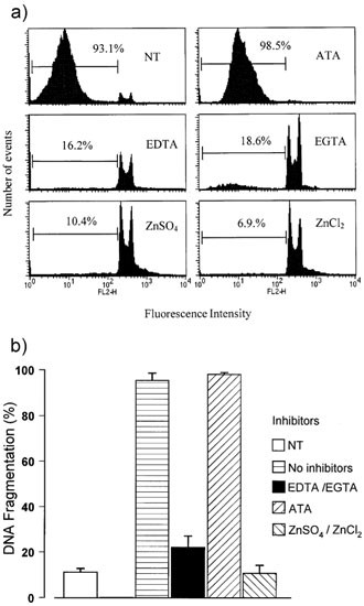
To further characterise the nature of the DNA fragmentation-factor in serum, we undertook a series of inhibition studies. The data from these experiments (Figure 10) demonstrate that EGTA, EDTA, and Zn2+ ions were capable of inhibiting the DNase activity in serum, but that aurintricarboxylic acid (ATA) was not. This inhibitor profile is similar to that of serum DNase I.26 To eliminate the possibility of a cytosolic DNase being involved during the death process, we isolated nuclei from the cells and repeated these studies. Similar results were achieved with isolated nuclei as in whole cells, such that DNA fragmentation was dependent on serum concentration, inhibited by EGTA/EDTA and Zn2+ and not by ATA (data not shown). Finally, to examine whether caspases were involved in the DNA fragmentation, three tetrapeptide caspase inhibitors (YVAD, ZVAD and DEVD) were used. All three inhibitors were ineffective even when applied at a concentration of 250 μM, implying that caspases are not involved in this process (data not shown). All of these data indicate that the DNA fragmentation which follows C attack is due to an extracellular endonuclease which is present in serum and that this endonuclease is likely to be DNase I.
Figure 10
Inhibitor profile of DNase activity following C attack of EHRB cells in 30% serum. Following C attack for 15 min at 37°C to allow maximal membrane permeability, various endonuclease inhibitors were added to the samples. In a series of experiments 1–5 mM final concentrations of ZnCl2, ZnSO4, EGTA, EDTA or ATA were used. Shown in (a) is an example of a typical set of results. In (b) two identical experiments are compared with the means shown
Discussion
We have shown that following C attack, various cell lines underwent an extensive and rapid apoptotic-like DNA fragmentation, which was dependent on high levels of serum. The DNA fragmentation was not seen in low levels of serum. However, low levels of serum (1–10%) were able to induce substantial membrane permeability. Titration experiments with C8-depleted serum and C8 protein confirmed that functional C was necessary for DNA fragmentation to occur and, more importantly, demonstrated that MAC formation was vital for the induction of DNA fragmentation. Subsequent experiments revealed that melittin pores could substitute for MAC in inducing membrane permeability, although high doses of serum were again necessary for the induction of DNA fragmentation. The requirement for high doses of serum in both of these systems indicated that DNA fragmentation was dependent upon a titratable factor. Furthermore, as isolated nuclei were similarly susceptible to the effects of high levels of serum (data not shown) this indicated that cytosolic endonucleases did not play an important role in the fragmentation process. Lack of a requirement for the cytosol also indicated that caspases were not involved in this process and this was supported by the fact that the tetrapeptides YVAD, ZVAD and DEVD did not inhibit the observed DNA fragmentation (data not shown).
The effects of high serum levels could not be mimicked by osmotic protectants either alone or in conjunction with elevated levels of Ca2+, indicating that the DNA fragmentation which occurred was not simply due to an influx of Ca2+ and activation of a nuclear-based Ca2+ dependent endonuclease such as that described by Wyllie.24 However, once initiated, it is possible that endogenous, cytosolic and nuclear endonucleases would become activated. In the absence of cytosolic and nuclear endonucleases, it must be supposed that the DNA fragmentation following C attack is due to the entry of an external DNase present in serum.
One possible source of external endonucleases would be mycoplasma infected cells, which have been shown to be capable of secreting endonucleases capable of inducing internucleosomal DNA fragmentation.29 However, in our study the cell lines used were routinely screened for mycoplasma and found to be uninfected. The FCS used in cell culture is screened for mycoplasma infection and can similarly be disregarded as a source of mycoplasma-derived external endonucleases.
DNase activity in serum and plasma has long been reported for a plethora of species, including rat,30 mouse23,31 and human.27,28,32 Various authors have investigated the nature of this serum DNase27,28,32 and although its origin is unclear, it would appear that the major endonuclease activity is due to isoforms of DNase I.27 In support of this suggestion, Polzar and Mannherz33 have identified a potential signal peptide in the rat DNase I gene, which would readily facilitate its secretion allowing its entry into the serum.
In addition to its role as the main endonuclease in serum, several groups have suggested that DNase I is the central endonuclease involved in apoptotic cell death2,26 and prior to the discovery of Caspase Activated DNase (CAD) in 1998,34 provided a number of convincing lines of evidence in support of this. However, one hurdle that proponents of DNase I face is that it is found predominantly within the endoplasmic reticulum (ER) and for DNA fragmentation to occur it must enter the nucleus. How it might do this is uncertain, although the rise in calcium seen during apoptosis has been suggested as a potential mechanism,26 as Ca2+ fluxes have been reported to induce dissolution of the ER and hence perturbation of the perinuclear membrane.35
Intriguingly, C attack also induces a rise in cytoplasmic calcium.19,20 Could it be that the endonuclease responsible for fragmentation of target cell DNA following C attack is also DNase I? Our study shows that fragmentation of the nuclear DNA occurs at neutral pH and is potently inhibited by Zn2+ ions, chelating agents EDTA and EGTA, but not ATA, an inhibitor profile which closely matches that reported for serum DNase I.26 In addition, DNase I is the only well known serum endonuclease23,27,28,41 and our fractionation data indicate that a protein of its size (32–38 kDa) is involved in this process. Furthermore, it has been established by Peitsch et al, that DNase I is capable of oligonucleosomal apoptotic DNA fragmentation in the presence of serum.23 For these reasons, we hypothesise that the observed DNA fragmentation of C attacked cells is a direct result of serum DNase and that this DNase is either DNase I, or a close homolog. Endogenous, intracellular DNase I may contribute to the DNA degradation in the manner suggested by Peitsch et al 23,26 but our data indicate a requirement for the external DNase present in serum. Moreover, we have recently carried out experiments that show that when increasing concentrations of purified DNase I are titrated into low (but lytic) levels of serum, a corresponding increase in DNA fragmentation is seen (data not shown). Ongoing work will attempt to define the nature of the external DNase more precisely.
Our proposed model then, for C attack of cells under near-physiological conditions, is as follows. Firstly, C5b-9 lesions are punched into the cell membrane allowing influx of small protein molecules, such as the serum DNase. Subsequently, the internalised DNase appears to access and degrade the target cell DNA. This process appears similar to that used by cytotoxic cells to kill their targets. During CTL- and NK-cell killing, cytotoxic granules are released which deliver perforin and granzymes to the target cell. Perforin forms pores in the target cell membrane, which facilitates the entry and activity of the granzymes.36 The granzymes then initiate the caspase cascades present within the target cell, which result in apoptosis and fragmentation of the resident DNA. Given this resemblance, it is intriguing to discover that perforin has substantial homology to the terminal component of C, C9.37 Furthermore, it is possible that defensins may operate similarly, given that they also are able to disrupt membrane integrity.38
One of the central predictions from our hypothesis is that all membrane damaging events should lead to similar modes of cell death. The preliminary results with the membrane pore forming polypeptide, melittin, seem to support this idea. Exposure to melittin in the presence of serum results in potent DNA fragmentation, in a dose-dependent manner similar to that reported here for C damage and in a range of cells including mouse LTK fibroblasts, Chinese Hamster Ovary cells and EBV-transformed B-cells (data not shown), as well as the lymphoma lines (Figure 8d). Membrane permeability induced with melittin in the absence of serum yields no observable DNA fragmentation, even in the presence of osmotic protectants and elevated levels of Ca2+, similar to C attack in low levels of serum (data not shown). If apoptotic DNA fragmentation is commonly seen following any form of membrane permeability, this has great relevance for the study of apoptosis both in vitro and in particular in vivo. In in vitro experiments, membrane damaging agents, such as residual C activity and perforin, may result in false detection of apoptotic death by DNA fragmentation criteria. Indeed, we have observed just such a phenomenon in the sensitive cell line Ramos-EHRB cultured in 10% non heat-inactivated FCS which contained enough residual C to facilitate lysis and DNA fragmentation with certain mAb (data not shown). This concern is even more relevant in vivo, where C and DNase activity will be maximal in circulating plasma. For this reason, criteria other than DNA fragmentation may have to be utilised to define apoptosis in vivo, or more likely a number of such criteria used in conjunction, as suggested by Kerr et al.39
Although we have demonstrated that the cellular integrity maintained following C attack in high concentrations of serum is not an important factor in initiating subsequent DNA fragmentation, its role in the death process should not be ignored. Under conditions of high serum, the external environment is at relatively high osmolarity and helps to prevent total cell lysis, allowing the cell to retain its integrity, particularly nuclear, for a longer period of time. These conditions mimic those seen in vivo more closely than those used in previous studies and the delay in lysis and retention of the nuclear material may be extremely important when considering C mediated death in vivo, since it should allow the DNA to be fragmented before its release into the surrounding environment.
In a multicellular organism, cells are targeted for death when they are either unwanted, infected or neoplastic. Therefore, maintaining nuclear integrity of a dying cell for a longer period of time allowing time for DNA to be degraded helps to prevent exposure of surrounding cells to potentially neoplastic or foreign DNA. The protection of neighbouring cells from so-called dangerous DNA has been suggested as the main role of the endonuclease during apoptosis.8 It seems that the serum DNase is well adapted for this role in several ways. Firstly, the single-strand breaks induced by the serum DNase halt transcription, thereby rendering any dangerous DNA transcriptionally inactive.40 Secondly, the induction of strand-breaks into target cell DNA facilitates phagocytic activity in neighbouring cells. In addition to aiding the clearance of potentially dangerous and immunogenic DNA, this function may also serve to prevent inflammation and subsequent auto-Ab generation.26 In particular, preventing chromatin, a highly immunogenic protein implicated in development of auto-immune disorders such as SLE, getting into the circulation may be important. It is interesting to note that SLE patients display diminished levels of DNase in their serum,41 perhaps implying that serum DNase may play a role in degrading DNA in serum and preventing auto-immune reactions from developing. Thirdly, DNA fragmentation prevents surrounding cells from having to cope with the huge volume of unpackaged genomic DNA which would be released after death.
All of these facets of DNA fragmentation are geared to reduce inflammation and remove potential immunogens: the primary goals of apoptosis. In recent years it has become apparent that the components of C link quite closely with those of apoptosis, certainly more so than was envisaged as little as 5 years ago. A spate of recent papers have demonstrated that C may be involved in the removal of apoptotic cells through tagging for phagocytosis via C1q or C3b fragments.42,43,44
In conclusion, this work demonstrates that C attack of mammalian cells can result in apoptotic-like condensation and fragmentation of nuclear DNA, which appears to arise from the influx of a serum DNase, which may be DNase I. These data contribute to the idea that C-induced death is not entirely necrotic in nature but may instead resemble a more rapid form of apoptosis which demonstrates certain necrotic-like cytosolic and membrane features. Furthermore, these data imply that other forms of membrane damage may also induce apoptotic-like cell death in vivo.
Materials and Methods
Materials
Daudi, Ramos, Ramos-EHRB, Raji, and Namalwa cells were all obtained from the European Collection of Cell Cultures (ECACC) and were maintained in RPMI-1640 medium supplemented with antibiotics and 10% FCS at 37°C in a 5% CO2 humidified incubator. Cells were maintained in log phase of growth for 24 h prior to experiments. WW2, EBV immortalised B-cells were a kind gift from Myf Spellerberg, HIT group, Southampton University Hospital. The sensitivity of these cell lines to C mediated cell lysis was directly related to the levels of C control proteins (CD55 and CD59) expressed at the cell surface (data not shown).
Serum from all species was prepared as follows: whole blood was allowed to clot at room temperature for 30–60 min. Serum was then collected and centrifuged at 900×g for 20 min. Serum was stored at −80°C. For experiments, 100% serum was taken to be the undiluted soluble fraction remaining after clotting. C8 depleted human serum and human C8 protein were generated and prepared as described previously.45 All human serum used in this study was from AB donors, although analogous results were achieved with serum from other donors (data not shown). Heat-inactivation of serum was carried out at 56°C for 30 min.
The Ab used for activation of the classical C pathway was anti-CD20 mAb [IF5;46], although similar results were achieved with the IgG fraction of rabbit anti-serum raised against human tonsil cells. The hybridoma line secreting IF5 was expanded in tissue culture and purified from culture supernatant using a Protein A column as detailed by Glennie et al.47 F(ab′)2 fragments of IgG were prepared by limited proteolysis with pepsin at pH 4.0–4.2 as detailed by Glennie et al.47 BSA (fraction V, 65 000 mw) was obtained from Wilfred Smith Ltd, Middlesex. HSA was obtained from BPL, Elstree, Herts. Dextran (65–85 000 mw) and melittin were purchased from Sigma, Poole, Dorset.
Electron microscopy (EM)
EM was performed using a protocol similar to that described by Papadimitriou et al.13 Briefly, after 30 min in serum, cells were fixed in the presence of their incubation media by the addition of an equal volume of 3% glutaraldehyde in 0.1 M sodium cacodylate buffer pH 7.4 for 2 h at room temperature. Cells were then washed several times in cacodylate buffer. Post-fixation with osmium was followed by en bloc staining with uranyl acetate before dehydration via graded alcohols and embedding in epon resin. Thick (0.5 μM) and thin (90 nM) sections were then cut, stained with toluene blue or lead citrate and viewed via light and electron microscopes respectively. EM was performed on a Hitachi H-7000.
Flow cytometric analysis of DNA fragmentation using PI
Samples were assessed rapidly for DNA fragmentation essentially as per the method of Nicoletti et al.17 Briefly, aliquots of cells (5–10×105) were centrifuged for 5 min at 500×g and then washed once in PBS. The cells were then resuspended in hypotonic fluorochrome solution (50 μg/ml PI, 0.1% (w/v) sodium citrate, 0.1% (w/v) Triton X-100) and incubated in the dark at 4°C overnight or 1 h at room temperature. PI staining of DNA from 10 000 cells was detected on a FACScan flow cytometer (Becton Dickinson) and the proportion of DNA giving fluorescence below the G1/0 peak was taken as a measure of apoptosis.
Analysis of DNA fragmentation by agarose gel electrophoresis
Following incubation of cells under appropriate conditions, samples containing 1–2×10⁶ cells were harvested. Twenty μl TE-SLS (10 mM EDTA, 50 mM Tris HCl pH 8.0, 0.5% (v/v) sodium lauryl sarkosinate (SLS, Sigma)) plus 0.5 mg/ml Proteinase K (Sigma) was then added, and the samples incubated for 1–2 h at 50°C. Ten μl 0.16 mg/ml RNase A (Sigma) was added and samples were incubated for a further 1–2 h at 50°C. The temperature of the samples was then increased to 70°C for addition of 10 μl of loading buffer (10 mM EDTA, 1% (w/v) low melting point agarose, 0.25% (w/v) bromophenol blue, 40% (w/v) sucrose). Samples were loaded into dry wells of 1% (w/v) agarose gels containing 0.5 mg/ml ethidium bromide and run either overnight at 30–40 V or for 2–3 h at 80 V, in TAE (89 mM Tris, 89 mM boric acid, 2 mM EDTA, pH 8.4). DNA was visualised under UV light illumination.
Nuclei extraction
Nuclei were isolated using the method of Nieto et al.48 Briefly, Ramos-EHRB cells were washed twice in PBS and lysed in a hypotonic solution of 1.5 mM MgCl2 for 30 min on ice, releasing intact nuclei. Nuclei were then harvested by centrifugation for 5 min at 500×g and washed twice in 1.5 mM MgCl2 before resuspension in a solution containing 10 mM Tris (pH 7.5), 200 mM sucrose and 60 mM NaCl.
Osmotic protection
In osmotic protection experiments, dextran, HSA, or BSA were used. Dextran was used at concentrations between 0.33 and 1.33 mM. HSA and BSA were used at 10–30% (w/v) final concentrations. These solutions allowed permeabilisation of the plasma membrane (as determined by PI entry) but delayed full cell lysis (as determined microscopically or via flow cytometry) and mimicked closely the osmotic protection effects of 30% human serum.
Membrane permeability assay
Membrane permeability was detected using a rapid and simple PI exclusion assay. Diminished membrane integrity facilitates entry of PI into cells where it binds DNA causing a large (1000-fold) increase in fluorescence in the red area of the spectrum. This assay was carried out by adding 100 μl of PI solution (10 μg/ml in PBS) to 150 μl of a given cell sample. The mixture was then assessed immediately by FACS, using a 488 nm argon laser for excitation and a 560 nm dichroic mirror and 600 nm band pass filter (bandwidth 35 nm) for detection. More traditional 51Cr release assays gave comparable results (data not shown).
Serum separation
The protein components of human serum were separated on a basis of size using an ACA44 gel filtration column. ACA44 gel (IBF, France) was packed by gravity flow into two 640 ml columns, which were connected in series and equilibrated with PBS. Human serum was then added to the column and run in PBS under pressure from gravity. Flow through was assessed by UV absorbance at 280 nm to detect protein. Minor fractions were collected, pooled into one of six major fractions, concentrated and adjusted back to the same volume as was loaded onto the column and subsequently assessed for DNA fragmentation activity. The approximate molecular weight of proteins in the different major fractions was then determined by comparison with the elution profile of known molecular weight standards.
