A conserved tetrapeptide motif: potentiating apoptosis through IAP-binding (original) (raw)
Alterations in apoptotic pathways have been implicated in many debilitating diseases such as cancer and neurodegenerative disorders.1,2 Thus, targeting cell death pathways has always been therapeutically attractive. In particular, as it is conceptually easier to kill than to sustain cells, abundant attention has been focused on anti-cancer therapies using pro-apoptotic agents such as conventional radiation and chemo-therapy. These treatments are generally believed to trigger activation of the mitochondria-mediated apoptotic pathways. However, these therapies lack molecular specificity. Over the last year or so, the discovery and structural characterization of an IAP-binding peptide motif have generated much enthusiasm in screening for an anti-cancer drug tailored for the caspase pathways.3
Apoptosis is primarily executed by activated caspases, a family of cysteine proteases with aspartate specificity in their substrates. Caspases are produced in cells as catalytically inactive zymogens and must be proteolytically processed to become active proteases during apoptosis. In normal surviving cells that have not received an apoptotic stimulus, most caspases remain inactive. Even if some caspases are aberrantly activated, their proteolytic activity can be fully inhibited by a family of evolutionarily conserved proteins called IAPs (inhibitors of apoptosis proteins).4 Each of the IAPs contains 1–3 copies of the so-called BIR (baculoviral IAP repeat) domains and directly interacts with and inhibits the enzymatic activity of mature caspases. Several distinct mammalian IAPs including XIAP, survivin and Livin/ML-IAP,5,6,7 have been identified, and they all exhibit anti-apoptotic activity in cell culture.4 As IAPs are expressed in most cancer cells,6,8,9 they may directly contribute to tumor progression and subsequent resistance to drug treatment.
In normal cells signaled to undergo apoptosis, however, the IAP-mediated inhibitory effect must be removed, a process at least in part performed by a mitochondrial protein named Smac10 (second mitochondria-derived activator of caspases) or DIABLO11 (direct IAP binding protein with low pI). Smac, synthesized in the cytoplasm, is targeted to the inter-membrane space of mitochondria. Upon apoptotic stimuli, Smac is released from mitochondria back into the cytosol, together with cytochrome c.10 Whereas cytochrome c directly activates Apaf-1 (apoptotic protease activating factor 1) to facilitate the formation of an apoptosome holoenzyme involving caspase-9, Smac interacts with multiple IAPs and relieves their inhibitory effect on both the initiator caspases, such as caspase-9, and the effector caspases, such as caspase-3 and -7.
The full-length Smac protein contains 239 amino acids, with the N-terminal 55 residues encoding the mitochondria-targeting sequence that is removed upon import.10 Thus the mature Smac has a fresh N-terminus Ala1-Val2-Pro3-Ile4 (AVPI). These N-terminal residues were found to play an indispensable role in binding IAPs and relieving IAP-mediated inhibition of caspases.12,13 A 7-residue peptide beginning with AVPI can efficiently remove XIAP-mediated inhibition of caspase-9.12 The specificity of this interaction was demonstrated by a complete loss of interactions with XIAP and concomitant loss of function associated with a single missense mutation of the N-terminal residue Ala (to Met) in the peptide.12 These findings suggest potential therapeutic applications using these peptides or peptidomimetics as prototypical anti-cancer drugs.
The potential of using Smac N-terminal peptide for drug design was significantly elevated by timely structural analyses, which reveal that the Smac N-terminal tetrapeptide (AVPI) recognizes a surface groove on the BIR3 domain of XIAP.14,15 The first residue Ala1 inserts itself into a hydrophobic pocket and makes several hydrogen bonds to neighboring XIAP residues. Stereochemical parameters indicate that replacement of Ala1 by any other residue except Gly will cause steric hindrance in this pocket, explaining the deleterious effect of the mutation Ala1 to Met in Smac. The surface groove on BIR3 contains multiple polar as well as hydrophobic groups, approximating an ideal drug-binding pocket.
Similar to mammals, flies contain two IAPs, DIAP1 and DIAP2, that bind and inactivate several Drosophila caspases.16 DIAP1 contains two BIR domains; the second BIR domain (BIR2) is necessary and sufficient to block cell death in many contexts. In Drosophila cells, the anti-death function of DIAP1 is removed by three pro-apoptotic proteins, Hid, Grim and Reaper, which physically interact with the BIR2 domain of DIAP1 and remove its inhibitory effect on caspases. Thus Hid, Grim and Reaper represent the functional homologues of the mammalian protein Smac/DIABLO. Curiously, however, except the N-terminal 10 residues, Hid, Grim and Reaper share no sequence homology. Moreover, there was no apparent homology between the Drosophila proteins and Smac/DIABLO.
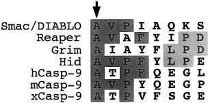
These puzzles are nicely accommodated by the structural information on the IAP-Smac complexes.14,15 The XIAP residues involved in binding the Smac tetrapeptide are highly conserved in the BIR2 domain of DIAP1. This observation and the realization that a mere tetrapeptide suffices IAP-recognition prompted a re-examination of the sequence homology between Smac and the Drosophila proteins Hid, Grim and Reaper. Indeed, the N-terminal four amino acids of the Drosophila proteins were found to share significant similarity with the mammalian protein Smac14,15,17 (Figure 1). This sequence conservation strongly suggests that the N-terminal sequences of Hid/Grim/Reaper may recognize a similarly conserved surface groove on DIAP1, a prediction recently confirmed by a structural analysis on the Drosophila complexes.18 Although the first four amino acids of Hid and Grim bind DIAP1 in the same manner as observed in the Smac-XIAP complex, the next three residues also make significant contribution to binding through hydrophobic interactions, consistent with the unique sequence features of the Drosophila proteins.18
Figure 1
An evolutionarily conserved family of IAP-binding motifs. The tetrapeptide motif has the consensus sequence A-(V/T/I)-(P/A)-(F/Y/I/V), where the invariable Ala is indicated by an arrow. The Drosophila proteins Reaper/Grim/Hid have an additional binding component18 (conserved residues 6–8, shaded in light gray)
The serendipitous nature for the discovery of these IAP-binding motifs suggests other yet-to-be-identified pro-apoptotic proteins bearing exposed AVPI-like sequences. Through sequence comparison, a Smac-like tetrapeptide (Ala316-Thr317-Pro318-Phe319) was discovered in procaspase-919 (Figure 1). Interestingly, the proteolytic cleavage that gives rise to the mature caspase-9 occurs after Asp315, thus releasing this potential IAP-binding motif. Subsequent experiments confirmed that this tetrapeptide motif in the p12 subunit of caspase-9 is primarily responsible for the interactions with the BIR3 domain of XIAP.19 Therefore, unlike other caspases, proteolytic processing of procaspase-9 serves as a mechanism for both inhibition and activation. In the absence of proteolytic processing, XIAP is unable to interact with procaspase-9. Upon apoptotic stimuli, procaspase-9 undergoes auto-catalytic processing after Asp315, exposing its internal tetrapeptide motif and resulting in the recruitment of and inhibition by XIAP. The release of the mature Smac from mitochondria presumably titrates XIAP, again using the same conserved N-terminal tetrapeptide in Smac. Thus, a conserved IAP-binding motif in caspase-9 and Smac mediates opposing effects on caspase activity and apoptosis. This fail-safe mechanism likely ensures protection against unwanted cell death resulting from accidental activation of caspases.
During apoptosis, the active caspase-9 can be further cleaved after Asp330 by downstream caspases such as caspase-3. This positive feedback not only removes XIAP-mediated inhibition of caspase-9 but also releases a 15-residue peptide that is freely available to relieve IAP-mediated inhibition of other caspases. Thus, this peptide represents an endogenous drug timed to release by the apoptotic cells themselves to facilitate death.
Cancer cells express elevated levels of IAPs. For example, the majority of melanoma cell lines express high levels of Livin/ML-IAP compared to primary melanocytes.6 The human survivin gene is expressed in most common forms of cancer but not in terminally differentiated normal adult tissues.8 In addition to serving as caspase inhibitors, some of these IAPs exhibit intrinsic ubiquitin ligase activity (E3) and tag active caspases for proteasome-mediated degradation.20,21 These activities may significantly reduce or eliminate the effects of pro-apoptotic treatments such as chemotherapy. Thus removing the negative effects of these IAPs may represent a promising approach to sensitize cancer cells for apoptosis. Supporting this strategy, reduction in survivin expression by antisense RNA led to apoptosis and sensitization to treatment by anticancer drugs.22 Given that the Smac-like tetrapeptide motif antagonizes IAP-mediated caspase inhibition, these peptides should also potentiate apoptosis in cells. Indeed, cells treated with the wild-type but not the mutant Smac tetrapeptide exhibited greater tendency to undergo apoptosis in response to UV radiation (unpublished data).
Although these peptides show some potency in cultured cell lines, they are likely to undergo rapid proteolytic degradation in living organisms. In addition, it is unclear how efficiently these peptides can cross the plasma membrane. Perhaps it is more advantageous to develop small chemical agents or to take a peptidomimetic approach to search for a synthetic, non-peptide compound with nanomolar potency. The available structural information will greatly facilitate any rational design approach, and the high-throughput screening is needed to ensure the discovery of multiple hits.
Present in seven proteins across several species (Figure 1), the IAP-binding tetrapeptide motif is destined to be discovered in more proteins. In each case, this motif is expected to antagonize the anti-apoptotic effects of IAPs.
