Rapid Dopamine Signaling in the Nucleus Accumbens during Contingent and Noncontingent Cocaine Administration (original) (raw)
INTRODUCTION
Dopaminergic neurotransmission in the mesolimbic system is associated with reward seeking (Everitt and Robbins, 2000; Salamone and Correa, 2002; Phillips et al, 2003b), prediction (Schultz, 1998; Waelti et al, 2001), and reinforcement (Wise, 1982, 2004). Owing to this, dopamine has been implicated in the neurobiological actions of many drugs of abuse, particularly those of cocaine (Di Chiara and Imperato, 1988; Di Chiara et al, 2004; Koob et al, 2004; Wise, 2004). Through its pharmacological actions, cocaine directly affects dopaminergic terminals by inhibiting dopamine uptake (Jones et al, 1995). Cocaine use leads to the development of conditioned effects through the pairing of the drug to environmental cues (Stewart, 1992; Volkow et al, 2003; Ciccocioppo et al, 2004; See et al, 2003), and these play a key role in the onset and maintenance of addiction (Gawin, 1991; O'Brien et al, 1992; Dackis and O'Brien, 2001). Microdialysis studies have revealed increases in extracellular nucleus accumbens dopamine during cocaine self-administration (Pettit and Justice, 1989, 1991; Wise et al, 1995; Bradberry, 2000). However, it has proved more difficult for microdialysis investigations of dopamine to resolve the pharmacological effects of cocaine from the conditioned effects because the sampling timescale is multiple minutes (Bradberry, 2000). Despite this, increases in dopamine on a minute timescale have been demonstrated by microdialysis in response to discriminative (Weiss et al, 2000) and conditioned (Ito et al, 2000) stimuli associated with cocaine self-administration.
Electrophysiological studies during operant behavior have shown that neuronal activity increases on a subsecond timescale during events associated with cocaine self-administration (Chang et al, 1994; Uzwiak et al, 1997; Carelli et al, 2000; Carelli, 2004). Thus, it is important to know whether dopamine can change on this timescale as well. Dopaminergic terminals form synapses on the neck of spines and dendrites of the projection neurons in the nucleus accumbens, and are in close proximity to glutametergic synapses originating from neurons in the hippocampus, basolateral amygdala, and prefrontal cortex (Totterdell and Smith, 1989; Sesack and Pickel, 1990; Johnson et al, 1994). Thus, dopamine is in a prominent position to modulate these inputs (O'Donnell et al, 1999; Nicola et al, 2000) and exert control over the activity of microcircuits within the nucleus accumbens that ultimately control behavior (Pennartz et al, 1994). Recently, we demonstrated that fast-scan cyclic voltammetry can be used to monitor dopamine changes during cocaine self-administration on a subsecond timescale (Phillips et al, 2003b). This technique is particularly well suited for measurement of transient dopamine release events (Rebec et al, 1997; Phillips et al, 2003a), defined here as signals greater than ∼15 nM and lasting less than 3 s (ie dopamine transients) that may arise from phasic firing of dopamine neurons (Venton et al, 2003a). Transient dopamine changes in the nucleus accumbens were found to occur during the ±10 s interval surrounding a rat's lever press to self-administer cocaine. The dopamine transients before the lever press appeared to promote cocaine seeking, whereas those that followed the lever press were closely time-locked to it and the accompanying cues.
In this work, we have furthered our investigation of subsecond dopamine changes that accompany cocaine administration. We first investigated the actions of noncontingent cocaine on both drug-naïve animals and animals with a history of cocaine self-administration. The high time resolution of the voltammetric technique allowed resolution of cocaine-induced dopamine transients under these conditions, which were not associated with any discernible behavioral responses. In rats trained to lever-press for cocaine, its self-administration caused similar transients due to the pharmacological effects of cocaine in addition to dopamine transients that were time-locked to each lever press. Thus, transients related to the pharmacological effects of cocaine accompany those that arise from the conditioned effects during self-administration.
MATERIALS AND METHODS
Animals
Male Sprague–Dawley rats (_n_=18; Harlan, Raleigh, NC; 280–300 g), individually housed on a 12:12 light cycle (lights on at 0700 h), were surgically implanted with an intravenous catheter under ketamine hydrochloride (100 mg/kg i.m.) and xylazine hydrochloride (20 mg/kg i.m.) anesthesia, as described previously (Caine and Koob, 1994; Carelli and Deadwyler, 1996). Following recovery, rats had ad libitum access to food, while water intake was restricted to 30 ml/day (above 85% of their presurgery weight) to maintain body weight and to remain consistent with our previous studies. All procedures were in accordance with the University of North Carolina Animal Care and Use Committee.
Cocaine Self-Administration Training
Following 1 week recovery from catheter surgery, some rats (_n_=8) were trained to self-administer cocaine during 2 h daily sessions. Rats were placed in a sound-attenuated operant chamber (Med Associates, St Albans, VT) and a cue light located 6.5 cm above the lever was illuminated to signal drug availability. Depression of the lever (FR1 schedule) resulted in simultaneous termination of the cuelight (20 s), onset of a tone-houselight stimulus (65 dB, 2900 Hz; 20 s) and intravenous cocaine infusion (0.33 mg in sterile heparinized saline over 6 s). During the 20 s post response period responding on the lever was recorded, but had no programmed consequences. Stable self-administration behavior (<10% variability in press number during a consecutive 3-day period) was typically achieved by 10–15 training sessions, and at this time rats were surgically prepared for voltammetric recording.
Voltammetry Surgery
Rats were anesthetized with ketamine hydrochloride (100 mg/kg i.m.) and xylazine hydrochloride (20 mg/kg i.m.) and placed in a stereotaxic frame. A guide cannula (Bioanalytical Systems, West Lafayette, IL) was positioned with its tip above the right nucleus accumbens core (+1.3 mm AP, +1.3 mm ML, −2.5 mm DV relative to bregma), an Ag/AgCl reference electrode was placed in the contralateral forebrain, and these were secured to the cranium with machine screws and cranioplastic cement. When cured, a detachable micromanipulator containing a carbon-fiber electrode (50–75 μm length cylinders, T-650; Amoco, Greenville, SC) was inserted into the guide cannula and the electrode was lowered into the nucleus accumbens core. A bipolar stimulating electrode was lowered into the ventral tegmental area (VTA; −5.2 mm AP, +1.0 mm ML, and −7.7 mm DV) until electrically evoked (60 biphasic pulses, 60 Hz, 120 μA, 2 ms/phase) dopamine release was detected at the carbon-fiber electrode. The stimulating electrode was then cemented, and the micromanipulator was removed and replaced with a stylet. Following surgery, rats were allowed to recover for at least 3 days.
Voltammetry Recording Sessions
On the experimental day, a new carbon-fiber electrode was lowered into the nucleus accumbens core. Voltammetric recordings were made every 100 ms by applying a triangular waveform (−0.6 to +1.4 V, 400 V/s). Data were digitized in a PC using software written in LabVIEW (National Instruments, Austin, TX). Dopamine release was optimized within the nucleus accumbens core by adjusting the vertical position of the working electrode (0.1 mm increments). Experiments were begun when a site that had a signal-to-noise ratio of electrically evoked (24 biphasic pulses, 60 Hz, 120 μA, 2 ms/phase) dopamine release of at least 30 was found. After the session, dopamine release was again electrically evoked to insure that carbon-fiber electrode was still in close proximity to a dopamine release site, and then the carbon-fiber electrode was removed and calibrated with dopamine (1 μM) in a flow-injection analysis system (Phillips et al, 2003a).
Noncontingent Cocaine Infusions
The response to noncontingent cocaine delivery was examined in rats that had been trained to self-administer cocaine (_n_=5) and a separate group of cocaine-naïve animals (_n_=5). Rats in the noncontingent group received identical treatment and surgery as rats self-administering cocaine. Rats were acclimated to the recording chamber (a different chamber to self-administration training) for 1 h, and then given four, 6-s infusions of saline (5 min apart), followed by four, 6-s infusions of cocaine (0.33 mg/infusion; the dose used in self-administration), while voltammetric recordings were made.
Cocaine Self-Administration
Self-administering rats were re-trained following voltammetry surgery for an additional 2–3 days to re-establish their behavior. On the experimental day, voltammetric data were collected and behavioral events were logged (Med Associates) on separate PCs that were digitally linked to allow synchronization of neurochemical and behavioral measurements during a cocaine self-administration session.
Signal Identification and Correction
Chemical identification of signals was carried out by examining background-subtracted cyclic voltammograms (Phillips and Wightman, 2003). The subtraction process yields changes in concentration that cannot be used to establish absolute levels (Phillips et al, 2003a). To isolate dopamine transients, a ‘sliding background’ subtraction was used. In this approach, each cyclic voltammogram in a 2-min file was subtracted from the average of the five cyclic voltammograms collected 1.0–0.5 s earlier. Each background-subtracted cyclic voltammogram was compared by linear regression to an in vivo dopamine template (average background-subtracted cyclic voltammogram recorded during electrically evoked dopamine release). Only voltammograms with an _r_2⩾0.75 were considered dopamine; the remainder of the signal was considered noise.
While the cyclic voltammograms for norepinephrine and dopamine are virtually identical, in vitro analysis shows that _r_2>0.75 distinguishes dopamine from other interferences (Heien et al, 2003) such as ascorbic acid, DOPAC, ionic changes in the extracellular space (Venton et al, 2003b), and noise. Norepinephrine is unlikely to be a significant contributor to the signal because its content in the nucleus accumbens is only 2% of that of dopamine (Garris et al, 1993), and immunohistochemical labeling of dopamine-_β_-hydroxylase is not detectable in this region (Baldo et al, 2003).
Examination of the cyclic voltammograms revealed that in addition to dopamine other chemical changes, such as changes in pH, were frequently detected. To remove their contribution, current measured at a potential that is not sensitive to dopamine (∼200 mV vs Ag/AgCl) was subtracted from the current at the peak oxidation potential for dopamine (∼650 mV vs Ag/AgCl). The current was then converted into concentration by calibrating the electrodes in vitro with 1 μM dopamine.
Characterization of the amplitude and duration of fluctuations in dopamine in self-administering animals within ±10 s of a lever press employed Mini-analysis software (Synaptosoft, Decatur, GA). Typical noise levels were equivalent to 6 nM dopamine. Only peaks with amplitudes that remained three times greater than the noise for at least 1.5 s were evaluated. Consecutive peaks were distinguished if they occurred after the prior event had decayed by at least 40%.
Histology
Following completion of experiments, rats were deeply anesthetized (ketamine hydrochloride, 150 mg/kg; xylazine hydrochloride, 20 mg/kg) and a stainless steel electrode (50-μm tip radius) was lowered to the recording site with the same micromanipulator, and an electrolytic lesion was made (50 μA, 10 s). Rats were then transcardially perfused with physiological saline, followed by 4% paraformaldehyde. Brains were stored in 4% paraformaldehyde for at least 3 days, frozen, and sectioned with a cryostat (50 μm). Brain sections were mounted on slides, stained with thionin, and coverslipped. Serial sections were examined under a light microscrope and all recording sites were verified to be located in the core region of the nucleus accumbens with the aid of a brain atlas (Paxinos and Watson, 1997).
RESULTS
Electrically and Behaviorally Evoked Dopamine Release
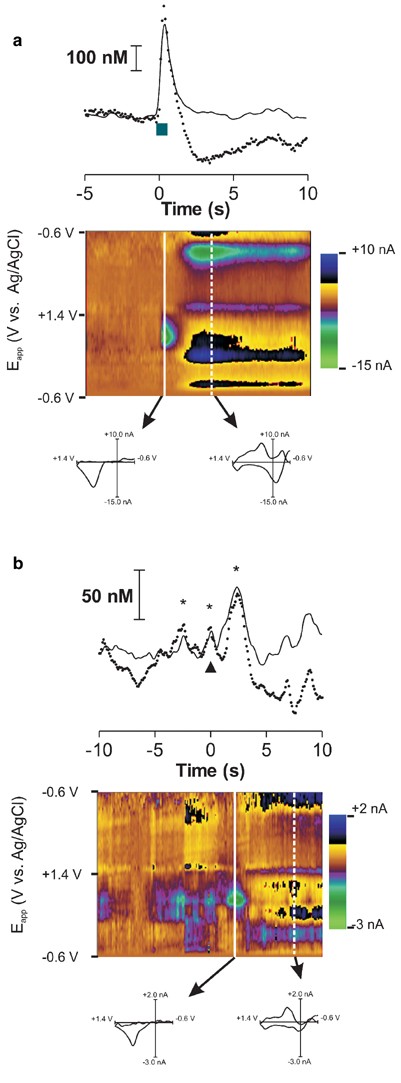
During insertion of the carbon fiber, electrical stimulation of the dopamine-containing cell bodies was used to evoke dopamine release. Prior studies have shown that electrical stimulation of the VTA and/or medial forebrain bundle causes a transient increase in extracellular dopamine in the nucleus accumbens (Kuhr et al, 1986; Garris et al, 1997), followed by a secondary basic change in local pH that correlates with increased local blood flow (Venton et al, 2003b). Both of these signals are readily detected electrochemically using fast-scan cyclic voltammetry with carbon-fiber microelectrodes. Figure 1a (top) shows electrically evoked dopamine release both before (dotted trace) and after (solid line) correction for the ionic changes that resemble pH. In this example, dopamine release was evoked using parameters that are analogous to burst firing of dopaminergic neurons (eight pulses, 30 Hz, 120 μA). Cyclic voltammograms taken immediately after the electrical stimulation confirms that the signal is dopamine, while those taken a few seconds later are consistent with the voltammograms for a basic pH change (Figure 1a bottom). The cyclic voltammogram obtained at the end of the stimulation was used as the template for dopamine identification during behaviorally evoked events in vivo (Phillips and Wightman, 2003).
Figure 1
Signal identification and correction of voltammetric data recorded in the NAc. (a) (Top): dopamine release (solid line) measured by cyclic voltammetry during electrical stimulation of the VTA (8p, 30 Hz, 120 μA; delivered at _t_=0 s) in a single trial. The signal before correction for ionic changes (dashed line) is also shown at the potential at which dopamine is oxidized. (a) Colored panel: the set of all background-subtracted cyclic voltammograms recorded over the 15 s interval. The ordinate is the applied potential (_E_app) during each voltammogram, the abscissa is the time at which it was recorded, and the current changes are encoded in color. Dopamine can be seen during stimulation by the features at 0.650 V (oxidation peak encoded as green) and at −0.20 V at the end of the voltage scan. Individual cyclic voltammograms from this set are shown for the end of the stimulation and at the time indicated by the white dashed line. The former is identical to that for authentic dopamine, while the latter is consistent with an ionic change at the electrode. (b) (Top): dopamine release (solid line) during a single lever-press response for cocaine (lever press at _t_=0 s). The signal before correction for ionic changes (dashed line) is also shown. The color plot reveals dopamine changes before and after the lever press, as well as ionic changes after the lever press. Dopamine transients are denoted in the trace by asterisks. The cyclic voltammogram 1 s after the lever press is consistent with dopamine (_r_2=0.89) and the cyclic voltammogram at 8 s after the lever press is consistent with an ionic change.
Behaviorally evoked dopamine transients had several similar characteristics. Figure 1b shows an example of phasic dopamine time-locked to the operant response for cocaine. The cyclic voltammogram was identical to that obtained during stimulated release. The time course, corrected for nondopaminergic components, showed an increase slightly before the lever press and a larger increase occurring immediately after the lever press (solid line). Without the correction for ionic changes (dotted trace), the increase appeared to be followed by a longer lasting decrease in current after the lever-press response that has cyclic voltammograms, which are also consistent with a basic pH change. In the remainder of the text, all traces have been pH corrected.
Pharmacological Effects of Cocaine on Dopaminergic Terminals in the Nucleus Accumbens
Cocaine is a competitive inhibitor of the dopamine transporter, and, as such, it prolongs the lifetime of dopamine in the extracellular fluid. As shown previously, (Wu et al, 2001) the prolonged lifetime in the nucleus accumbens can be directly observed with fast-scan cyclic voltammetry following evoked release caused by electrical stimulation of dopamine-containing cell bodies in anesthetized animals.
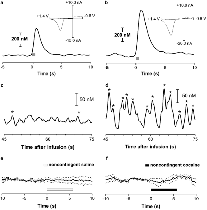
In self-administering animals, similar effects on evoked release were seen. At the beginning of self-administration sessions, the position of the carbon-fiber electrode within the nucleus accumbens was optimized to a site where robust dopamine release was evoked with electrical stimulation of the VTA. During the stimulation, the current arising from dopamine oxidation, corrected for interference from other substances, increased (an example is shown in Figure 2a). Dopamine rapidly returned to baseline when the stimulation terminated. At the end of the 2-h self-administration sessions, consisting of 19±1.1 lever presses in eight animals, the stimulation was repeated. In all cases, dopamine release was observed, verifying that the recording site still supported dopamine release (an example is shown in Figure 2b). Typically, evoked dopamine release was greater after the session, and it returned to baseline in 5.3±0.39 s, significantly slower than the 2.6±0.16 s observed before the session (_p_⩽0.01; Student's _t_-test).
Figure 2
Pharmacological responses to cocaine measured with in vivo voltammetry. (a) Example of electrically evoked (24 pulses, 60 Hz, 120 μA; gray box) dopamine release before a cocaine self-administration session. The dopamine signal was extracted from the voltammetric recordings. The cyclic voltammogram shown (insert) was recorded at the maximum response and confirms that dopamine is the substance detected. (b) Electrically stimulated release in the same animal after a cocaine self-administration session. The cyclic voltammogram shown (inset) was recorded at the maximum and has a high correlation with authentic dopamine. (c) A 30 s trace from an individual animal after a noncontingent i.v. infusion of saline. (d) Trace from the same animal after a noncontingent i.v. infusion of cocaine. In (c, d) peaks with an asterisk were due to dopamine since their cyclic voltammograms correlated with electrically evoked dopamine with an _r_2>0.75. (e) Average trace±SEM (dotted lines) (_n_=5 animals) for dopamine changes during saline infusion. (f) Average trace (_n_=5 animals) for dopamine changes during noncontingent cocaine infusion. Black bars show the infusion duration.
Cocaine administration also caused the frequency of naturally occurring dopamine transients to increase. Recent improvements in the sensitivity of the voltammetric approach (Heien et al, 2003) allow resolution of periodic fluctuations in dopamine in the nucleus accumbens extracellular fluid (Robinson et al, 2002). The example shown in Figure 2c, recorded in a cocaine-naïve rat after a saline infusion, illustrates these naturally occurring transients. The asterisks denote maxima whose voltammograms matched those of dopamine with an _r_2⩾0.75. However, 45 s after noncontingent infusion of 0.33 mg cocaine to this animal, the dopamine transients increased in frequency and amplitude (Figure 2d). The changes were neither immediate nor synchronized with the infusion: averaged traces collected for 10 s immediately following either saline or cocaine infusion do not show dopamine changes (Figure 2e and f; averaged across five animals).
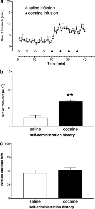
When examined on a slower timescale, the frequency of dopamine transients from recordings in five animals that were naïve to cocaine correlated closely (_r_=0.8) to the computed local concentration of cocaine (Pan et al, 1991; Figure 3a). While the frequency of dopamine transients was unchanged by saline infusions, in the first minute after cocaine infusion, the frequency increased and then declined as the local cocaine concentration declined. The baseline also increased in the first minute following cocaine infusion, and it reached 20% of its final value 38.1±17.0 s after initiation of cocaine infusion. The frequency of dopamine transients increased with subsequent cocaine infusions as well, although the relative increase was not as large as the relative change in local cocaine concentration. Overall, the frequency of dopamine transients during saline infusions was 1.0±0.3 min−1 and this increased almost eight-fold following noncontingent cocaine (frequency of dopamine transients after cocaine was 7.7±2.4 min−1, Figure 3b; t(4)=2.94; _p_⩽0.05). The mean amplitude of dopamine transients also significantly increased from 39.5±6.9 nM during saline infusions to 74.1±13.6 nM during cocaine infusions (t(4)=4.49; _p_⩽0.01; Figure 3c).
Figure 3
Noncontingent cocaine increases the frequency and amplitude of dopamine transients detected in naïve animals. (a) Upper: local cocaine concentration in the brain as calculated from equation (3) in Pan et al, 1991. Lower: mean rate of dopamine transients with SEM (_n_=5 animals) following four noncontingent infusions of saline followed by four infusions of cocaine (0.33 mg/inf). The arrowheads indicate the infusion time. (b) Frequency of transients before and after noncontingent cocaine in cocaine-naïve animals. (c) Amplitude of transients before and after noncontingent cocaine in cocaine-naïve animals. *_p_⩽0.05, **_p_⩽0.01 vs saline.
Similar results were obtained in animals with a history of cocaine self-administration (Figure 4a). In animals that had trained for 2 weeks to self-administer cocaine, the frequency of dopamine transients during saline infusions was 2.3±0.72 min−1 and it increased to 6.8±0.34 min−1 following noncontingent cocaine infusion (Figure 4b; t(4)=9.3; _p_⩽0.001). When the frequency of dopamine transients from these two groups was compared, there was no significant main effect of cocaine history (cocaine experienced vs cocaine naïve; _p_>0.05) or an interaction (_p_>0.05). However, during the cocaine infusion period in cocaine-experienced animals, dopamine transients did not significantly increase in amplitude (46.1±6.0 nM during saline administration and 51.6±4.6 nM during cocaine infusion; _p_>0.05, Figure 4c). Thus, cocaine increased the frequency of dopamine transients in both naïve and cocaine-experienced animals, but the increase in transient amplitude was only seen in naïve animals.
Figure 4
Noncontingent cocaine increases the frequency of dopamine transients detected in animals with a history of self-administration. (a) Mean rate of dopamine transients with SEM (_n_=5 animals) following four noncontingent infusions of saline, followed by four infusions of cocaine (0.33 mg/inf). The arrowheads indicate the infusion time. (b) Frequency of transients before and after noncontingent cocaine in cocaine-experienced animals. (c) Amplitude of dopamine transients before and after noncontingent cocaine in cocaine-experienced animals. **_p_⩽0.001 vs saline.
Dopamine Transients during Self-Administration of Cocaine
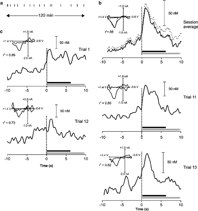
During self-administration sessions, rats exhibited stable self-administration behavior as seen in the behavioral response pattern for a representative animal (Figure 5a). Dopamine transients were observed throughout the 2-h self-administration session, with a mean frequency of 5.8±1.1 min−1. This frequency is not significantly different (_p_>0.05) from that observed following noncontingent cocaine in rats with a self-administration history. Most transients occurred at random times and were not correlated with any specific behavior. However, as we have shown previously, dopamine transients were always seen around the lever press (Phillips et al, 2003b). The averaged result from all bar presses in this animal shows dopamine transients in the time interval (±10 s) around the lever press (Figure 5b). Dopamine began to rise 0.8 s before the lever press, reached a peak concentration of 69±12 nM at 1.5 s after the lever press, and returned to baseline at 6.5 s after the press. The peak concentration is not significantly different (_p_>0.05) from that for the random transients following cocaine in rats with a cocaine self-administration history. The averaged cyclic voltammogram (Figure 5, upper right inset) confirmed dopamine as the contributing species (_r_2=0.88).
Figure 5
Examples of rapid dopaminergic signaling during cocaine self-administration. (a) A lever-press response record from a single animal during a 2-h self-administration session. Each vertical tick represents a single lever press for cocaine. (b) Average dopamine concentration change at ±10 s around the lever press for cocaine (solid line) and SEM (dashed line) from each of the 16 presses in this rat. _t_=0 indicates the lever press, the black bar represents the duration of the infusion, and the gray bar represents the duration of drug-associated cues (which extend for an additional 10 s). The average cyclic voltammogram (solid line, inset) shows a high correlation (_r_2=0.88) with that recorded during stimulated dopamine release (dashed line, inset). (c) Individual examples of phasic dopamine time-locked to the operant response to cocaine for specific trials from an individual rat showing the variability in responses. In all four panels, the cyclic voltammograms shown were recorded after the lever press and show a high correlation with that for dopamine (_r_2 values when compared to stimulated dopamine release ranged between 0.73 and 0.89). Correlation coefficients for the events before the lever press were in the same range, but the dopamine transients occur at variable times relative to the lever press.
Aspects of individual trials (Figure 5, lower four panels) are masked in the average. The dopamine transients before the lever press vary in their timing relative to the lever press, which causes them to be washed out in the average. The dopamine transients following the lever press are more time-locked and thus are not diluted in the average. The dopamine transients associated with the lever press for cocaine (±10 s around the press) are summarized in Table 1. A total of 343 dopamine transients were observed during 152 lever presses in eight animals. Of these, 122 (36%) occurred in the 10 s before the lever press, while 221 (64%) were observed within 10 s after the lever press. There are more transients than lever presses because, in some cases, two or three dopamine transients in close proximity followed the lever press. Among the dopamine transients before the lever press, 59 (42%) occurred within 3 s before the operant response. Of the 221 dopamine transients observed post lever press, 115 (52%) reached a maximum within 3 s after the response. Thus, in the ±3 s around the lever press, the frequency of dopamine transients exceeds 11 min−1, almost double the rate observed during the remainder of the self-administration session.
Table 1 Distribution of Dopamine Transients Relative to the Lever Presses (±10 s) in Rats Trained to Press a Lever for Cocaine
DISCUSSION
The primary pharmacological effect of cocaine on dopamine-containing neurons is inhibition of uptake (Giros et al, 1996). In this study, uptake inhibition is observed by the decreased rate of dopamine clearance following its stimulated release. In addition, the subsecond measurements reveal an additional consequence of cocaine—the transient changes in extracellular dopamine in the nucleus accumbens that normally occur in awake rats are increased in amplitude and frequency following its administration. The increase in frequency of dopamine release transients is a pharmacological effect, as demonstrated by its induction following noncontingent cocaine on a timescale that is similar to that for cocaine transport to the brain (Pan et al, 1991). During lever pressing for cocaine, similar dopamine transients occur, but the subset is closely time-locked to the lever press. These are evident before cocaine reaches the brain and exerts its pharmacological effect. Previously, we demonstrated that these dopamine transients are due to the learned association between cocaine, the operant response, and the environmental cues that predict drug delivery (Phillips et al, 2003b).
Fast-scan cyclic voltammetry provides high temporal resolution of the chemical dynamics of dopaminergic neurotransmission and allowed the first observation of naturally occurring, transient, release events (Robinson et al, 2002). While in vivo electrochemical techniques vary in their chemical selectivity (Di Chiara, 2002; Wightman and Robinson, 2002), fast-scan cyclic voltammetry has the best chemical resolution because the cyclic voltammograms for most electroactive species are distinct (Heien et al, 2003). As illustrated in Figure 1, both high temporal resolution and considerable chemical selectivity are required because dopamine changes are rapid and ionic changes, resembling changes in extracellular pH, accompany and overlap both evoked and spontaneous dopamine transients. Other electrochemical studies of cocaine self-administration had insufficient time resolution to observe these changes (Gratton and Wise, 1994; Kiyatkin and Stein, 1995). Furthermore, they did not resolve the simultaneous chemical changes, and thus, their signals are likely a composite of several substances (eg including pH). In this work, the chemical selectivity was higher; nonetheless, some transient dopamine changes may be beneath the threshold (18 nM), set by the electrical noise of the system.
The rapid measurements of dopamine concentrations reveal the pharmacological effects of cocaine on dopamine neurons in two ways. First, stimulated dopamine release obtained after cocaine self-administration sessions show prolonged dopamine clearance (Figure 2a and b), consistent with results following cocaine administration either in vitro (Jones et al, 1995) or in vivo (Wu et al, 2001). Uptake inhibition increases the lifetime of dopamine in the extracellular space, allowing diffusion over greater distances and the opportunity to interact with remote receptors (Venton et al, 2003a), and leads to a rise in dopamine concentration when sampled on a minute timescale with microdialysis (Wise et al, 1995). Second, dopamine transients increase in frequency following noncontingent cocaine (Figures 2c, d and 3). This occurred both in drug-naïve animals and those with a history of cocaine self-administration, and the frequency following cocaine is similar to that in the nucleus accumbens of rats lever-pressing for cocaine. The cocaine-induced transients in the drug-naïve rats were larger than in the two other groups. This suggests that both contingent and noncontingent cocaine cause an adaptation in the mechanism(s) responsible for these transients. As noncontingent administration of nomifensine, another dopamine uptake inhibitor, also causes these effects (Robinson and Wightman, 2004), they appear to be a general consequence of dopamine uptake inhibition. The increase in amplitude of the spontaneous, transient dopamine release events is quite similar to the increase in electrically stimulated release that is seen after uptake inhibition (Wu et al, 2001).
The naturally occurring, spontaneous, dopamine transients occur at relatively low frequency, are not associated with specific external cues or behavioral responses, and their function is currently unknown. However, their amplitude is sufficient to activate high-affinity dopaminergic receptors located on post-synaptic cells in the nucleus accumbens (Richfield et al, 1989). Electrophysiological studies in behaving rats (Hyland et al, 2002) have demonstrated that dopamine-containing neurons burst fire on a timescale similar to the observed dopamine transients. Burst firing, in turn, is driven by excitatory glutamatergic and cholinergic inputs to dopamine-containing cells (Kitai et al, 1999; Floresco et al, 2003; Paladini et al, 2004). Dopamine release evoked with parameters that mimic a dopaminergic burst (Figure 1a) has a similar time course as the naturally occurring signals. Another pharmacological challenge that causes dopamine release transients is CB1 cannabinoid receptor activation (Cheer et al, 2004), a process that causes increased burst firing of dopamine-containing neurons (Gessa et al, 1998).
Both noncontingent and self-administered cocaine cause these naturally occurring release events to increase in frequency. The mechanism responsible for this is unclear, since cocaine partially inhibits VTA dopaminergic cell firing both in brain slices (Bunney et al, 2000) and in anesthetized rats (Einhorn et al, 1988). Cocaine and other pyschostimulants can induce low-frequency oscillations in the firing rate of dopamine-containing neurons; however, this has only been demonstrated in anesthetized rats following dopamine receptor blockade with raclopride (Shi et al, 2004). Alternatively, local activation in the nucleus accumbens by excitatory afferents may evoke these transients (Parkinson et al, 2000).
Despite the presence of ongoing dopamine release events following cocaine self-administration, there is a subset of dopamine transient release events that are tightly linked to the animal behavior at the time of the lever press (Figures 1 and 5), a time removed by several minutes from that of the maximal brain cocaine levels. In particular, the transients immediately following the lever press, occasionally exhibiting multiple peaks, always occur within a few seconds of the press. In contrast, the pharmacologically induced dopamine transients during noncontingent cocaine administration are absent in this time interval (compare Figures 2f and 5). Our previous results implicate that transients can code reward expectation because they occur in response to drug-predictive cues in the absence of cocaine (Phillips et al, 2003b). Thus, dopamine transients associated with reward are consistent with the increased burst firing of VTA neurons in primates during reward-related events (Waelti et al, 2001). Increases in dopamine in response to cocaine-associated cues can also be observed by microdialysis (Ito et al, 2000; Weiss et al, 2000), but more complex experimental designs must be used to separate temporally the contingent from the pharmacological effects. The dopamine transients immediately preceding the lever press are more variable in their occurrence (Table 1) and are not so tightly time-locked to it, but are more closely correlated with the initiation of appetitive behaviors. We have noted previously that dopamine transients preceding the lever press occur when the animal initiates the approach to the lever. As there is a high degree of variability in the time between when the animal approaches the lever, and when they actually press it, this may explain why the pre-response transients are variable. Indeed, evoking dopamine release on this timescale promotes drug seeking (Phillips et al, 2003b). Consistently, similar dopamine transients are seen in trained rats at the onset of a cue that promotes lever pressing for sucrose (Roitman et al, 2004).
Previous studies have implicated an important role of dopamine in both the reinforcing (Wise et al, 1995) and conditioned effects (Ito et al, 2000; Weiss et al, 2000) of cocaine. In those studies, dopamine was measured on a relatively long timescale (over minutes) using microdialysis. The rapid dopamine signaling reported here results from both pharmacological and conditioned effects of cocaine. Although dopamine alone is not responsible for the manifestation of specific behaviors, it can modulate neural signals underlying behavior (Carelli, 2004). Here we have revealed a new temporal domain of dopaminergic chemical transmission that is relevant to behavior and is highly susceptible to modification by psychoactive drugs and associated conditioning effects.
References
- Baldo BA, Daniel RA, Berridge CW, Kelley AE (2003). Overlapping distributions of orexin/hypocretin- and dopamine-beta-hydroxylase immunoreactive fibers in rat brain regions mediating arousal, motivation, and stress. J Comp Neurol 464: 220–237.
Article PubMed Google Scholar - Bradberry CW (2000). Acute and chronic dopamine dynamics in a nonhuman primate model of recreational cocaine use. J Neurosci 20: 7109–7115.
Article CAS PubMed PubMed Central Google Scholar - Bunney EB, Appel SB, Brodie MS (2000). Cocaine potentiates ethanol-induced excitation of dopaminergic reward neurons in the ventral tegmental area. J Pharmacol Exp Ther 293: 383–389.
CAS PubMed Google Scholar - Caine SB, Koob GF (1994). Effects of dopamine D-1 and D-2 antagonists on cocaine self-administration under different schedules of reinforcement in the rat. J Pharmacol Exp Ther 270: 209–218.
CAS PubMed Google Scholar - Carelli RM (2004). Nucleus accumbens cell firing and rapid dopamine signaling during goal-directed behaviors in rats. Neuropharmacology 47(Suppl 1): 180–189.
Article CAS PubMed Google Scholar - Carelli RM, Deadwyler SA (1996). Dose-dependent transitions in nucleus accumbens cell firing and behavioral responding during cocaine self-administration sessions in rats. J Pharmacol Exp Ther 277: 385–393.
CAS PubMed Google Scholar - Carelli RM, Ijames SG, Crumling AJ (2000). Evidence that separate neural circuits in the nucleus accumbens encode cocaine versus ‘natural’ (water and food) reward. J Neurosci 20: 4255–4266.
Article CAS PubMed PubMed Central Google Scholar - Chang JY, Sawyer SF, Lee RS, Woodward DJ (1994). Electrophysiological and pharmacological evidence for the role of the nucleus accumbens in cocaine self-administration in freely moving rats. J Neurosci 14: 1224–1244.
Article CAS PubMed PubMed Central Google Scholar - Cheer JF, Wassum KM, Heien ML, Phillips PE, Wightman RM (2004). Cannabinoids enhance subsecond dopamine release in the nucleus accumbens of awake rats. J Neurosci 24: 4393–4400.
Article CAS PubMed PubMed Central Google Scholar - Ciccocioppo R, Martin-Fardon R, Weiss F (2004). Stimuli associated with a single cocaine experience elicit long-lasting cocaine-seeking. Nat Neurosci 7: 495–496.
Article CAS PubMed Google Scholar - Dackis CA, O'Brien CP (2001). Cocaine dependence: a disease of the brain's reward centers. J Subst Abuse Treat 21: 111–117.
Article CAS PubMed Google Scholar - Di Chiara G (2002). Nucleus accumbens shell and core dopamine: differential role in behavior and addiction. Behav Brain Res 137: 75–114.
Article CAS PubMed Google Scholar - Di Chiara G, Bassareo V, Fenu S, De Luca MA, Spina L, Cadoni C et al (2004). Dopamine and drug addiction: the nucleus accumbens shell connection. Neuropharmacology 47(Suppl 1): 227–241.
Article CAS PubMed Google Scholar - Di Chiara G, Imperato A (1988). Drugs abused by humans preferentially increase synaptic dopamine concentrations in the mesolimbic system of freely moving rats. Proc Natl Acad Sci USA 85: 5274–5278.
Article CAS PubMed PubMed Central Google Scholar - Einhorn LC, Johansen PA, White FJ (1988). Electrophysiological effects of cocaine in the mesoaccumbens dopamine system: studies in the ventral tegmental area. J Neurosci 8: 100–112.
Article CAS PubMed PubMed Central Google Scholar - Everitt BJ, Robbins TW (2000). Second-order schedules of drug reinforcement in rats and monkeys: measurement of reinforcing efficacy and drug-seeking behaviour. Psychopharmacology (Berl) 153: 17–30.
Article CAS Google Scholar - Floresco SB, West AR, Ash B, Moore H, Grace AA (2003). Afferent modulation of dopamine neuron firing differentially regulates tonic and phasic dopamine transmission. Nat Neurosci 6: 968–973.
Article CAS PubMed Google Scholar - Garris PA, Christensen JR, Rebec GV, Wightman RM (1997). Real-time measurement of electrically evoked extracellular dopamine in the striatum of freely moving rats. J Neurochem 68: 152–161.
Article CAS PubMed Google Scholar - Garris PA, Collins LB, Jones SR, Wightman RM (1993). Evoked extracellular dopamine in vivo in the medial prefrontal cortex. J Neurochem 61: 637–647.
Article CAS PubMed Google Scholar - Gawin FH (1991). Cocaine addiction: psychology and neurophysiology. Science 251: 1580–1586.
Article CAS PubMed Google Scholar - Gessa GL, Melis M, Muntoni AL, Diana M (1998). Cannabinoids activate mesolimbic dopamine neurons by an action on cannabinoid CB1 receptors. Eur J Pharmacol 341: 39–44.
Article CAS PubMed Google Scholar - Giros B, Jaber M, Jones SR, Wightman RM, Caron MG (1996). Hyperlocomotion and indifference to cocaine and amphetamine in mice lacking the dopamine transporter. Nature 379: 606–612.
Article CAS PubMed Google Scholar - Gratton A, Wise RA (1994). Drug- and behavior-associated changes in dopamine-related electrochemical signals during intravenous cocaine self-administration in rats. J Neurosci 14: 4130–4146.
Article CAS PubMed PubMed Central Google Scholar - Heien ML, Phillips PE, Stuber GD, Seipel AT, Wightman RM (2003). Overoxidation of carbon-fiber microelectrodes enhances dopamine adsorption and increases sensitivity. Analyst 128: 1413–1419.
Article CAS PubMed Google Scholar - Hyland BI, Reynolds JN, Hay J, Perk CG, Miller R (2002). Firing modes of midbrain dopamine cells in the freely moving rat. Neuroscience 114: 475–492.
Article CAS PubMed Google Scholar - Ito R, Dalley JW, Howes SR, Robbins TW, Everitt BJ (2000). Dissociation in conditioned dopamine release in the nucleus accumbens core and shell in response to cocaine cues and during cocaine-seeking behavior in rats. J Neurosci 20: 7489–7495.
Article CAS PubMed PubMed Central Google Scholar - Johnson LR, Aylward RL, Hussain Z, Totterdell S (1994). Input from the amygdala to the rat nucleus accumbens: its relationship with tyrosine hydroxylase immunoreactivity and identified neurons. Neuroscience 61: 851–865.
Article CAS PubMed Google Scholar - Jones SR, Garris PA, Wightman RM (1995). Different effects of cocaine and nomifensine on dopamine uptake in the Caudate-Putamen and nucleus accumbens. J Pharmacol Exp Ther 274: 396–403.
CAS PubMed Google Scholar - Kitai ST, Shepard PD, Callaway JC, Scroggs R (1999). Afferent modulation of dopamine neuron firing patterns. Curr Opin Neurobiol 9: 690–697.
Article CAS PubMed Google Scholar - Kiyatkin EA, Stein EA (1995). Fluctuations in nucleus accumbens dopamine during cocaine self-administration behavior: an in vivo electrochemical study. Neuroscience 64: 599–617.
Article CAS PubMed Google Scholar - Koob GF, Ahmed SH, Boutrel B, Chen SA, Kenny PJ, Markou A et al (2004). Neurobiological mechanisms in the transition from drug use to drug dependence. Neurosci Biobehav Rev 27: 739–749.
Article CAS PubMed Google Scholar - Kuhr WG, Bigelow JC, Wightman RM (1986). In vivo comparison of the regulation of releasable dopamine in the caudate nucleus and the nucleus accumbens of the rat brain. J Neurosci 6: 974–982.
Article CAS PubMed PubMed Central Google Scholar - Nicola SM, Surmeier J, Malenka RC (2000). Dopaminergic modulation of neuronal excitability in the striatum and nucleus accumbens. Annu Rev Neurosci 23: 185–215.
Article CAS PubMed Google Scholar - O'Brien CP, Childress AR, McLellan AT, Ehrman R (1992). Classical conditioning in drug-dependent humans. Ann NY Acad Sci 654: 400–415.
Article CAS PubMed Google Scholar - O'Donnell P, Greene J, Pabello N, Lewis BL, Grace AA (1999). Modulation of cell firing in the nucleus accumbens. Ann NY Acad Sci 877: 157–175.
Article CAS PubMed Google Scholar - Paladini CA, Mitchell JM, Williams JT, Mark GP (2004). Cocaine self-administration selectively decreases noradrenergic regulation of metabotropic glutamate receptor-mediated inhibition in dopamine neurons. J Neurosci 24: 5209–5215.
Article CAS PubMed PubMed Central Google Scholar - Pan HT, Menacherry S, Justice Jr JB (1991). Differences in the pharmacokinetics of cocaine in naive and cocaine-experienced rats. J Neurochem 56: 1299–1306.
Article CAS PubMed Google Scholar - Parkinson JA, Cardinal RN, Everitt BJ (2000). Limbic cortical–ventral striatal systems underlying appetitive conditioning. Prog Brain Res 126: 263–285.
Article CAS PubMed Google Scholar - Paxinos G, Watson C (1997). The Rat Brain in Stereotaxic Coordinates. Academic Press: San Diego.
Google Scholar - Pennartz CM, Groenewegen HJ, Lopes da Silva FH (1994). The nucleus accumbens as a complex of functionally distinct neuronal ensembles: an integration of behavioural, electrophysiological and anatomical data. Prog Neurobiol 42: 719–761.
Article CAS PubMed Google Scholar - Pettit HO, Justice Jr JB (1989). Dopamine in the nucleus accumbens during cocaine self-administration as studied by in vivo microdialysis. Pharmacol Biochem Behav 34: 899–904.
Article CAS PubMed Google Scholar - Pettit HO, Justice Jr JB (1991). Effect of dose on cocaine self-administration behavior and dopamine levels in the nucleus accumbens. Brain Res 18: 94–102.
Article Google Scholar - Phillips PE, Robinson DL, Stuber GD, Carelli RM, Wightman RM (2003a). Real-time measurements of phasic changes in extracellular dopamine concentration in freely moving rats by fast-scan cyclic voltammetry. Methods Mol Med 79: 443–464.
CAS PubMed Google Scholar - Phillips PE, Stuber GD, Heien ML, Wightman RM, Carelli RM (2003b). Subsecond dopamine release promotes cocaine seeking. Nature 422: 614–618.
Article CAS PubMed Google Scholar - Phillips PE, Wightman RM (2003). Critical guidelines for validation of the selectivity of in-vivo chemical microsensors. Trends Anal Chem 22: 509–514.
Article CAS Google Scholar - Rebec GV, Christensen JR, Guerra C, Bardo MT (1997). Regional and temporal differences in real-time dopamine efflux in the nucleus accumbens during free-choice novelty. Brain Res 776: 61–67.
Article CAS PubMed Google Scholar - Richfield EK, Penney JB, Young AB (1989). Anatomical and affinity state comparisons between dopamine D1 and D2 receptors in the rat central nervous system. Neuroscience 30: 767–777.
Article CAS PubMed Google Scholar - Robinson DL, Heien ML, Wightman RM (2002). Frequency of dopamine concentration transients increases in dorsal and ventral striatum of male rats during introduction of conspecifics. J Neurosci 22: 10477–10486.
Article CAS PubMed PubMed Central Google Scholar - Robinson DL, Wightman RM (2004). Nomifensine amplifies subsecond dopamine signals in the ventral striatum of freely-moving rats. J Neurochem 90: 894–903.
Article CAS PubMed Google Scholar - Roitman MF, Stuber GD, Phillips PE, Wightman RM, Carelli RM (2004). Dopamine operates as a subsecond modulator of food seeking. J Neurosci 24: 1265–1271.
Article CAS PubMed PubMed Central Google Scholar - Salamone JD, Correa M (2002). Motivational views of reinforcement: implications for understanding the behavioral functions of nucleus accumbens dopamine. Behav Brain Res 137: 3–25.
Article CAS PubMed Google Scholar - Schultz W (1998). Predictive reward signal of dopamine neurons. J Neurophysiol 80: 1–27.
Article CAS PubMed Google Scholar - See RE, Fuchs RA, Ledford CC, McLaughlin J (2003). Drug addiction, relapse, and the amygdala. Ann NY Acad Sci 985: 294–307.
Article CAS PubMed Google Scholar - Sesack SR, Pickel VM (1990). In the rat medial nucleus accumbens, hippocampal and catecholaminergic terminals converge on spiny neurons and are in apposition to each other. Brain Res 527: 266–279.
Article CAS PubMed Google Scholar - Shi WX, Pun CL, Zhou Y (2004). Psychostimulants induce low-frequency oscillations in the firing activity of dopamine neurons. Neuropsychopharmacology 29: 2160–2167.
Article CAS PubMed Google Scholar - Stewart J (1992). Neurobiology of conditioning to drugs of abuse. Ann NY Acad Sci 654: 335–346.
Article CAS PubMed Google Scholar - Totterdell S, Smith AD (1989). Convergence of hippocampal and dopaminergic input onto identified neurons in the nucleus accumbens of the rat. J Chem Neuroanat 2: 285–298.
CAS PubMed Google Scholar - Uzwiak AJ, Guyette FX, West MO, Peoples LL (1997). Neurons in accumbens subterritories of the rat: phasic firing time-locked within seconds of intravenous cocaine self-infusion. Brain Res 767: 363–369.
Article CAS PubMed Google Scholar - Venton BJ, Michael DJ, Wightman RM (2003b). Correlation of local changes in extracellular oxygen and pH that accompany dopaminergic terminal activity in the rat caudate-putamen. J Neurochem 84: 373–381.
Article CAS PubMed Google Scholar - Venton BJ, Zhang H, Garris PA, Phillips PE, Sulzer D, Wightman RM (2003a). Real-time decoding of dopamine concentration changes in the caudate-putamen during tonic and phasic firing. J Neurochem 87: 1284–1295.
Article CAS PubMed Google Scholar - Volkow ND, Fowler JS, Wang GJ (2003). The addicted human brain: insights from imaging studies. J Clin Invest 111: 1444–1451.
Article CAS PubMed PubMed Central Google Scholar - Waelti P, Dickinson A, Schultz W (2001). Dopamine responses comply with basic assumptions of formal learning theory. Nature 412: 43–48.
Article CAS PubMed Google Scholar - Weiss F, Maldonado-Vlaar CS, Parsons LH, Kerr TM, Smith DL, Ben Shahar O (2000). Control of cocaine-seeking behavior by drug-associated stimuli in rats: effects on recovery of extinguished operant-responding and extracellular dopamine levels in amygdala and nucleus accumbens. Proc Natl Acad Sci USA 97: 4321–4326.
Article CAS PubMed PubMed Central Google Scholar - Wightman RM, Robinson DL (2002). Transient changes in mesolimbic dopamine and their association with ‘reward’. J Neurochem 82: 721–735.
Article CAS PubMed Google Scholar - Wise RA (1982). Common neural basis for brain stimulation reward, drug reward, and food reward. In BG Hoebel, D Novin (eds). The Neural Basis of Feeding and Reward. Haer Institute: Brunswick, ME. pp 445–454.
Google Scholar - Wise RA (2004). Dopamine, learning and motivation. Nat Rev Neurosci 5: 483–494.
Article CAS PubMed Google Scholar - Wise RA, Newton P, Leeb K, Burnette B, Pocock D, Justice Jr JB (1995). Fluctuations in nucleus accumbens dopamine concentration during intravenous cocaine self-administration in rats. Psychopharmacology (Berl) 120: 10–20.
Article CAS Google Scholar - Wu Q, Reith ME, Kuhar MJ, Carroll FI, Garris PA (2001). Preferential increases in nucleus accumbens dopamine after systemic cocaine administration are caused by unique characteristics of dopamine neurotransmission. J Neurosci 21: 6338–6347.
Article CAS PubMed PubMed Central Google Scholar
Acknowledgements
The authors thank MLAV Heien, SE Brooks, JH Wondolowski, and the UNC Electronics Facility. This work was supported by grants from the National Institute on Drug Abuse; DA10900 (RMW), DA17318 (RMC & RMW), and DA015923 (GDS).
Author information
Authors and Affiliations
- Curriculum in Neurobiology, University of North Carolina at Chapel Hill, Chapel Hill, NC, USA
Garret D Stuber, Regina M Carelli & R Mark Wightman - Department of Psychology, University of North Carolina at Chapel Hill, Chapel Hill, NC, USA
Mitchell F Roitman & Regina M Carelli - Department of Psychiatry and Behavioral Sciences, University of Washington, Seattle, WA, USA
Paul E M Phillips - Department of Pharmacology, University of Washington, Seattle, WA, USA
Paul E M Phillips - Department of Chemistry and Neuroscience Center, University of North Carolina at Chapel Hill, Chapel Hill, NC, USA
R Mark Wightman
Authors
- Garret D Stuber
You can also search for this author inPubMed Google Scholar - Mitchell F Roitman
You can also search for this author inPubMed Google Scholar - Paul E M Phillips
You can also search for this author inPubMed Google Scholar - Regina M Carelli
You can also search for this author inPubMed Google Scholar - R Mark Wightman
You can also search for this author inPubMed Google Scholar
Corresponding author
Correspondence toR Mark Wightman.
Rights and permissions
About this article
Cite this article
Stuber, G., Roitman, M., Phillips, P. et al. Rapid Dopamine Signaling in the Nucleus Accumbens during Contingent and Noncontingent Cocaine Administration.Neuropsychopharmacol 30, 853–863 (2005). https://doi.org/10.1038/sj.npp.1300619
- Received: 27 August 2004
- Revised: 11 October 2004
- Accepted: 13 October 2004
- Published: 17 November 2004
- Issue Date: 01 May 2005
- DOI: https://doi.org/10.1038/sj.npp.1300619


