Wilson's disease: ‘face of giant panda’ and ‘trident’ signs together (original) (raw)
Journal Article
Department of Neurology
,
S.M.S. Medical College and Hospital
,
Jaipur 302004
,
India
Search for other works by this author on:
Department of Neurology
,
S.M.S. Medical College and Hospital
,
Jaipur 302004
,
India
Search for other works by this author on:
Received:
28 February 2014
Revision received:
10 March 2014
 PDF
PDF Cite
Jigar R. Parekh, Preetesh R. Agrawal, Wilson's disease: ‘face of giant panda’ and ‘trident’ signs together, Oxford Medical Case Reports, Volume 2014, Issue 1, April 2014, Pages 16–17, https://doi.org/10.1093/omcr/omu005
Close
Navbar Search Filter Mobile Enter search term Search
Abstract
Wilson's disease is an inborn error of metabolism characterized by inability to excrete copper into the bile, with excessive deposition of copper into the eyes, liver and brain. Lentiform nuclei are involved most commonly, but involvement of thalamus, midbrain and pons results in certain characteristic radiological signs on neuroimaging. Atrophy of cerebral and cerebellar cortex is also common yet under-recognized. Identification of these signs helps in the diagnosis in appropriate clinical setting.
INTRODUCTION
Wilson's disease is an inborn error of metabolism characterized by inability to excrete copper into the bile, with excessive deposition of copper into the eyes, liver and brain. Within the central nervous system, basal ganglia are common sites of affection, but there are characteristic signs on neuroimaging identified due to involvement of brainstem. Of these signs, ‘face of giant panda’ and ‘trident’ signs are remarkably seen and point towards the diagnosis in appropriate clinical setting, especially when occur simultaneously.
CASE REPORT
A 42-year-old gentleman presented with history of tremors of both upper limbs (left > right) since 1 year, postural instability since 8 months and slurring of speech since 3 months. Neurological examination showed Kayser–Fleischer (KF) rings in both eyes, rest and postural tremors of both upper limbs (proximal and distal), ataxic dysarthria, rigidity in all four limbs and gait ataxia. There was no evidence of jaundice or cirrhosis to suggest hepatic involvement.
Slit-lamp examination revealed KF rings in both eyes and 24 h urinary copper was diagnostic of Wilson's disease. Liver function tests were normal. Magnetic resonance imaging (MRI) of brain revealed:
- Diffuse hyperintensities in pons with ‘trident sign’ (Fig. 1, left).
- Symmetrical hyperintensities in both halves of midbrain and periaqueductal gray matter with hypointense red nuclei and subtantia nigra forming ‘face of giant panda’ sign (Fig. 1, right) and hyperintensities in bilateral thalami.
- Abnormal hypointensities seen in bilateral globus pallidus.
- Hyperintesities in the claustrum and external capsule bilaterally.
- Cerebral and cerebellar atrophy.
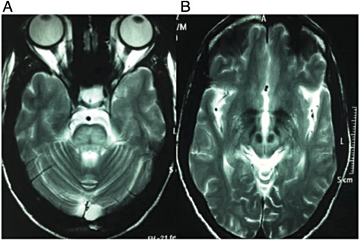
Figure 1:
MRI brain T2-weighted image shows abnormal hyperintensity in pons with characteristic hypointense ‘trident’ sign (A) and characteristic ‘face of giant panda’ sign in midbrain (B).
DISCUSSION
Wilson's disease is an inherited disorder in which defective biliary excretion of copper leads to its accumulation, particularly in the liver and brain. Wilson's disease is due to mutations of the ATP7B gene on chromosome 13, which encodes a copper-transporting P-type ATPase (ATP7B) residing in the _trans_-Golgi network of hepatocytes. Copper is collected in the liver, and after hepatic binding sites are saturated, it is released. Systemic disease then develops and there is an abnormal accumulation of copper in the brain. Neuroimaging findings in Wilson's disease vary from normal to symmetric bilateral areas of T2 hyperintensity involving subcortical white matter, basal ganglia, external capsules, thalami, midbrain and pons [1]. On T1W images, these lesions are normal to hypointense but hyperintense lesions especially in globus pallidus may also be seen [2]. Hypointensity on T2-weighted images may be seen sometimes, secondary to copper deposition or iron deposition [3]. Yousaf et al. [4] reported a case of Wilson's disease with minimal involvement of the lentiform nuclei and marked lesions involving the thalami, midbrain and pons.
The midbrain ‘face of giant panda sign’ [5] is due to high signal in the tegmentum, normal signals in the red nuclei and lateral portion of the pars reticulata of the substantia nigra, and hypointensity of the superior colliculus. The ‘face of the miniature panda’ [6], or the ‘panda cub’ sign seen in dorsal pons, is delineated by the relative hypointensity of the medial longitudinal fasciculi and central tegmental tracts (‘eyes of the panda’) in contrast to the hyperintensity of the aqueduct opening into the fourth ventricle (‘nose and mouth of the panda’) bounded inferiorly by the superior medullary velum. Central pontine myelinolysis (CPM)-like changes have been described with three distinct patterns: (i) characteristic round shape, (ii) bisected and (iii) trisected [7]. The trisected pattern with CPM-like changes is also known as ‘trident’ or ‘Mercedes-Benz’ sign.
T2-weighted confluent hyperintensities in the cortical gray matter and subcortical white matter of frontal, parietal and temporal lobes are also uncommonly seen in Wilson's disease [8]. The high signal intensity on T2-weighted images is believed to be due to oedema, gliosis, necrosis and cystic degeneration [9]. In contrast to gray matter lesions, which are mostly symmetrical, those in the white matter are usually asymmetrical. Cerebral atrophy with ventricular dilatation, especially of the frontal horns and cerebellar atrophy, are also frequently observed in Wilson's disease [10].
In our patient, the brain MRI shows the tegmental hyperintensity and ‘face of giant panda’ sign in midbrain, CPM-like changes with ‘trident’ sign in pons, bilateral thalamic T2 hyperintensities, abnormal hypointensities in bilateral globus pallidus, cerebral cortical and cerebellar atrophy. As mentioned, these neuroimaging signs have been reported previously in literature, but simultaneous occurrence of the ‘giant panda’ sign in midbrain and ‘trident’ sign in pons is a rare occurrence. Both these signs, which are characteristics of Wilson's disease, when occur simultaneously, significantly increase the likelihood of this diagnosis if the clinical setting is appropriate.
REFERENCES
1
, , . ,
Faces of the giant panda and her cub: MRI correlates of Wilson's disease
,
J Neurol Neurosurg Psychiatry
,
2003
, vol.
74
pg.
682
2
, . ,
Atypical MRI features of Wilson's disease: high signal in globus pallidus on T1-weighted images
,
Neuroradiology
,
1997
, vol.
39
(pg.
171
-
4
)
3
, . ,
Unusual MR findings in CNS Wilson disease (letter)
,
AJR Am J Roentgenol
,
1988
, vol.
151
pg.
834
4
, , , , . ,
Atypical MRI features involving the brain in Wilson's disease
,
Radiol Case Rep
,
2009
, vol.
4
pg.
312
5
, . ,
Teaching NeuroImages: face of the giant panda and her cub: MRI correlates of Wilson disease
,
Neurology
,
2009
, vol.
72
pg.
e50
6
, , , . ,
The “double panda sign” in Wilson's disease
,
Neurology
,
2003
, vol.
61
pg.
969
7
, , , , . ,
Central pontine signal changes in Wilson's disease: distinct MRI morphology and sequential changes with de-coppering therapy
,
J Neuroimaging
,
2007
, vol.
17
(pg.
286
-
91
)
8
, , , . ,
Extensive gray & white matter abnormalities in Wilson's disease: a case report
,
Indian J Radiol Imaging
,
2006
, vol.
16
(pg.
91
-
4
)
9
. ,
Wilson's disease: MRI demonstration of cavitations in basal ganglia and thalami
,
Pediatr Radiol
,
1993
, vol.
23
pg.
157
10
van Wassenaer-van Hall
HN
, , , , . ,
Wilson disease: findings at MR imaging and CT of the brain with clinical correlation
,
Radiology
,
1996
, vol.
198
(pg.
531
-
6
)
Published by Oxford University Press and JSCR Publishing Ltd. All rights reserved. © The Author 2014.
This is an Open Access article distributed under the terms of the Creative Commons Attribution Non-Commercial License (http://creativecommons.org/licenses/by-nc/3.0/), which permits non-commercial re-use, distribution, and reproduction in any medium, provided the original work is properly cited. For commercial re-use, please contact [email protected]
Topic:
- neuroimaging
- atrophy
- bile fluid
- cell nucleus
- cerebellar cortex
- hepatolenticular degeneration
- midbrain
- inborn errors of metabolism
- pons
- thalamus
- brain
- copper
- diagnosis
- eye
- liver
- copper deposition
Advertisement intended for healthcare professionals
Advertisement intended for healthcare professionals