Severe diabetes, age-dependent loss of adipose tissue, and mild growth deficiency in mice lacking Akt2/PKBβ (original) (raw)
Targeted disruption of the Akt2 gene. Disruption of the Akt2 gene was accomplished by homologous recombination and consisted of the replacement of 260 bp of the catalytic domain (corresponding to bp 788–1047 of the murine cDNA; U22445) with the neomycin gene (Figure 1a). Southern blot analysis of genomic DNA isolated from DBA/1lacJ ES cells confirmed the correct recombination (Figure 1b), and PCR genotyping analysis of F2-generation mice confirmed the generation of Akt2-null mice (Figure 1c). Western blot analysis of protein lysates derived from brains isolated from Akt20+/+ and Akt20–/– mice shows no detectable level of Akt2 protein in Akt20–/– mice (Figure 1d). Thus, the targeted disruption resulted in a functionally null allele.
Generation of Akt2-deficient mice. (a) Illustration depicting strategy for homologous recombination in DBA/1_lac_J ES cells. The locations of the 3′ probe and XbaI recognition sites used for Southern blot screening of the ES cells are indicated. The PGK-neo cassette was inserted in the same orientation as the Akt2 gene. (b) Southern blot analysis of DNA isolated from ES cells transfected with the Akt2 targeting vector. Genomic DNA from six ES cell clones was isolated and digested with XbaI restriction endonuclease and hybridized with the external 1.0-kb BamHI/XbaI 3′ probe. This probe will recognize a 7.0-kb endogenous, 3.0-kb targeted, and 2.7-kb Akt2 pseudogene XbaI fragment. The probe recognizes the pseudogene due to the presence of a 3′ exon, also contained in the Akt2 pseudogene sequence. Note that only clone 2 contains the targeted allele. (c) PCR genotyping analysis of F2-generation mice. Two PCR reactions are performed on each sample; one is specific for a targeted allele (top panel) and the other is specific for the Akt2 locus within the knockout region (bottom panel). Taken together, all three genotypes, wild-type, heterozygous, and homozygous (+/+, +/–, and –/–), can be determined. (d) Western blot analysis of protein extracts from brains isolated from Akt20+/+ and Akt20–/– mice. The blot to the left was hybridized with an anti-Akt1 Ab and the one to the right with an anti-Akt2 Ab.
Phenotype of Akt2 KO mice. The Akt20–/– mice (genetic background: DBA/1lacJ) are viable, and examination of 49 pups (ten litters) from matings between two Akt2 heterozygous mice showed a Mendelian ratio among Akt20+/+ (wild-type), Akt20+/– (heterozygous), and Akt20–/– (homozygous) mice. In contrast to a previous report, both male and female Akt2-null mice exhibited a mild growth deficiency. At birth, Akt2-null mice were 8% smaller than control mice (1.37 ± 0.03 g versus 1.48 ± 0.03 g, respectively; P < 0.05). This decrease in body weight persisted throughout life, averaging 13% and 16% for male and female Akt2-null mice, respectively, over 6 months (P < 0.01 for both from 5 to 24 weeks of age; n = 9–13 per group). Akt2-null mice also exhibit a modest but significant (P < 0.01) decrease in length as compared with control mice, with a difference averaging 5% evident from 6 to 11 weeks of age.
Comparison of body and selected organ weights of 7-week-old Akt2-null and wild-type mice revealed several interesting differences (Table 1). While body weight was reduced by 16% in both males and females, the relative weight of brain and liver were increased by approximately 10% in Akt2-deficient mice of both sexes (Table 1). In contrast, the relative amount of brown adipose tissue was reduced significantly in both males (13%) and females (20%), whereas white adipose tissue, as assessed by weight of the gonadal fat pad, was reduced by more than 50% in females, but not significantly in males (Table 1).
Body and organ weights in Akt20–/– and Akt20+/+ mice
To further explore the observed differences in fat-pad weights, micro-CT scanning was used to assess the size of multiple adipose depots. Akt2-null animals (22 weeks of age) were found to exhibit significant lipoatrophy, with all adipose depots in both males and females being dramatically reduced in size (Figure 2 and Figure 3). The reduction in size of the fat depots in females was 80–90% (Figure 2, c and d, and Figure 3a). In 22-week-old males, which had normal fat pad mass at 7 weeks (Table 1), a reduction of 65–75% in the inguinal-subcutaneous and epididymal depots was observed, and the retroperitoneal and mesenteric depots were almost completely absent (Figure 2, a and b, and Figure 3b). The more significant reduction in size of the gonadal fat pads in both males and females at 22 weeks (Figure 3) relative to 7 weeks (Table 1) suggests that loss of adipose tissue is progressive with age. Consistent with this, the weight of the epididymal fat pad was found to be similar in male control and Akt2-null mice at 9 weeks of age (0.21 ± 0.04 versus 0.18 ± 0.02 g, control and Akt2-null mice, respectively; P > 0.05) but was reduced 24% in Akt2-null mice at 12 weeks of age (0.21 ± 0.11 versus 0.158 ± 0.017, control and Akt2-null mice, respectively; P < 0.05). Morphometric analysis of adipose tissue from Akt2-null and control mice indicated that adipocyte size was not significantly different at either 9 or 12 weeks (9 week: 9.8 ± 1.9 versus 11.0 ± 3.9 adipocytes per unit area; 12 week: 9.0 ± 1.7 versus 11.1 ± 3.4 adipocytes per unit area, control versus Akt2-null, respectively). This indicates that the decrease in adipose mass is due to a decrease in cell number. As observed in other lipoatrophic syndromes, plasma triglycerides were elevated 60% in male Akt2-null mice (248 ± 21 mg/dl versus 154 ± 24 mg/dl, Akt2-null and control, respectively; P < 0.05). The decrease in adipose tissue was also reflected in a decrease of 30% in plasma leptin concentration in male Akt2-null mice (2.5 ± 0.1 versus 3.5 ± 0.5 ng/ml for Akt2-null and wild-type, respectively; P < 0.05) and a trend toward lower leptin concentration in females (2.3 ± 0.1 versus 3.1 ± 0.4 ng/ml, Akt2-null and wild-type, respectively; P = 0.09).
Adipose tissue mass is decreased in Akt2-null mice. Representative cross-sectional images of wild-type (WT) and Akt2-null (KO) male (a and b) and female (c and d) mice subjected to micro-CT analysis of in situ adipose tissue mass. (a and c) The inguinal subcutaneous (red) and epididymal/gonadal (green) depots and (b and d) the retroperitoneal (red) and mesenteric (green) depots are demarcated for illustration of the gross effect of the Akt2 deficiency on adipose tissue mass.
Adipose tissue mass is decreased in Akt2-null mice. Adipose tissue mass of wild-type and Akt2-null mice was determined by micro-CT scanning in four regional depots, the inguinal subcutaneous (Ing), epididymal/gonadal (Epi/Gon), retroperitoneal (RP), and mesenteric (Mes) regions. Adipose tissue mass was significantly (P < 0.05) reduced in both female (a) and male (b) Akt2-null mice. In female Akt2-null mice adipose depot mass was reduced 80–90% in all depots measured. In male Akt2-null mice adipose depot mass was reduced 65–75% in the inguinal and epididymal depots, and more than 95% in the retroperitoneal and mesenteric depots.
Both male and female Akt2-null mice exhibited fasting hyperglycemia and glucose intolerance (Figure 4). Fed hyperglycemia was observed in 5-week-old male Akt2-null mice (220 mg/dl versus 170 mg/dl for Akt2 null and wild-type, respectively; P < 0.001) and became more severe with age (Figure 5a). Female mice exhibited a milder fed hyperglycemia that did not become significantly elevated until 10 weeks of age (Figure 5a; 185 mg/dl versus 160 mg/dl for wild-type, P = 0.001) and remained stable until 1 year of age (182 mg/dl versus 160 mg/dl; P = 0.024). Plasma insulin levels, however, were elevated in both males and females at all ages (Figure 5b). In 5-week-old male and female Akt2-null mice, plasma insulin was elevated 2.6- and 3.6-fold, respectively (males, 4.5 versus 1.7 ng/ml, and females, 5.8 versus 1.6 ng/ml, Akt2-null and control, respectively). While insulin levels in female Akt2-null mice remained stable for the duration of the 6-month-long study (Figure 5b), average insulin levels in male Akt2-null mice increased further with age, suggestive of deteriorating insulin sensitivity. Furthermore, insulin levels in Akt2-null males were more heterogeneous than in females, due in part to mice exhibiting two distinct patterns of insulinemia and glycemia over the 6-month-period of observation. Three mice (25%) from this group exhibited a transient hyperinsulinemia that peaked at 8 weeks of age, followed by a decline in plasma insulin to undetectable levels by 15–18 weeks of age, suggestive of β cell failure (Figure 6a). This was accompanied by progression to extreme hyperglycemia (Figure 6a), with blood glucose values greater than 500 mg/dl evident by 12 weeks of age. The remaining mice exhibited a more stable and milder, albeit significant, hyperglycemia in the face of steadily increasing plasma insulin levels (Figure 6b), consistent with deteriorating insulin sensitivity. Notably, in three separate cohorts of male Akt2-null mice, a high percentage exhibited hypoinsulinemia with accompanying extreme hyperglycemia: 100% (n = 6) of 8-month-old mice (Figure 7), 80% (n = 5) of 20-week-old mice, and 92% (n = 25) of 7- to 8-month-old mice (data not shown). Thus, 75% of 48 male Akt2-null mice progressed to this extreme diabetic phenotype between 5 and 8 months of age. The two groups of mice depicted in Figure 6 may reflect temporal differences in the progression of the phenotype in the population or, conversely, different susceptibilities to development of more extreme diabetes, possibly due to differences in prenatal or perinatal nutrition (37, 38).
Seven-week-old Akt2-null mice exhibit fasting hyperglycemia and glucose intolerance in an oral glucose-tolerance test. Blood samples were taken from overnight-fasted Akt2-null (open symbols) and wild-type (filled symbols) mice at time zero. Mice were immediately given an oral dose of glucose (1 g/kg), and blood was sampled at the indicated times. Plasma glucose levels were significantly elevated in both male and female Akt2-null mice (open circles and diamonds, respectively) relative to wild-type male and female mice (filled circles and diamonds, respectively) at time zero and 30 minutes following the glucose load.
Hyperglycemia and hyperinsulinemia in male and female Akt2-null mice. Plasma glucose (a) and insulin (b) levels were determined every 14 days in male wild-type (filled circles, n = 11), male Akt2-null (open circles, n = 12), female wild-type (filled diamonds, n = 9), and female Akt2-null (open diamonds, n = 13) mice.
Diabetic phenotype of male Akt2-null mice. (a) Plasma glucose (filled symbols) and insulin (open symbols) levels in three male Akt2-null mice from Figure 5 exhibiting β cell failure (see Figure 9). (b) The remaining nine male Akt2-null mice were mildly hyperglycemic (filled diamonds) while becoming increasingly hyperinsulinemic (open squares), or insulin resistant, with age.
Male Akt2-null mice become severely hypoinsulinemic and hyperglycemic by 8 months of age. A group (n = 6) of 8-month-old male Akt2-null (KO) mice were hypoinsulinemic (a) and extremely hyperglycemic (b) relative to age-matched wild-type males.
To determine whether impaired glucose disposal into skeletal muscle contributed to the hyperglycemia and insulin resistance of Akt2-null mice, glucose uptake into isolated soleus muscles was examined (Figure 8). No difference was observed in basal glucose uptake into muscles from control and Akt2-null mice. Submaximal (1 nM) insulin, however, failed to increase glucose uptake above basal level, and uptake in response to maximal (100 nM) insulin was reduced in muscles from Akt2-null mice (Figure 8). Thus, the lack of Akt2 in skeletal muscle decreased both the insulin sensitivity and responsiveness of glucose transport.
Muscle glucose uptake is impaired in Akt2-null mice. The 2-deoxyglucose (2–DG) uptake into isolated soleus muscles from male control and Akt2-null mice was determined in the absence of insulin (basal, white bars) or in the presence of a submaximal (1 nM, gray bars) or maximal (100 nM, black bars) concentration of insulin. *P < 0.05 versus corresponding basal; **P < 0.01 versus corresponding basal; 0#P < 0.05 versus corresponding insulin-treated control sample.
Regulation of enzymes involved in glucose production and storage in liver was also abnormal in Akt2-null mice. Expression of PEPCK, the rate-limiting enzyme of gluconeogenesis, was elevated prior to β cell failure in livers of diabetic, 7-week-old, fed Akt2-null mice 1.9-fold relative to control (P < 0.05; Table 2). At 24 weeks of age, the level of PEPCK expression was consistent with the prevailing glycemia, being elevated 4.1-fold (P < 0.01) in the subset of hypoinsulinemic, hyperglycemic (Figure 6a) Akt2-null mice, although it was not different between control and hyperinsulinemic (Figure 6b) Akt2-null mice that exhibited only mild hyperglycemia (Table 2). The G6Pase gene expression was not elevated in Akt2-null mice either at 7 or 24 weeks of age, except in the subset of hypoinsulinemic Akt2-null mice, in which the level of G6Pase mRNA was elevated 2.2-fold (P < 0.05; Table 2). The proportion of liver GS in the active state did not differ between fed control and Akt2-null mice at 21 weeks of age (activity ratio, 0.055 ± 0.009 versus 0.070 ± 0.011, control and Akt2 null, respectively, P = 0.312). Total GS activity measured in the presence of high (10 mM) glucose-6-phosphate was reduced 46% in severely diabetic Akt2-null mice (P < 0.05), however, suggesting that the absolute amount of active GS was decreased. Nine-week-old Akt2-null mice that were still hyperinsulinemic, did not exhibit this decrease in total liver GS activity.
Expression of gluconeogenic enzymes in liver of male control and Akt2-null mice
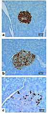
Pancreas morphometry and insulin immunohistochemistry. At 7 and 24 weeks of age, no significant difference in either the number or size of pancreatic islets was observed in either male or female hyperinsulinemic Akt2-null mice relative to control mice (data not shown). Pancreata from the hypoinsulinemic/hyperglycemic male Akt2-null mice (Figure 6a and Figure 7), however, were characterized by a variable (10–59%) decrease in the total number of islets as compared with their wild-type controls (data not shown). The vast majority of the remaining islets in this cohort of mice were distorted and contained only a few β cells scattered within the exocrine pancreas (Figure 9c). The percentage of islets containing apoptotic cells, as indicated by caspase-3 staining, was increased in pancreata from 24-week-old, hypoinsulinemic, male Akt2-null mice (37% of the remaining islets or islet remnants from hypoinsulinemic Akt2-null mice contained one or more apoptotic cells versus less than 2% of islets from control or hyperinsulinemic Akt2-null mice). Occasionally, inflammatory and necrotic cells or mitotic figures were observed within those remaining islets. Islets from wild-type mice had abundant intracytoplasmic insulin staining in β cells (Figure 9a). Insulin staining was diffuse and uniform, except for cells at the periphery of the islets, which stained positive for glucagon (data not shown). A decrease, predominantly in males, in the intensity of staining for insulin in the cytoplasm of β cells was observed in islets from Akt2-null mice (Figure 9b). The remaining β cells contained variable amounts of intracytoplasmic insulin interspersed with areas lacking immunohistochemical staining. The decreased staining for insulin in the Akt20–/– mice was already evident at 7 weeks but did not progress in incidence or severity by 24 weeks of age. The islets from the hypoinsulinemic/hyperglycemic males (Figure 6a and Figure 7) were characterized by loss of normal islet architecture and severe loss of insulin staining with only occasional, weak, intracytoplasmic staining of a few remaining cells, consistent with the very low levels of plasma insulin in these animals (Figure 9c).
Insulin immunohistochemical analysis of β cells in male Akt2-null mice. Representative pancreatic islets from (a) a 7-week-old wild-type DBA/1lacJ mouse, (b) a 7-week-old Akt2-null mouse, and (c) a 24-week-old Akt2-null mouse displaying the hypoinsulinemic/hyperglycemic phenotype (Figure 6a). The remaining β cells of the latter mouse were interspersed between the exocrine pancreatic cells and exhibited little to no cytoplasmic staining.


