HIV-1 nef suppression by virally encoded microRNA (original) (raw)
- Research
- Open access
- Published: 15 December 2004
- Masafumi Ito1,3,
- Yutaka Tsutsumi4,
- Yuko Ichikawa2,
- Harumi Okuyama2,
- Ebiamadon Andi Brisibe5,
- Nitin K Saksena6 &
- …
- Yoichi R Fujii1
Retrovirology volume 1, Article number: 44 (2004)Cite this article
- 15k Accesses
- 195 Citations
- 3 Altmetric
- Metrics details
Abstract
Background
MicroRNAs (miRNAs) are 21~25-nucleotides (nt) long and interact with mRNAs to trigger either translational repression or RNA cleavage through RNA interference (RNAi), depending on the degree of complementarity with the target mRNAs. Our recent study has shown that HIV-1 nef dsRNA from AIDS patients who are long-term non-progressors (LTNPs) inhibited the transcription of HIV-1.
Results
Here, we show the possibility that _nef_-derived miRNAs are produced in HIV-1 persistently infected cells. Furthermore, nef short hairpin RNA (shRNA) that corresponded to a predicted nef miRNA (~25 nt, miR-N367) can block HIV-1 Nef expression in vitro and the suppression by shRNA/miR-N367 would be related with low viremia in an LTNP (15-2-2). In the 15-2-2 model mice, the weight loss, which may be rendered by nef was also inhibited by shRNA/miR-N367 corresponding to suppression of nef expression in vivo.
Conclusions
These data suggest that nef/U3 miRNAs produced in HIV-1-infected cells may suppress both Nef function and HIV-1 virulence through the RNAi pathway.
Background
The human immunodeficiency virus (HIV), which infect humans cause acquired immunodeficiency syndrome (AIDS), which has reached pandemic levels in some societies, especially those in Southern Africa and Southeast Asia [1]. Given the immensity of HIV pandemic, the development of a rather safe and cheap, effective therapeutics, has become the main focus [2]. Several strategies attempted to control the spread of AIDS have not shown major breakthrough and the vaccines have shown little promise as far as their efficacy is concerned. However, one approach used extensively in other diploid organisms, which now has tremendous potential to encourage antiviral defense against HIV appears to be double stranded RNA-dependent post-transcriptional gene silencing or RNA interference (RNAi).
RNAi is a defense mechanism against aberrant transcripts that may be produced during viral infection and mobilization of transposons [3, 4]. The RNAi pathway has been implicated in silencing transposons in the C. elegans germline [5, 6], silencing stellate repeats in the Drosophila germline, and the response against invading viruses in plants [7]. Post-transcriptional regulation by RNAi is mediated by small non-coding RNAs (~25-nucleotides; nt). Small interfering RNAs (siRNAs) are short RNA duplexes that direct the degradation of homologous transcripts [8]. In contrast, the single stranded microRNAs (miRNAs) bind to 3' untranslated regions of mRNA with complementarity of 50 to 85% to give translational repression without target degradation [9]. The mature miRNA (~25-nt) is produced by processing of ~70-nt precursor stem-loop hairpin RNAs (Pre-miRNA) by Dicer [10, 11]. At the moment several human diseases, including spinal muscular atrophy (Paushkin et al., 2002), fragile X mental retardation [13, 14] and chronic lymphocytic leukemia [15] have been identified as illnesses in which miRNAs or their machinery might be implicated. However, up until now there has been no clear-cut scientific proof that establishes the exact correlation between miRNAs and human infectious diseases such as AIDS.
One of the human immunodeficiency virus type 1 (HIV-1) coding accessory genes, nef, is located at the 3' end of the viral genome and partially overlaps the 3'-long terminal repeat (LTR). The nef gene is uniquely conserved in HIV-1, HIV-2 and simian immunodeficiency virus (SIV) and is not essential but important for viral replication in vivo [16]. The nef gene is expressed during HIV infection and often accounts for up to 80% of HIV-1 specific RNA transcripts during the early stages of viral replication [2]. Our own investigations have shown that defective variants of nef dsRNA containing the 3'-LTR regions, obtained from long-term non-progressor (LTNP) AIDS patients, actually inhibited the transcription of HIV-1 [17]. Furthermore, _cis_-expression of mutated F12-HIV-1 nef inhibits replication of highly productive NL43-HIV-1 strain, which is not related to down-regulation of CD4 [18, 19]. It has been demonstrated that F12 nef gene cloned from the provirus of naturally occurring HUT-78 T cells infected with the supernatant of the peripheral blood mononuclear cells (PBMCs) of an HIV-seropositive non-producer patient, induces a block of viral replication [19]. Thus, it has been suggested that nef RNAs may be a _cis_-regulatory factor for HIV-1 replication [20].
In the current study, we have established the link between miRNAs and HIV infections by demonstrating that _nef_-derived miRNAs are produced in HIV-1-infected cells. The results presented here show that nef short hairpin RNAs (shRNAs) corresponding to the nef miRNAs efficiently block RNA stability or mRNA translation, perhaps an indication that HIV-1 regulates its own replication by using nef miRNAs.
Results and Discussion
Identification of a candidate of miRNAs in HIV-1-infected cells
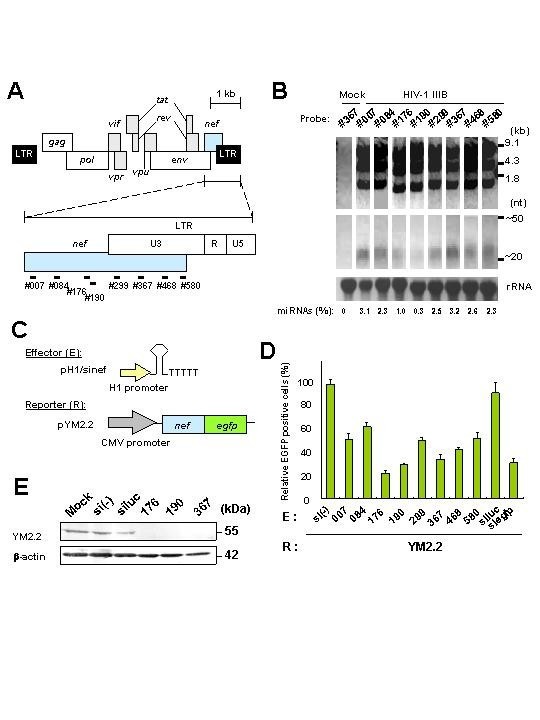
Very recently, the Epstein-Barr virus (EBV)-encoded miRNAs were identified. Thus, during the preliminary stages of this study, our curiosity was fixed on the need to find out if indeed there was any relationship between nef miRNAs and HIV-1-infected cells. To achieve this purpose, we extracted total RNA from HIV-1 IIIB strain persistently infected MT-4 T cells and northern blot analysis was performed using eight probes against the nef coding region, as shown in Figure 1A. Analyses using several anti-sense probes, small RNA molecules approximately ~25-nt in size were detected as well as HIV-1 major transcripts, 9.1, 4.3 and 1.8 kb bands (Fig. 1B). Similar results were obtained with total RNA from HIV-1 SF2 strain infected MT-4 T cells (data not shown). RNA samples treated with a mixture of the single stranded specific RNases A and T1 also generated ~25-nt RNAs that hybridized in northern blots with the sense probes against the same nef region. However HIV-1 major transcripts were not detected (data not shown), indicating that the structure of the small RNA molecules could be double-stranded RNAs (miRNAs). Some variability was observed when the quantity of the miRNAs was compared with the total of the major transcripts. A maximum of 3.2% of miRNAs was detected by using #367 probe when compared with total HIV-1 transcripts, and the minimum of 0.3% was detected by using probe# 90 (Fig. 1B).
Figure 1
Detection of HIV-1 nef miRNAs and inhibition of Nef expression by nef RNAs. (A) Schematic representation of HIV-1 nef in its genome and synthetic DNA probes (#) used in this study. (B) Total RNAs were extracted from MT-4 T cells persistently infected with HIV-1 IIIB, separated on a 15% polyacrylamide-7 M urea gel, and subjected to northern blot analysis. The approximate sizes of the three classes of HIV-1 transcripts and small RNAs are indicated on the right. The loading control was rRNA stained with ethidium bromide. Relative expression (%) of nef small RNAs to the three classes of HIV-1 transcripts is at the bottom of figure. (C) Schematic representation of effector plasmids (E) H1 promoter-driven shRNA expression plasmids. Reporter (R) Nef-EGFP expression plasmid (pYM2.2) is also shown. (D) Inhibition by sinefs in pH1 plamids of Nef-EGFP expression. Either sinef, siluc or siegfp in each plasmid was transfected into Jurkat T cells in the presence of either pYM2.2 or control pEGFP-N1. At 36 h after transfection EGFP-positive cells were counted by flow cytometry. Data represent the relative activity of EGFP-positive cells where the percentage of positive cells in the sample transfected with pYM2.2 or pEGFP-N1 plus pcDNA3.1 or si(-) in pH1 plasmid was scored as 100%. Data are averages of three independent experiments + SD. Bars, SD. (E) Immunoblot analysis showing inhibition of YM2.2 expression by different nef shRNAs. Jurakat T cells were transfected with pYM2.2 and pH1/sinefs or siluc plasmid, cellular lysates were prepared 48 h after transfection, and immunoblotted with rabbit serum against Nef (upper panel) and anti-β-actin antibody (lower panel). The β-actin expression shows equal loading of all samples.
To randomly clone the nef miRNA, ~25-nt RNAs were gel purified, cloned and sequenced. The sequences from the nef miRNA clones were 5'-acugaccuuuggauggugcuucaa-3' or similar ones, corresponded to the nucleotides approximately 420 to 443 conserved region of nef (miR-N367). The most notable feature of this analysis is that it has proven beyond reasonable doubts that _nef_-derived miRNAs are produced in HIV-1 infected cells.
Inhibition of Nef by plasmids-encoding siRNA/miRNA
To examine inhibition of nef expression by the nef miRNA, we constructed eight shRNAs homologous to the native miRNA or probes used in Figure 1A[21]. Although it has been reported that three to four mutations in the sense strand derived from miRNA could have the potential to control unmutated 21-nt stem loop [22], we investigated whether the native shRNA-expressing plasmid can effectively reduce nef gene expression or not (Fig. 1C and 1D). We used egfp or luc gene (pH1/siegfp or luc) as a positive or negative control. All the shRNA-expressing plasmids including the controls were co-transfected into Jurkat T cells with pYM2.2 and cell fluorescence resulting from the expression of EGFP reporter gene was quantified by flow cytometry. The sinef176, 190, 367/miR-N367 and control siegfp all showed efficient reduction, but the sinef 007, 084, 299, 468 and 580 constructs gave only modest reductions, and no suppression was observed with si(-) and luc (Fig. 1D and Table 1). Immunoblot analysis using anti-Nef rabbit serum also confirmed the inhibition of Nef-EGFP expression by sinef 176, 190 and 367/miR-N367 (Fig. 1E).
Table 1 Relation between AIDS clinical courses and nef dsRNA or siRNA/miRNA in suppression of nef gene expression
Inhibition of Nef expression by STYLE vector-encoding nef siRNA/miR-N367
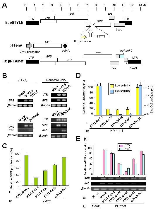
To assess the effects of nef miR-N367 in vivo, we constructed a prototype foamy virus (PFV)-based live vector. The PFV vector expressed HIV-1 SF2 nef gene as a reporter and the STYLE vector expressed shRNA as effectors. The full-length nef gene was inserted into the bel-2 portion of a PFV clone (22) in frame to obtain pPFV/nef (Fig. 2A). The pPFV/nef was transfected into BHK cells and treated with the histone deacetylase inhibitor, trichostatin A (TA) [23]. The viral supernatant, which contained approximately 5 × 10⁶ infectious units (IFU), was collected at 72 h after transfection. For preparation of the STYLE vectors to deliver the shRNAs, the env gene portion of pSKY3.0 was replaced with the shRNA expression cassette under the control of the H1 promoter. The pSTYLE was produced (Fig. 2A), and transfected into the FFV envelope-expressing packaging cells, CRFK sugi clone # 6, in the presence of TA. The transfected CRFKsugi clone #6 had 99% FFV Env positive cells when analyzed by flow cytometry. The viral supernatant with a titer of ca. 1 × 10⁵ IFU was collected at 72 h post-transfection.
Figure 2
Inhibition of nef expression in human T cells by nef siRNAs. (A) Schematic representation of shRNA-expressing STYLE vector (E) and HIV-1 SF2 nef gene expressing pPFV/nef vector (R). The helper plasmid, pFFenv expresses FFV envelope protein under the control of CMV promoter. (B) Detection of expression of nef mRNA and integration of vectors. STYLE, SKY3.0, PFV/nef, PFV or mock was used to infect Jurkat T cells and the infected cells were cultured for 2 weeks. After 2 weeks, gag and nef mRNA expression was measured by RT-PCR. Genomic DNA of LTR, gag and nef of STYLE or PFV/nef were also detected by PCR. β-actin was used as a control. (C) Inhibition of Nef-EGFP expression by nef siRNA-expressing STYLE in Jurkat T cells. The pYM2.2 was transfected into each of the STYLE or mock-infected Jurkat T cells and EGFP-expressing cells were counted by flow cytometry at 48 h after transfection. Data represent the relative activity of EGFP-positive cells, where the percentage of positive cells in the sample transfected with pYM2.2 upon the STYLE-si(-) infected cells was scored as 100%. (D) Inhibition of HIV-1 transcription and replication by nef STYLE-367. HIV-1 IIIB persistently infected MT-4 T cells were transfected with the pLTRSF2 reporter and β-gal expressing control pCMVβ plasmids at 72 h after infection with STYLE. At 48 h post-transfection, Luc activity was measured and normalized as Luc values (Luc/β-gal). Absolute levels of Luc activity in the samples of pLTRSF2 plus SRYLE-si(-) were 16,311 + 1,253 or 783 + 87 light units for STYLE-367/miR-N367 transfectants. Data represent the relative Luc activities where the percentage of positive cells in the samples infected with the STYLE-si(-) was scored as 100%. After 48 h, p24 antigen was also measured in the cell culture supernatant of STYLE-infected Jurkat T cells. Data are averages of three independent experiments + SD. Bars, SD. (E) Inhibition of nef expression by nef siRNA in Jurkat T cells. Cells were infected with PFV/nef 48 h after infection with the STYLE and then subjected to semi-quantitative RT-PCR analysis. Data represent the relative expression of mRNA, where the percentage of positive cells in the sample of mock-infected cells (E: Mock) relative to the PFV/nef (R: PFV/nef) infected cells was scored as 100%. Data averages were derived from three independent experiments + SD. Bars, SD.
The expression of viral mRNAs and integrated DNAs from either the PFV/nef or STYLE vectors was confirmed by infection of Jurkat T cells. The mRNAs and genomic DNA were extracted from the infected cells at 2 weeks post-infection. The PFV/nef-expressed gag and nef mRNAs and the STYLE-expressed gag mRNA were detected after amplification of these regions using reverse transcription (RT)-PCR. The integration of the DNAs into the genome of Jurkat T cells was also confirmed by PCR of the LTR, gag and/or nef regions (Fig. 2B). The control SKY3.0 and PFV-infected cells were both negative for nef mRNA and integrated DNA (Fig. 2B). The integrated DNA was also detected by southern blot analysis with genomic DNA of either PFV/nef or STYLE-infected cells (data not shown). Expression of shRNAs (~22-nt) was also confirmed in STYLE-infected cells by northern blot analysis (data not shown). Expression of Nef protein in PFV/nef-infected cells was also detected with specific rabbit anti-Nef serum in immunoblots (data not shown).
To evaluate whether the STYLE encoding siRNA could inhibit the expression of the nef gene in cultured human T cells, pYM2.2 was transfected into each of the STYLE-infected Jurkat T cells (m.o.i. = ca. 0.1). The most efficient sinef176, 190 and 367/miR-N367 vectors for reduction in nef expression (Fig. 1D and 1E) were selected for this experiment. The EGFP-positive cells were counted by flow cytometry at 48 h after transfection. Expression of Nef-EGFP fusion protein was reduced drastically following treatment with either the STYLE-176 (74 + 3.2) or 190 (51 + 4.2) and also reduced with 367/miR-N367 (32 + 2.3%). Reduction was insignificant with either the STYLE-si(-) (0 + 0.7) or STYLE-luc (7 + 0.9%) controls (Fig. 2C).
The in vitro inhibitory effects of STYLE encoding nef siRNA on HIV-1-infected cells were evaluated in Luc assays and using MT-4 T cells persistently infected with HIV-1 IIIB. Cultivation of the STYLE infected cells for 72 h followed by transfection with the pLTRSF2 and culture for another 48 h showed that STYLE-176, 190 and 367/miR-N367 all significantly (p < 0.005) suppressed Luc activity when compared to controls (Fig. 2D). HIV-1 p24 Gag was also significantly inhibited in the culture supernatant by infection with STYLE-176, 190 and 367/miR-N367 when compared to controls (p < 0.001) (Fig. 2D). These data suggested that shRNA/miR-N367 could inhibit HIV-1 transcription and replication in intact HIV-1-infected human T cells.
Jurkat T cells that had been transduced with nef shRNA for 48 h were infected with the PFV/nef. Semi-quantitative RT-PCR analysis revealed that while treatment with STYLE-190 dramatically reduced the expression of both nef and gag mRNAs of the PFV/nef, the expression of nef mRNA was also drastically suppressed by STYLE-176 and 367/miR-N367 (Fig. 2E). However the STYLE-si(-) and luc controls showed ~10% suppression of nef and ~20% suppression of gag mRNAs (Fig. 2E), which was probably a result of interference following super-infection. Nonetheless, both nef transcription and PFV/nef replication were substantially inhibited by STYLE-176, 190 and 367/miR-N367.
Inhibition of Nef expression by siRNA/miR-N367 in mice
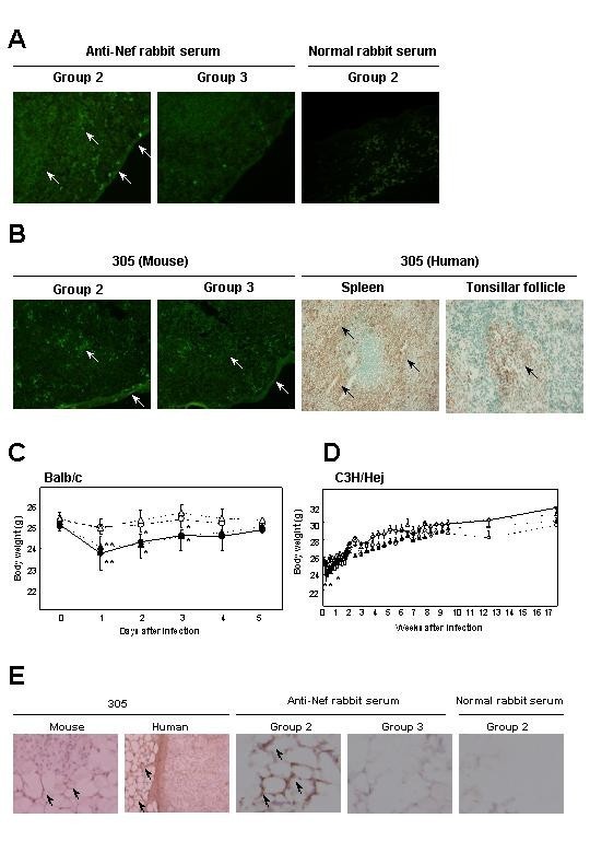
Since different host gene products are required for siRNA-mediated RNAi and miRNA-mediated translational repression with let-7 and lin-4 in C. elegans, the two RNAs may not have the same functions in vivo [24]. To test this point, we investigated the efficacy of miR-N367 using STYLE-367 in mammalian tissues. The study mice were group 1 = PFV/nef-infected (n = 6); group 2 = PFV/nef and control STYLE-luc infected (n = 6); group 3 = PFV/nef and STYLE-367-infected (n = 8); and group 4 = STYLE-367-infected (n = 6). Identical study groups were used for both Balb/c and C3H/Hej mouse strains. Nef protein expressing lymphocytes were quantified by histochemical analysis using F3 Nef monoclonal antibody (mAb) or anti-Nef rabbit serum 2 days after PFV/nef infection. Nef protein was detected by immunofluoresence assay in the subcapsular area of the spleens of groups 1 or 2 Balb/c mice, but not groups 3 or 4 (Fig. 3A and Table 2). No positive cell staining was observed using normal rabbit serum as a primary antibody (Fig. 3A). To test the expression of nef, nested RT-PCR was also done on day 2 to evaluate the degree of nef mRNA expression in the spleen, liver, adipose tissues and hematopoietic cells in groups 1–4. The nef mRNA was significantly expressed in liver and hematopoietic cells of Balb/c mice in groups 1 and 2, but not in the group 3 animals that were STYLE-367 infected (Table 2). Tissues from group 4 did not show any nef bands after RT-PCR (data not shown).
Figure 3
In vivo effects of miR-N367. (A) Distribution of Nef positive staining cells in the subcapsular area of groups 2 or 3 mouse spleens at 2 days after infection with PFV/nef. Anti-Nef rabbit serum or normal rabbit serum was used as a primary antibody. (B) Immunofluorescence for 305 mAb positive staining cells in the subcapsular area of groups 2 or 3 mouse spleens at 2 days after infection with PFV/nef and immunoperoxidase staining by 305 mAb in cells of interfollicular area of HIV-1 uninfected human spleen and tonsillar follicle. (C) Short term body weights of PFV/nef-infected Balb/c mice. The body weights of the PFV/nef-infected mice (group 1, n = 6, solid circle), the PFV/nef-infected followed by the STYLE-luc-infected mice (group 2, n = 6, solid triangle), the PFV/nef-infected followed by the STYLE-367-infected mice (group 3, n = 8, open triangle) and the STYLE-367-infected mice (group 4, n = 6, open circles) were measured from days 0 to 5. (D) Long term body weights of PFV/nef-infected C3H/Hej mice. Treatment of each group and numbers of mice were same as (C). Bars, SD. *; p < 0.05, **; p < 0.01 (relative to group 3). (E) Immunoperoxidase staining by 305 mAb and anti-Nef rabbit serum in cells of mouse or human adipose tissue. Arrows show positively stained areas. Magnification, X 20 (A and B); X 20 and X 200 (E).
Table 2 Histochemical detection and RT-PCR amplification of Nef from human and mouse tissues
Because extracellular Nef is internalized into human and mouse lymphocytes and macrophages [25–27], we examined putative Nef receptor molecule (Ner) expression with 305 mAb [27] in both mouse and human tissues by histochemical analysis. In mice, 305 mAb positive lymphocytes were detected in the subcapsular area of the spleens by immunofluoresence assay (Fig. 3B) and liver and hematopoietic cells (Table 2) in groups 1, 2 and 3, indicating that detection of antigen by 305 mAb was not altered by Nef expression. In HIV-1 uninfected humans, the 305 mAb positive cells were detected by immunoperoxidase staining in spleen (red pulp), tonsillar follicle (germinal center), liver (Kupffer cells), salivary gland (germinal center and adipose cells), bronchi (smooth muscle cells), lung (stroma cells), thyroid gland (colloid), heart muscle (smooth muscle cells), prostate gland (smooth muscle cells), colon mucosa (intestinal absorptive and muscle cells), testis (basement membrane of tubuli seminiferi), adrenal gland (adipose cells), and brain (cerebrum cortex and cortical cells) (Fig. 3B and Table 2).
Since Nef suppressed PPARγ expression and reduced fatty acid levels in vitro [29–32], we monitored the expression of PPARγ mRNA and body weights of mice. Significant PPARγ mRNA expression in intestinal adipose tissue of group 3, but not group 1 and 2, was detected on day 2 (Table 2). All Balb/c mice in group 1 showed sedation and a drastic loss of weight from days 1 to 3 (day 1, p = 0.003; day 2, p = 0.021; day 3, p = 0.032 relative to mice in group 3) (Fig. 3C). Similar results were obtained in group 2 (Fig. 3C). However, group 3 mice infected with STYLE-367 did not appear to be sedated and had no drastic loss of weight (Fig. 3C). The group 4 animals, which were not infected with the PFV/nef but treated with STYLE-367, had no changes in either behavior or weight (Fig. 3C). In longitudinal examinations done during the post-infection period, the animals in groups 1 and 2 had recovered the lost weight (Fig. 3D). Similar results were obtained in group 2 from day 1 to 5 (day 1, p = 0.037; day 3, p = 0.044; day 5, p = 0.048 relative to mice in group 3) in the C3H/Hej mouse groups (Fig. 3D). To assess the above in vivo results, expression of nef mRNA was examined in adipose tissues (Table 2). As shown in Table 2, although mRNAs of nef and gag were not detected in mouse adipose tissues, 305 mAb and anti-Nef rabbit serum positive staining cells were detected in mouse group 1 and 2 adipose tissues (Fig. 3E and Table 2). Considering that the 305 mAb positive staining adipocytes appeared in mouse as well as human tissues (Fig. 3E and Table 2), these data suggest that the interaction between 305 and soluble Nef detected in adipose tissues may be responsible for the weight loss observed in mice.
In this study, whereas siRNA has been reported to inhibit hepatitis B virus replication in vivo (33–34), our results show that nef_-derived miRNAs are produced in HIV-1 infected cells, and support the possibility that miRNA and siRNA may be functionally identical, at least in a retrotransposon such as HIV. Recent studies have revealed that miRNAs and siRNAs could block mRNA expression by similar mechanisms [9] and that siRNAs could function as miRNAs [35] and EBV-encoded miRNAs were found [36]. Our results reported here are consistent with these previous observations and are suggestive of the fact that nef miR-N367 could regulate nef expression even in vivo. In our unpublished data, HIV-1 LTR promoter activity was inhibited by miR-N367 (nt number 379 to 449 of SF2 nef, 71-nt) expression, of which activity was dependent on negative responsive element (NRE) of U3 region (our unpublished data). Although no mismatch shRNA against region #367 was active, the miR-N367 from HIV-1 genome may have some mismatches and effectively inhibit HIV-1 transcription. Further the effects of siRNAs of Tc_1, in particular those to the terminal inverted repeats derived from read-through transcription of entire transposable elements, were presented for silencing transposase gene expression by RNAi machinery in germ lines of C. elegans, [37]. Taken together, it could be implied from these and our other results that miRNAs produced in HIV-1-infected cells can efficiently block not only Nef function but also HIV-1 replication through RNAi, which renders persistently low pathogenic infection latent as observed in an LTNP of 15-2-2 (see Table 1). It is equally important that although the weight loss reported here occurred only temporarily in vivo, however the inserted nef gene in the foamy retrotransposon may represent miRNAs which could inhibit nef mRNA expression by presumably an identical mechanism to that observed of siRNAs. Thus, RNAi might serve as a new sequence-specific therapeutic arsenal in AIDS prevention and possibly treatment.
Overall, our results indicate that nef shRNA transduced into T cell line inhibited HIV-1 transcription. Further, nef miRNAs could be produced from infected T cells and can block the trans-activity of Nef as well as HIV-1 replication on its own via the cis-action of nef. These functions of nef via RNAi pathways may allow persistently low pathogenic or latent infection as observed in HIV-infected non-progressors. Cumulatively, these data suggest that Nef may be involved in both viral replication and the disease progression, the findings, which may facilitate new strategies for HIV control in vivo.
Materials and Methods
Patient details
Patient selection is showed in Table 1. These SF2 (HIV-1 subtype B prototype) was included as a control nef sequence, because of the inclusion of viruses, which were also subtype B. The SF2 contained full-length nef reading frame as indicated in Table 1. Patients 1-3-3, 4-2-1 (Table 1) are rapid progressors infected with HIV-1. These patients were infected in 1984–1985 and died within 3 years of primary infection with >1 × 10⁶ viral copies and CD4+ T cell count of 75 and 110/ml blood. Patients 15-2-2 and 16-1-1 (Table 1) are slow progressors, who were infected in 1984 and have survived HIV-1 infection with high and stable CD4+ T cell counts (690 and 760/ml blood) with low (<5000 copies) plasma viremia. All these patients acquired virus through homosexual sex. JW95-1 (Table 1) is a boy who was infected from his mother via breast feeding. The child was infected in 1983 and has survived disease free with high CD4+ T cell count (890/ml blood) with undetectable viremia. Human samples were obtained from a donor after informed consent.
Cells and viruses
HeLa and BHK cells were grown in Dulbecco's modified Eagle Medium (DMEM) (GIBCO, Grand Island, NY) supplemented with 10% heat-inactivated fetal bovine serum (FBS) and antibiotics. CRFK cells were grown in Iscove's Modified Dulbecco's Medium (IMDM) (GIBCO) with 10% FBS and antibiotics. Jurkat T cells and MT-4 T cells persistently infected with HIV-1 IIIB strain were cultured in RPMI-1640 medium (GIBCO) supplemented with 10% FBS and antibiotics. The packaging cells (CRFKsugi) were made by transfecting CRFK cells with 10 μg of the pFFenv with Lipofectin Reagent (Invitrogen) and selecting transformants after culture for 14 days with 25 μg/ml of hygromycine B (Invitrogen). After 14 days, FFV Env protein expression was measured by flow cytometry and immunoblot analyses with FFV-infected cat B serum [34]. The pPFV/nef (10 μg) was transfected into BHK cells and pSTYLE/si (10 μg) was transfected into CRFKsugi cells with Lipofectin Reagent. The transfected cells were cultured for 72 hr, and the viral supernatant was collected and filtered through a 0.45 μm pore size Millex-GP filter (Millipore, Bedford, MA). Vector stocks were stored at -70°C prior to use. Viral titers were determined as described previously [21]. Cells were infected with PFV/nef and/or SKY/si at an m.o.i. of ca. 0.1 in the presence of 4 μg/ml of polybrene and infected cells were cultured at a density of 1 × 10⁶ cells per ml for 3 days.
The details of plasmid constructs and the primer sequences used in cloning strategies are shown in supplementary file (see Additional file: 1).
Flow cytometric analysis
Flow cytometry was performed with a FACS Calibur (Becton Dickson, San Jose, CA) as described previously [17].
Luc assay and Immunoblotting
Firefly Luc assay was performed using the Luciferase Assay System (Promega) as described previously [17]. Immunoblotting was performed essentially as described previously by Otake et al. [28].
P24 ELISA
The concentration of p24 supernatant was determined by an antigen capture assay (Beckman Coulter, Fullerton, CA) according to the manufacturer's instructions.
Confocal laser microscopy analysis
Confocal laser microscopy analysis was performed as described previously [28].
Northern blot analysis
Total RNAs were extracted from HIV-1 IIIB or SF2 persistently infected or uninfected MT-4 T cells using TRIzol reagent (Invitrogen). Approximately 40 μg of total RNA was treated with RNase A and T1 (Sigma, St. Louis, MO) as described previously [17], subjected to electrophoresis on a 15% polyacrylamide-7 M urea gel and electroblotted to HybondN+ (Pharmacia, Uppsala, Sweden) for 4 hr at 400 mA. RNAs were immobilized by UV crosslinking and baking for 1 hr at 80°C. Hybridization was done with an ECL direct Kit (Pharmacia). Synthetic DNA probes were labeled with horseradish peroxidase. The sequence for synthetic sense DNA probes for northern blot analysis are as follows: #007 (5'-gcgtcgacggcaagtggtcaaaacgta-3'); #084 (5'-gcgtcgacgccagcagcagatggggtg-3'); #176 (5'-gcgtcgacgtgcctggctagaagcaca-3'); #190 (5'-gcgtcgacgcacaagaggaggaga-3'); #299 (5'-gcgtcgacgactggaagggctaatttg-3'); #367 (5'-gctcgacggctacttccctgattggc-3'); #468 (5'-gcgtcgacggtagaagaggccaatgaa-3'); #580 (5'-gcgtcgacgcatttcatcacatggccc-3'). RNAs were cloned by 5'RACE System (Invitrogen, CA., USA) with a slight modification in that primers were used that were the same as the synthetic DNA probes as described above that are abbreviated as #primer. In brief, gel purified small RNAs were annealed with #primer and first strand cDNA was synthesized with SuperScript II RT (Invitrogen, CA., USA). Afrer RNase H and T1 treatment, a homopolymeric tail was added to the 3'-end of the cDNA using terminal deoxynucleotidyl transferase and dCTP. After ethanol precipitation, PCR amplification was done with abridged anchor primer and #primer. Then the PCR products were obtained using abridged universal amplification primer and #primer. The PCR fragments were digested with Sal I and cloned into Sal I site of pBluescript SK(-), followed by sequence analysis. The secondary structures of RNAs were predicted by GENETYX-MAC program (Software Development Co. Ltd, Tokyo, Japan).
Semi-quantitative RT-PCR analysis
Semi-quantitative RT-PCR analysis was performed using the ThermoScript RT-PCR System (Invitrogen, CA., USA) according to the manufacturer's protocol with the following primers: III (5'-atcatgggccaaagagaattc-3') and IV (5'-aaatttcactcaatcgagcc-3') for FFV LTR, VI (5'-aggacctgaaaggcatg-3') and VII (5'-ttgttgagatcgtcccg-3') for FFV gag, VIII (5'-tgtggtggaatgccactag-3') and IX (5'-attgtcatggaattttgta-3') for PFV LTR, XI (5'-tcttacagaccagtaacaa-3') and XII (5'-gtcaatcattacatctgca-3') for PFV gag, XIII (5'-aactactagtacccttcagg-3') and XIV (5'-aaaactcttgctttatggcc-3') for HIV-1 gag, XV (5'-atgggtggcaagtcaaaacg-3') and XVI (5'-tcagcagtctttgtagtactccg-3') for HIV-1 nef, XVII (5'-gttatgggtgaaactctgggagat-3') and XVIII (5'-atgttcctgaacataatcgtc-3') for PPARγ, XIX (5'-gacaacggctccggcatgtgcaaag-3') and XX (5'-ttcacggttggccttagggttcag-3') for β-actin, respectively. The nested PCR followed RT reaction was performed as described previously [34]. PCR products were quantified with the NIH image program. Relative mRNA expression was calculated as percentage expression using the following formula: integrating number of nef or gag bands/integrating number of β-actin X 100.
In vivo studies and tissue analyses
Balb/c and C3H/Hej mice were raised under specific pathogen-free (SPF) conditions. Mice were infected with 1 ml of 105 IFU of PFV/nef and STYLE-367 by intravenous (i.v.) injection. RT-PCR analyses were performed 2 days after infection. For histological analysis, cryostat sections were prepared from both human and mouse tissues. The fixed sections were rinsed with PBS and incubated with 5% BSA for at least 1 hr to inhibit nonspecific binding of antibodies. Sections were incubated overnight at 4°C with anti-Nef rabbit serum, F3 or 305 mAb, and incubated with peroxidase or FITC conjugated secondary antibodies. The washed sections were incubated in 0.03% 3,3'diaminobenzidine (Sigma) solution in 0.05 M Tris buffer with 0.01% H2O2 for development of peroxidase activity. After counterstaining with hematoxylin or methylgreen, the sections were dehydrated and mounted.
Statistical methods
Data were analysed using a one-way ANOVA analysis with a post-hoc Fisher's test. P values of 0.05 or more were determined for that of cut off.
Abbreviations
HIV-1:
human immunodeficiency virus type 1
miRNA:
microRNA
nt:
nucleotides
LTNP:
long-term non-progressors
shRNA:
short hairpin RNA
AIDS:
acquired immunodeficiency syndrome
RNAi:
RNA interference
siRNA:
small interfering RNA
LTR:
long terminal repeat
SIV:
simian immunodeficiency virus
PBMCs:
peripheral blood mononuclear cells
EBV:
Epstein-Barr virus
EGFP:
enhanced green fluorescence protein
PFV:
prototype foamy virus
TA:
trichostatin
A IFU:
infectious units
FFV:
feline foamy virus
RT:
reverse transcription
m.o.i.:
multiplicity of infectionm
Ab:
monoclonal antibody
Ner:
Nef receptor molecule
NRE:
negative responsive element.
References
- Weidle PJ, Mastro TD, Grant AD, Nkengasong J, Macharia D: HIV/AIDS treatment and HIV vaccines for Africa. Lancet. 2002, 359: 2261-2267. 10.1016/S0140-6736(02)09297-8.
Article PubMed Google Scholar - Brisibe EA, Okada N, Mizukami H, Okuyama H, Fujii YR: RNA interference: potentials for the prevention of HIV infections and the challenges ahead. Trends Biotechnol. 2003, 21: 306-311. 10.1016/S0167-7799(03)00118-5.
Article CAS PubMed Google Scholar - Fire A, Xu S, Montgomery MK, Kostas SA, Driver SE, Mello CC: Potent and specific genetic interference by double-stranded RNA in Caenorhabditis elegans. Nature. 1998, 391: 806-811. 10.1038/35888.
Article CAS PubMed Google Scholar - Tuschl T, Zamore PD, Lehmann R, Bartel DP, Sharp PA: Targeted mRNA degradation by double-stranded RNA in vitro. Genes Dev. 1999, 13: 3191-3197. 10.1101/gad.13.24.3191.
Article PubMed Central CAS PubMed Google Scholar - Ketting RF, Haverkamp TH, van Luenen HG, Plasterk RH: mut-7 of C. elegans, required for transposon silencing and RNA interference, is a homolog of Werner syndrome helicase and RNaseD. Cell. 1999, 99: 133-141. 10.1016/S0092-8674(00)81645-1.
Article CAS PubMed Google Scholar - Tabara H, Sarkissian M, Kelly WG, Fleenor J, Grishok A, Timmons L, Fire A, Mello CC: The rde-1 gene, RNA interference, and transposon silencing in C. elegans. Cell. 1999, 99: 123-132. 10.1016/S0092-8674(00)81644-X.
Article CAS PubMed Google Scholar - Aravin AA, Naumova NM, Tulin AV, Vagin VV, Rozovsky YM, Gvozdev VA: Double-stranded RNA-mediated silencing of genomic tandem repeats and transposable elements in the D. melanogaster germline. Curr Biol. 2001, 11: 1017-1027. 10.1016/S0960-9822(01)00299-8.
Article CAS PubMed Google Scholar - Elbashir SM, Lendeckel W, Tuschl T: RNA interference is mediated by 21- and 22-nucleotide RNAs. Gene Dev. 2001, 15: 188-200. 10.1101/gad.862301.
Article PubMed Central CAS PubMed Google Scholar - Zeng Y, Yi R, Cullen BR: MicroRNAs and small interfering RNAs can inhibit mRNA expression by similar mechanisms. Proc Natl Acad Sci U S A. 2003, 100: 9779-9784. 10.1073/pnas.1630797100.
Article PubMed Central CAS PubMed Google Scholar - Lee Y, Ahn C, Han J, Choi H, Kim J, Yim J, Lee J, Provost P, Radmark O, Kim S, Kim VN: The nuclear RNase III Drosha initiates microRNA processing. Nature. 2003, 425: 415-419. 10.1038/nature01957.
Article CAS PubMed Google Scholar - Lee Y, Jeon K, Lee JT, Kim S, Kim VN: MicroRNA maturation: stepwise processing and subcellular localization. EMBO J. 2002, 21: 4663-4670. 10.1093/emboj/cdf476.
Article PubMed Central CAS PubMed Google Scholar - Paushkin S, Gubitz AK, Massenet S, Dreyfuss G: The SMN complex, an assemblyosome of ribonucleoproteins. Curr Opin Cell Biol. 2002, 14: 305-312. 10.1016/S0955-0674(02)00332-0.
Article CAS PubMed Google Scholar - Caudy AA, Myers M, Hannon GJ, Hammond SM: Fragile X-related protein and VIG associate with the RNA interference machinery. Genes Dev. 2002, 16: 2491-2496. 10.1101/gad.1025202.
Article PubMed Central CAS PubMed Google Scholar - Ishizuka A, Siomi MC, Siomi H: A Drosophila fragile X protein interacts with components of RNAi and ribosomal proteins. Genes Dev. 2002, 16: 2497-2508. 10.1101/gad.1022002.
Article PubMed Central CAS PubMed Google Scholar - Calin GA, Dumitru CD, Shimizu M, Bichi R, Zupo S, Noch E, Aldler H, Rattan S, Keating M, Rai K, Rassenti L, Kipps T, Negrini M, Bullrich F, Croce CM: Frequent deletions and down-regulation of micro-RNA genes miR15 and miR16 at 13q14 in chronic lymphocytic leukemia. Proc Natl Acad Sci U S A. 2002, 99: 15524-15529. 10.1073/pnas.242606799.
Article PubMed Central CAS PubMed Google Scholar - Kestler HW, Ringler DJ, Mori K, Panicali DL, Schgal PK, Daniel MD, Desrosiers RC: Importance of the nef gene for maintenance of high virus loads and for development of AIDS. Cell. 1991, 65: 651-662. 10.1016/0092-8674(91)90097-I.
Article CAS PubMed Google Scholar - Yamamoto T, Omoto S, Mizuguchi M, Mizukami H, Okuyama H, Okada N, Saksena NK, Brisibe EA, Otake K, Fujii YR: Double-stranded nef RNA interferes with human immunodeficiency virus type 1 replication. Microbiol Immunol. 2002, 46: 809-817.
Article CAS PubMed Google Scholar - D'Aloja PE, Olivetta R, Bona F, Nappi D, Pedacchia K, Pugliese G, Ferrari P, Verani P, Federico M: gag, vif, and nef genes contribute to the homologous viral interference induced by a nonproducer human immunodeficiency virus type 1 (HIV-1) variant: identification of novel HIV-1-inhibiting viral protein mutants. J Virol. 1998, 72: 4308-4319.
PubMed Central PubMed Google Scholar - Olivetta E, Pugliese K, Bona R, D'Aloja P, Ferrantelli F, Santarcangelo AC, Mattia G, Verani P, Federico M: cis expression of the F12 human immunodeficiency virus (HIV) Nef allele transforms the highly productive NL4-3 HIV type 1 to a replication-defective strain: involvement of both Env gp41 and CD4 intracytoplasmic tails. J Virol. 2000, 74: 483-492.
Article PubMed Central CAS PubMed Google Scholar - Jacque JM, Triques K, Stevenson M: Modulation of HIV-1 replication by RNA interference. Nature. 2002, 418: 435-438. 10.1038/nature00896.
Article CAS PubMed Google Scholar - Das AT, Brummelkamp TR, Westerhout EM, Vink M, Madiredjo M, Bernards R, Berkhout B: Human immunodeficiency virus type 1 escapes from RNA interference-mediated inhibition. J Virol. 2004, 78: 2601-2605. 10.1128/JVI.78.5.2601-2605.2004.
Article PubMed Central CAS PubMed Google Scholar - Miyagishi M., Sumimoto H, Miyoshi H, Kawakami Y, Taira K: Optimization of an siRNA-expression system with an improved hairpin and its significant suppressive effects in mammalian cells. J Gene Med. 2004, 6: 715-723. 10.1002/jgm.556.
Article CAS PubMed Google Scholar - Fujii Y, Murase Y, Otake K, Yokota Y, Omoto S, Hayashi H, Okada H, Okada N, Kawai M, Okuyama H, Imakawa K: A potential live vector, foamy virus, directed intra-cellular expression of ovine interferon-τ exhibited the resistance to HIV infection. J Vet Med Sci. 2004, 66: 115-121. 10.1292/jvms.66.115.
Article CAS PubMed Google Scholar - Hatama S, Otake K, Ohta M, Kobayashi M, Imakawa K, Ikemoto A, Okuyama H, Mochizuki M, Miyazawa T, Tohya Y, Fujii Y, Takahashi E: Reactivation of feline foamy virus from a chronically infected feline renal cell line by trichostatin A. Virology. 2001, 283: 315-323. 10.1006/viro.2000.0861.
Article CAS PubMed Google Scholar - Grishok A, Pasquinelli AE, Conte D, Li N, Parrish S, Ha I, Baillie DL, Fire A, Ruvkun G, Mello CC: Genes and mechanisms related to RNA interference regulate expression of the small temporal RNAs that control C. elegans developmental timing. Cell. 2001, 106: 23-34. 10.1016/S0092-8674(01)00431-7.
Article CAS PubMed Google Scholar - Okada H, Takei R, Tashiro M: Nef protein of HIV-1 induces apoptotic cytolysis of murine lymphoid cells independently of CD95 (Fas) and its suppression by serine/threonine protein kinase inhibitors. FEBS Lett. 1997, 417: 61-64. 10.1016/S0014-5793(97)01255-6.
Article CAS PubMed Google Scholar - Quaranta MG, Camponeschi B, Straface E, Malorini W, Viora M: Induction of interleukin-15 production by HIV-1 Nef protein: a role in the proliferation of uninfected cells. Exp Cell Res. 1999, 250: 112-121. 10.1006/excr.1999.4494.
Article CAS PubMed Google Scholar - Otake K, Ohta M, Minowada J, Hatama S, Takahashi A, Ikemoto A, Okuyama H, Fujii Y: Extracellular Nef of HIV-1 can target CD4 memory T population. AIDS. 2000, 14: 1662-1664. 10.1097/00002030-200007280-00026.
Article CAS PubMed Google Scholar - Olivetta E, Percario Z, Fiorucci G, Mattia G, Schiavoni I, Dennis C, Jager J, Harris M, Romeo G, Affabris E, Federico M: HIV-1 Nef induces the release of inflammatory factors from human monocyte/macrophages: Involvement of Nef endocytotic signals and NF-κB activation. J Immunol. 2003, 170: 1716-1727.
Article CAS PubMed Google Scholar - Hanna Z, Kay DG, Rebai N, Guimond A, Jothy S, Jolicoeur P: Nef harbors a major determinant of pathogenicity for an AIDS-like disease induced by HIV-1 in transgenic mice. Cell. 1998, 95: 163-175. 10.1016/S0092-8674(00)81748-1.
Article CAS PubMed Google Scholar - Brisibe EA, Okada N, Mizukami H, Okuyama H, Fujii YR: RNA interference: potentials for the prevention of HIV infections and the challenges ahead. Trends Biotechnol. 2003, 21: 306-11. 10.1016/S0167-7799(03)00118-5.
Article CAS PubMed Google Scholar - Otake K, Omoto S, Yamamoto T, Okuyama H, Okada H, Okada N, Kawai M, Wang B, Saksena NK, Fujii YR: HIV-1 Nef in the nucleus influences adipogenesis as well as viral transcription through the peroxisome proliferator-activated receptors. AIDS. 2004, 18: 189-198. 10.1097/00002030-200401230-00007.
Article CAS PubMed Google Scholar - Klein C, Bock CT, Wedemeyer H, Wüstefeld T, Locarnini S, Dienes HP, Kubicka S, Manns MP, Trautwein C: Inhibition of hepatitis B virus replication in vivo by nucleoside analogues and siRNA. Gastroenterology. 2003, 125: 9-18. 10.1016/S0016-5085(03)00720-0.
Article CAS PubMed Google Scholar - Giladi H, Ketzinel-Gilad M, Rivkin L, Felig Y, Nussbaum O, Galun E: Small interfering RNA inhibits hepatitis B virus replication in mice. Mol Ther. 2003, 8: 769-776. 10.1016/S1525-0016(03)00244-2.
Article CAS PubMed Google Scholar - Doench JG, Petersen CP, Sharp PA: siRNAs can function as miRNAs. Gene Dev. 2003, 17: 438-442. 10.1101/gad.1064703.
Article PubMed Central CAS PubMed Google Scholar - Pfeffer S, Zavolan M, Grasser FA, Chien M, Russo JJ, Ju J, John B, Enright AJ, Marks D, Sander C, Tuschl T: Identification of virus-encoded microRNAs. Science. 2004, 304: 734-736. 10.1126/science.1096781.
Article CAS PubMed Google Scholar - Sijen T, Plasterk RH: Transposon silencing in the Caenorhabditis elegans germ line by natural RNAi. Nature. 2003, 426: 310-314. 10.1038/nature02107.
Article CAS PubMed Google Scholar
Acknowledgements
We thank S. Hatama, T. Yamamoto, R. Shimizu, M. Sugiyama, Y. Mitsuki and Y. Yasui for excellent technical assistance; T. Kawamura, N. Okada and H. Okada for financial supports; K. Otake for technical support of flow cytometric analysis; Y. Murase and N. Takeo, for primary experiments; M. Kameoka for gift of Gal4 plasmid; and K. Imakawa for supplement of technical advices of Luc assay.
Author information
Authors and Affiliations
- Molecular Biology and Retroviral Genetics Group, Graduate School of Pharmaceutical Sciences, Nagoya City University, Nagoya, 467-8603, Japan
Shinya Omoto, Masafumi Ito & Yoichi R Fujii - Division of Nutritional Sciences, Graduate School of Pharmaceutical Sciences, Nagoya City University, Nagoya, 467-8603, Japan
Yuko Ichikawa & Harumi Okuyama - Department of Molecular Diagnostics, Fields of Pathology, Nagoya University Graduate School of Medicine, Nagoya, 464-8550, Japan
Masafumi Ito - Department of Pathology, Fujita Health University School of Medicine, Toyoake, Aichi, 470-1192, Japan
Yutaka Tsutsumi - Research and Scientific Developments Division, Molecular Bio/Sciences Limited, 124 MCC Road, Calabar, Cross River State, Nigeria
Ebiamadon Andi Brisibe - Retroviral Genetics Division, Center for Virus Research, Westmead Millennium Institute, Westmead Hospital, Westmead, NSW 2145, Sydney, Australia
Nitin K Saksena
Authors
- Shinya Omoto
You can also search for this author inPubMed Google Scholar - Masafumi Ito
You can also search for this author inPubMed Google Scholar - Yutaka Tsutsumi
You can also search for this author inPubMed Google Scholar - Yuko Ichikawa
You can also search for this author inPubMed Google Scholar - Harumi Okuyama
You can also search for this author inPubMed Google Scholar - Ebiamadon Andi Brisibe
You can also search for this author inPubMed Google Scholar - Nitin K Saksena
You can also search for this author inPubMed Google Scholar - Yoichi R Fujii
You can also search for this author inPubMed Google Scholar
Corresponding author
Correspondence toYoichi R Fujii.
Additional information
Competing interests
The authors declare that they have no competing interests.
Authors' contributions
S.O. carried out northern analyses, immunoblot analyses, RNAi assays and was involved in the construction of plasmids. M.I. and Y.T. participated in in vivo studies and tissue analyses. Y.I. and H.O. participated in data validation and overall experimental design. E.A.B. and N.K.S. carried out the clinical, sequencing, and virological studies and the writing of the manuscript. Y.R.F. participated in the design of the study and coordinated it. All authors read and approved the final manuscript.
Electronic supplementary material
Authors’ original submitted files for images
Rights and permissions
This article is published under license to BioMed Central Ltd. This is an Open Access article distributed under the terms of the Creative Commons Attribution License (http://creativecommons.org/licenses/by/2.0/), which permits unrestricted use, distribution, and reproduction in any medium, provided the original work is properly cited.
About this article
Cite this article
Omoto, S., Ito, M., Tsutsumi, Y. et al. HIV-1 nef suppression by virally encoded microRNA.Retrovirology 1, 44 (2004). https://doi.org/10.1186/1742-4690-1-44
- Received: 24 August 2004
- Accepted: 15 December 2004
- Published: 15 December 2004
- DOI: https://doi.org/10.1186/1742-4690-1-44


