Lipopolysaccharide Induces Both a Primary and a Secondary Phase of Sensitization in the Developing Rat Brain (original) (raw)
- Article
- Published: 01 July 2005
Pediatric Research volume 58, pages 112–116 (2005)Cite this article
- 1915 Accesses
- 182 Citations
- Metrics details
Abstract
Data indicate that bacterial products in combination with other antenatal or postnatal exposures increase the risk of perinatal brain injury. We have previously shown that administration of lipopolysaccharide (LPS) 4 h before hypoxia-ischemia (HI) increases brain injury in 7-d-old rats. The mechanisms behind such sensitization are unclear, but contrasts against a preconditioning effect of LPS given 1–3 d before ischemia in adult animals. To investigate how the effects of LPS depend on the time interval between administration and HI in the developing brain, we evaluated the effect of varying time interval (2–72 h) between LPS and HI, the duration of HI (20 or 50 min), and age of the rat pups (postnatal d 4 or 7). Outcome was assessed by brain injury scoring of specific regions. We found that LPS reduced brain injury (by 78%) when administered 24 h before 50 min of HI. However, when LPS was administered 6 h before either 20 or 50 min of HI, brain injury was increased by 2026% and 137%, respectively. Even LPS given 72 h before HI increased injury, both when LPS was administered at postnatal d 4 (by 446%) and 7 (by 77%). In conclusion, LPS enhanced vulnerability in the developing brain both in the acute (4–6 h) and the chronic (72 h) phase after administration, whereas an intermediate interval between LPS and HI had the opposite effect. The long-term sensitizing effect of LPS has not been previously described.
Main
The etiology of newborn encephalopathy is complex and many causal pathways have been suggested to operate antenatally and to interact with intrapartal and postnatal factors (1,2). Antenatal infection has been identified as one cause of encephalopathy and cerebral palsy (1,3,4), acting alone or in combination with other events such as potentially asphyxiating conditions during birth (5).
We previously showed that the combination of a low-dose LPS and a sub-threshold period of HI results in increased brain damage in 7-d-old rats, implicating that bacterial products sensitize the immature brain (6). We also detected an increase in CD14 mRNA in the neonatal brain, suggesting activation of the innate immune system. Recently, we explored the effect of peripheral LPS on global gene expression in the immature CNS (unpublished observations). Multiple genes were regulated in a sequential manner with specific patterns of expression at different time points after LPS, suggesting that CNS vulnerability may depend on the time interval between LPS and the subsequent insult. Indeed, previous studies in the adult setting show that a single dose of LPS before permanent MCAO reduced brain injury when LPS was injected 2–4 d before MCAO (7,8). Furthermore, it is well recognized that the neurodevelopmental stage is important for brain injury, therefore, speculatively, the age at the induction of infection could also be of importance. There is an urgent need to further explore how the sensitizing or preconditioning effects of LPS depend on the time interval between its administration and HI and age, specifically in the developing brain.
Therefore, we evaluated the effect of LPS on the immature brain by varying 1) the time interval (2–72 h) between LPS and HI; 2) the duration of HI (20 or 50 min), and 3) the age of the rat pups (postnatal d 4 or 7). Outcome was assessed 2 wk after LPS exposure by both neuropathological quantification of tissue loss and injury scoring of specific brain regions.
MATERIALS AND METHODS
Wistar rat pups (Mollegaard Breeding and Research Centre A/S, Skensved, Denmark) were used in all experiments. The Animal Ethics Committee of the University of Göteborg approved all experiments. Animals were housed in accordance with the guidelines of the Animal Ethics Committee of the University of Göteborg.
Model for inducing HI.
Animals were anesthetized with a combination of Enflurane, N2O, and O2. Thereafter, the left common carotid artery was identified and cut between ligatures (9). Two hours after ischemia, pups were exposed to either 20 or 50 min of hypoxia (7.7% oxygen in nitrogen) in a chamber at 36°C. After exposure to hypoxia, pups were returned to their dams.
Protocols.
LPS (LPS O55:B5, 0.3 mg/kg, Sigma Chemical Co., St. Louis, MO) or NaCl (0.9%) were administered as a single dose intraperitoneally at PND 4 or PND 7. The different study groups are shown in Table 1. At 2, 6, 24, or 72 h after LPS or NaCl, pups were subjected to 20 or 50 min of HI, according to procedures described above. Two weeks after the induction of HI, animals were deeply anesthetized with Pentothal Natrium, 50 mg/mL, and thereafter perfused intracardially with 0.9% NaCl followed by 4% buffered formaldehyde. Brains were embedded in paraffin and sectioned (5 μm) in the coronal plane.
Table 1 Study groups
Immunohistochemistry.
Every 200th section was selected for neuropathological analysis, resulting in approximately seven brain levels per animal. Sections were stained with thionin/acid fuchsin (10) for morphologic analysis. Adjacent sections were stained for MAP-2 (mouse-anti-rat MAP-2, 1:2000, Sigma Chemical Co.) and immunoreactivity was visualized using DAB, as previously described (11) and used for area calculations.
Neuropathological analysis.
A qualitative morphologic scoring system was used to evaluate brain damage at the seven different levels. Injury in the cerebral cortex was graded from 0 to 4 (with 0 meaning no observable damage and 4 being confluent infarction encompassing most of the hemisphere). The damage in the hippocampus, striatum, and thalamus was assessed with consideration taken to both atrophy (0–3) and cell injury/infarction (0–3) (12). The evaluation was made by an observer blinded to the different study groups. The brain tissue loss was measured on MAP-2–stained sections using the Olympus Micro Image analysis software system, version 4.0 (Olympus Optical, Tokyo, Japan). MAP-2–positive areas in both the contralateral and ipsilateral hemisphere were outlined and areas were calculated by the program. The proportion of infarction, expressed as percentage of the contralateral hemisphere, was calculated by subtracting the MAP-2–positive area of the ipsilateral hemisphere from the contralateral hemisphere and dividing by the area of the contralateral hemisphere.
The volume of tissue loss was calculated according to the Cavalieri principle, using the formula V = ΣAPt, where V is the total volume expressed as mm3, ΣA is the sum of the areas measured, P is the inverse of the section sampling fraction (1/200), and t is the section thickness (5 μm) (13).
Statistics.
All data are expressed as mean ± SEM. Comparison of area measurements between groups were performed using the Mann-Whitney U test for unpaired groups. Fisher's exact test was used for brain injury scoring. For correction of multiple comparisons, the Bonferroni method was used. Significant changes were accepted at the level of p < 0.05.
RESULTS
Induction of HI 2 and 6 h after LPS Administration
LPS administered at PND 7 in combination with 20 min HI.
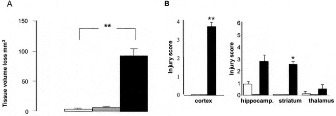
The volume of brain damage was substantially increased in the LPS 6 h/20 min HI (91.4 ± 10.6 mm3) group versus NaCl/20 min HI (4.3 ± 1.7 mm3) (p < 0.01) (Fig. 1A), and injury scores in the cerebral cortex and striatum were higher in LPS 6 h/20 min HI animals (Fig. 1B). LPS pretreatment given 2 h before 20 min HI did not result in brain injury (Fig. 1). There was an increased mortality in the LPS 6 h/20 min HI group (6/11) compared with controls (0/11) (p < 0.01). No difference in mortality was found between LPS 2 h/20 min HI and the control group.
Figure 1
Time interval of 2 and 6 h between LPS and 20 min of hypoxia. Brain injury in rats pretreated with LPS (0.3 mg/kg) at PND 7, 2 and 6 h before 20 min of HI compared with NaCl. (A) Brain damage was quantified as volume of tissue loss according to the Cavalieri principle, where the total volume is expressed as cubic millimeters. (B) Neuropathological scoring of HI brain injury in cerebral cortex (0–4), hippocampus, striatum, and thalamus (0–6). □ NaCl/20minHl _n_=10; LPS2h/20minHl _n_=11; ▪ LPS6h/20minHl _n_=11.Values are given as mean ± SEM (**p < 0.01, *p < 0.05).
LPS administered at PND 7 in combination with 50 min HI.
The volume tissue loss was more pronounced in the group exposed to LPS 6 h/50 min HI (189.5 ± 29.6 mm3) compared with NaCl/50 min HI (79.8 ± 14.9 mm3) (p < 0.01), and a similar trend was found in the evaluations of infarction area at different section levels and brain regions. No significant differences were found in the examined brain regions comparing LPS 2 h/50 min HI and NaCl/50 min HI. In animals subjected to 50 min of HI, there was an increased mortality rate in the LPS 6 h/50 min HI group (8/12) compared with PND 7 controls (1/11) (p < 0.01).
Induction of HI 24 h after LPS Administration
LPS administered at PND 7 in combination with 20 min HI.
The brain tissue volume loss was not different in the group pretreated with LPS and subjected to 20 min of HI (2.3 ± 1.1 mm3) compared with NaCl/20 min HI (4.7 ± 2.5 mm3), and there were no differences in regional damage between the groups.
LPS administered at PND 7 in combination with 50 min HI.
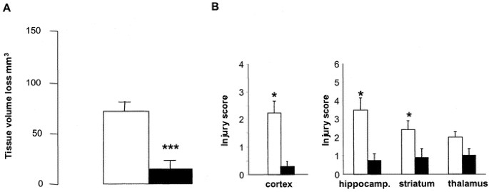
Brain damage was significantly reduced in the LPS 24 h/50 min HI group (15.6 ± 7.6 mm3) versus NaCl-treated controls (71.6 ± 10.1 mm3) (p < 0.001) (Fig. 2A). There was also a regional reduction in brain injury in the cortex (p < 0.05), hippocampus (p < 0.05), and striatum (p < 0.05) in LPS 24 h/50 min HI compared with controls (Fig. 2B). There was no difference in mortality rate between study groups.
Figure 2
Time interval of 24h between LPS and 50 min, of hypoxia. Brain injury in rats pretreated with LPS (0.3 mg/kg) at PND 7, 24 h before 50 min of HI compared with NaCl. (A) Brain damage was quantified as volume of tissue loss according to the Cavalieri principle, where the total volume is expressed as cubic millimeters. (B) Neuropathological scoring of HI brain injury in cerebral cortex (0–4), hippocampus, striatum, and thalamus (0–6). □ NaCl24h/50minHl _n_=11; ▪ LPS24h/50minHl _n_=11. Values are given as mean ± SEM (***p < 0.001, *p < 0.05).
Induction of HI 72 h after LPS Administered at PND 7
LPS administered at PND 7 in combination with 20 min HI.
There were no differences with respect to total brain tissue loss between the LPS 72 h/20 min HI (12.3 ± 3.9 mm3) and the NaCl 72 h/20 min HI (18.3 ± 5.5 mm3) groups. Neither were there any differences with regard to regional injury scores.
LPS administered at PND 7 in combination with 50 min HI.
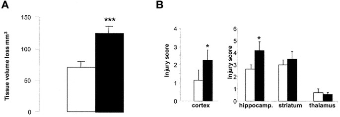
On the other hand, in rat pups subjected to 50 min of HI, there was an increased tissue volume loss in the LPS- versus NaCl-pretreated group (125.3 ± 11.9 mm3 and 70.5 ± 9.9 mm3, respectively) (p < 0.001) (Fig. 3A). There was also a regional difference with increased injury scores in the cortex (p < 0.05) and hippocampus (p < 0.05) in the LPS 72 h/50 min HI group compared with NaCl 72 h/50 min HI (Fig. 3B). There were no differences in mortality rate in the study groups.
Figure 3
Time intervals of 72h between LPS at PND 7 and 50 min of hyposia. Brain injury in rats pretreated with LPS (0.3 mg/kg) at PND 7, 72 h before 50 min of HI compared with NaCl. (A) Brain damage was quantified as volume of tissue loss according to the Cavalieri principle, where the total volume is expressed as cubic millimeters. (B) Neuropathological scoring of HI brain injury in cerebral cortex (0–4), hippocampus, striatum, and thalamus (0–6). □ NaCI72h/50minHI, n = 12; ▪ LPS72h/50minHI, n = 12.Values are given as mean ± SEM (***p < 0.001, *p < 0.05).
Induction of HI 72 h after LPS Administered at PND 4
LPS administered at PND 4 in combination with 20 min HI.
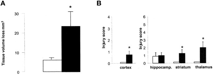
Brain tissue volume loss was more pronounced in the LPS 72 h/20 min HI group (27.3 ± 7.7 mm3) compared with NaCl/20 min HI (5.0 ± 1.1 mm3) (p < 0.05) (Fig. 4A). There was an increased injury in the cortex, striatum, and thalamus after LPS pretreatment compared with controls (p < 0.05) (Fig. 4B).
Figure 4
Time interval of 72h between LPS at PND 4 and 20 min of hyposia. Brain injury in rats pretreated with LPS (0.3 mg/kg) at PND 4, 72 h before 20 min of HI, compared with NaCl. (A) Brain damage was quantified as volume of tissue loss according to the Cavalieri principle, where the total volume is expressed as cubic millimeters. (B) Neuropathological scoring of HI brain injury in cerebral cortex (0–4), hippocampus, striatum, and thalamus (0–6). □ NaCI72h/20minHI, n = 10; ▪ LPS72h/20minHI, n = 11. Values are given as mean ± SEM (*p < 0.05).
LPS administered at PND 4 in combination with 50 min HI.
After 50 min of HI, there were no differences in total brain tissue loss between the LPS 72 h/50 min HI (83.4 ± 12.1 mm3) and the NaCl 72 h/50 min HI (78.9 ± 10.6 mm3) groups or in injury score in any brain region. There was no difference in mortality rate in study or control groups exposed to 20 or 50 min of HI.
DISCUSSION
This is the first study in neonatal animals investigating CNS responses when both the interval between LPS and HI, and the duration of the HI insult were varied. The results demonstrate that the temporal relationship between LPS and the subsequent HI is critical. When LPS was administered 24 h before HI, brain injury was reduced, which agrees with several experimental studies on preconditioning in the adult brain (8,14,15). Interestingly, a 72-h interval between LPS and HI resulted in enhanced brain injury, which is a similar response as when HI is induced shortly (4–6 h) after LPS. Such a long-term sensitizing effect of LPS has not previously been described for the developing brain and disagrees with reports in adult animals (7,8,16,17).
We found that a 2-h delay between LPS and HI did not affect CNS susceptibility, whereas a 4- to 6-h interval efficiently sensitized the neonatal brain to injury. LPS induces systemic inflammation, which triggers NF-κB activation in the CNS with subsequent production of chemokines and cytokines (18–20). Similarly, in a recently performed microarray analysis of gene expression in the neonatal brain after LPS, we found an early inflammatory response with a marked up-regulation of chemokines (IP-10, MCP-1, and interferon-inducible protein variant 10), cytokines (IL-4 and IL-6 receptor and LPS-induced TNF-α) and other inflammatory mediators like CD14 (unpublished observations). In addition, we found an early up-regulation of several pro-apoptotic genes, such as TNF receptor superfamily, member 1a (Tnfr1), galectin-9, and STAT1 at 6 h, but to a much lesser extent at 2 h. Such an early inflammatory and apoptotic response could help to explain the primary sensitizing effect of LPS found in the immature brain in vivo (6,21–23).We also found that LPS sensitized the immature brain if given 72 h before HI either at PND 4 or PND 7. This is interesting in that the animals have completely recovered from the systemic effects of LPS after 3 d. Early physiologic changes found after LPS injection, related to blood glucose, heart rate, temperature, and behavior, returned to normal after 3 d (24–26). We found no mortality after HI in this group. A persistently increased expression of TNF-α, nitric oxide, and prostaglandin E2 has been observed in microglia cultures 72 h after LPS (27). In addition, a secondary inflammatory gene response was detected 72 h after LPS in neonatal rats (unpublished observations). There was a difference in the sensitizing effect of LPS between PND 4 and PND 7 rats in that the younger animals demonstrated increased brain injury after 20 min HI, whereas sensitization was observed after 50 min HI in PND 7 rats. These results emphasize the complexity of the interaction between LPS and HI and demonstrate the importance of both the intensity of the HI insult and the developmental stage of the animals.
Hypothetically, LPS, in addition to the initial primary phase of inflammation, also induces a secondary chronic state of inflammation in the developing brain, which could confer heightened vulnerability. The long-lasting effect of a low dose of endotoxin is intriguing and further studies are needed to investigate the duration of this sensitizing effect. Hence, it was recently demonstrated that intrauterine LPS can induce a progressive degeneration of dopamine neurons that leads to permanent brain injury still present in 16-mo-old rats (28) Such long-lasting adverse CNS effects of bacterial products may have important implications in understanding the full consequences of intrauterine infections.
The preconditioning effect that was achieved when LPS was administered 24 h before HI was in agreement with previous studies on hypoxic preconditioning in 7-d-old rats that also require a 24-h interval between the preconditioning stimulus and HI (29), as well as rat and mouse adult models of CNS preconditioning using LPS, TNF-α, or ischemia (14). The mechanisms are unclear but induction of hypoxia-inducible factor (30), nitric oxide (16,31), heat shock proteins (32), inhibition of caspases (33), and induction of the anti-apoptotic system (34) seem important in the immature brain. In our microarray analysis we found, in agreement with previous studies, an up-regulation of heat shock protein in association with LPS-induced preconditioning (unpublished observations). There was also an up-regulation of granulin and allograft inflammatory factor 1, which is induced by cytokines and interferon.
In the adult rat, LPS preconditioning in focal cerebral ischemia could be due to its stimulation of cytokines, such as TNF-α expression. Hence, the brain protective effect of LPS-induced preconditioning was completely lost after neutralizing TNF-α by administration of TNF-binding protein (7). Furthermore, ceramide, a downstream messenger of TNF-α signaling, contributes to LPS-induced tolerance (14,35). Preconditioning induced by LPS is also associated with the induction of TGF-β1, which has neuroprotective properties (15). Other studies suggest that up-regulation of antioxidants (superoxide dismutase) after low doses of endotoxin may be important in LPS-induced tolerance (36). To conclude, LPS demonstrates a biphasic sensitizing effect on the immature brain including a primary phase at 4–6 h and a secondary response at 72 h after administration. In contrast, LPS confers a preconditioning effect when administered 24 h before HI. The secondary sensitizing effect has not previously been described and may have important clinical implications.
Abbreviations
DAB:
3,3-diaminobenzidine
HI:
hypoxia-ischemia
LPS:
lipopolysaccharide
MAP-2:
microtubule-associated protein-2
MCAO:
middle cerebral artery occlusion
NF-κB:
nuclear factor-kappaB
PND:
postnatal day
TGF-β1:
transforming growth factor-β1
TNF-α:
tumor necrosis factor-α
References
- Badawi N, Kurinczuk JJ, Keogh JM, Alessandri LM, O'Sullivan F, Burton PR, Pemberton PJ, Stanley FJ 1998 Intrapartum risk factors for newborn encephalopathy: the Western Australian case-control study. BMJ 317: 1554–1558
Article CAS Google Scholar - Badawi N, Kurinczuk JJ, Keogh JM, Alessandri LM, O'Sullivan F, Burton PR, Pemberton PJ, Stanley FJ 1998 Antepartum risk factors for newborn encephalopathy: the Western Australian case-control study. BMJ 317: 1549–1553
Article CAS Google Scholar - Grether JK, Nelson KB 1997 Maternal infection and cerebral palsy in infants of normal birth weight. JAMA 278: 207–211
Article CAS Google Scholar - Dammann O, Leviton A 2000 Role of the fetus in perinatal infection and neonatal brain damage. Curr Opin Pediatr 12: 99–104
Article CAS Google Scholar - Nelson KB, Grether JK 1998 Potentially asphyxiating conditions and spastic cerebral palsy in infants of normal birth weight. Am J Obstet Gynecol 179: 507–513
Article CAS Google Scholar - Eklind S, Mallard C, Leverin AL, Gilland E, Blomgren K, Mattsby-Baltzer I, Hagberg H 2001 Bacterial endotoxin sensitizes the immature brain to hypoxic–ischaemic injury. Eur J Neurosci 13: 1101–1106
Article CAS Google Scholar - Tasaki K, Ruetzler CA, Ohtsuki T, Martin D, Nawashiro H, Hallenbeck JM 1997 Lipopolysaccharide pre-treatment induces resistance against subsequent focal cerebral ischemic damage in spontaneously hypertensive rats. Brain Res 748: 267–270
Article CAS Google Scholar - Dawson DA, Furuya K, Gotoh J, Nakao Y, Hallenbeck JM 1999 Cerebrovascular hemodynamics and ischemic tolerance: lipopolysaccharide-induced resistance to focal cerebral ischemia is not due to changes in severity of the initial ischemic insult, but is associated with preservation of microvascular perfusion. J Cereb Blood Flow Metab 19: 616–623
Article CAS Google Scholar - Rice JE 3rd, Vannucci RC, Brierley JB 1981 The influence of immaturity on hypoxic-ischemic brain damage in the rat. Ann Neurol 9: 131–141
Article Google Scholar - Mallard EC, Williams CE, Gunn AJ, Gunning MI, Gluckman PD 1993 Frequent episodes of brief ischemia sensitize the fetal sheep brain to neuronal loss and induce striatal injury. Pediatr Res 33: 61–65
Article CAS Google Scholar - Gilland E, Bona E, Hagberg H 1998 Temporal changes of regional glucose use, blood flow, and microtubule-associated protein 2 immunostaining after hypoxia-ischemia in the immature rat brain. J Cereb Blood Flow Metab 18: 222–228
Article CAS Google Scholar - Hedtjarn M, Leverin AL, Eriksson K, Blomgren K, Mallard C, Hagberg H 2002 Interleukin-18 involvement in hypoxic-ischemic brain injury. J Neurosci 22: 5910–5919
Article CAS Google Scholar - Mallard EC, Rehn A, Rees S, Tolcos M, Copolov D 1999 Ventriculomegaly and reduced hippocampal volume following intrauterine growth-restriction: implications for the aetiology of schizophrenia. Schizophr Res 40: 11–21
Article CAS Google Scholar - Zimmermann C, Ginis I, Furuya K, Klimanis D, Ruetzler C, Spatz M, Hallenbeck JM 2001 Lipopolysaccharide-induced ischemic tolerance is associated with increased levels of ceramide in brain and in plasma. Brain Res 895: 59–65
Article CAS Google Scholar - Boche D, Cunningham C, Gauldie J, Perry VH 2003 Transforming growth factor-beta 1-mediated neuroprotection against excitotoxic injury in vivo. J Cereb Blood Flow Metab 23: 1174–1182
Article CAS Google Scholar - Puisieux F, Deplanque D, Pu Q, Souil E, Bastide M, Bordet R 2000 Differential role of nitric oxide pathway and heat shock protein in preconditioning and lipopolysaccharide-induced brain ischemic tolerance. Eur J Pharmacol 389: 71–78
Article CAS Google Scholar - Bastide M, Gele P, Petrault O, Pu Q, Caliez A, Robin E, Deplanque D, Duriez P, Bordet R 2003 Delayed cerebrovascular protective effect of lipopolysaccharide in parallel to brain ischemic tolerance. J Cereb Blood Flow Metab 23: 399–405
Article CAS Google Scholar - Cai Z, Pan ZL, Pang Y, Evans OB, Rhodes PG 2000 Cytokine induction in fetal rat brains and brain injury in neonatal rats after maternal lipopolysaccharide administration. Pediatr Res 47: 64–72
Article CAS Google Scholar - Bell MJ, Hallenbeck JM 2002 Effects of intrauterine inflammation on developing rat brain. J Neurosci Res 70: 570–579
Article CAS Google Scholar - Reyes TM, Walker JR, DeCino C, Hogenesch JB, Sawchenko PE 2003 Categorically distinct acute stressors elicit dissimilar transcriptional profiles in the paraventricular nucleus of the hypothalamus. J Neurosci 23: 5607–5616
Article CAS Google Scholar - Coumans AB, Middelanis JS, Garnier Y, Vaihinger HM, Leib SL, Von Duering MU, Hasaart TH, Jensen A, Berger R 2003 Intracisternal application of endotoxin enhances the susceptibility to subsequent hypoxic-ischemic brain damage in neonatal rats. Pediatr Res 53: 770–775
Article CAS Google Scholar - Lehnardt S, Massillon L, Follett P, Jensen FE, Ratan R, Rosenberg PA, Volpe JJ, Vartanian T 2003 Activation of innate immunity in the CNS triggers neurodegeneration through a Toll-like receptor 4-dependent pathway. Proc Natl Acad Sci U S A 100: 8514–8519
Article CAS Google Scholar - Yang L, Sameshima H, Ikeda T, Ikenoue T 2004 Lipopolysaccharide administration enhances hypoxic-ischemic brain damage in newborn rats. J Obstet Gynecol Res 30: 142–147
Article CAS Google Scholar - Wu CC, Yen MH 1997 Beneficial effects of dantrolene on lipopolysaccharide-induced haemodynamic alterations in rats and mortality in mice. Eur J Pharmacol 327: 17–24
Article CAS Google Scholar - Cabrera R, Korte SM, Lentjes EG, Romijn F, Schonbaum E, De Nicola A, De Kloet ER 2000 The amount of free corticosterone is increased during lipopolysaccharide-induced fever. Life Sci 66: 553–562
Article CAS Google Scholar - Choi JS, Park HJ, Cha JH, Chung JW, Chun MH, Lee MY 2003 Induction and temporal changes of osteopontin mRNA and protein in the brain following systemic lipopolysaccharide injection. J Neuroimmunol 141: 65–73
Article CAS Google Scholar - Ajmone-Cat MA, Nicolini A, Minghetti L 2003 Prolonged exposure of microglia to lipopolysaccharide modifies the intracellular signaling pathways and selectively promotes prostaglandin E2 synthesis. J Neurochem 87: 1193–1203
Article CAS Google Scholar - Carvey PM, Chang Q, Lipton JW, Ling Z 2003 Prenatal exposure to the bacteriotoxin lipopolysaccharide leads to long-term losses of dopamine neurons in offspring: a potential, new model of Parkinson's disease. Front Biosci 8: s826–s837
Article CAS Google Scholar - Gidday JM, Fitzgibbons JC, Shah AR, Park TS 1994 Neuroprotection from ischemic brain injury by hypoxic preconditioning in the neonatal rat. Neurosci Lett 168: 221–224
Article CAS Google Scholar - Bergeron M, Gidday JM, Yu AY, Semenza GL, Ferriero DM, Sharp FR 2000 Role of hypoxia-inducible factor-1 in hypoxia-induced ischemic tolerance in neonatal rat brain. Ann Neurol 48: 285–296
Article CAS Google Scholar - Gidday JM, Shah AR, Maceren RG, Wang Q, Pelligrino DA, Holtzman DM, Park TS 1999 Nitric oxide mediates cerebral ischemic tolerance in a neonatal rat model of hypoxic preconditioning. J Cereb Blood Flow Metab 19: 331–340
Article CAS Google Scholar - Ikeda T, Ikenoue T, Xia XY, Xia YX 2000 Important role of 72-kd heat shock protein expression in the endothelial cell in acquisition of hypoxic-ischemic tolerance in the immature rat. Am J Obstet Gynecol 182: 380–386
Article CAS Google Scholar - McLaughlin B, Hartnett KA, Erhardt JA, Legos JJ, White RF, Barone FC, Aizenman E 2003 Caspase 3 activation is essential for neuroprotection in preconditioning. Proc Natl Acad Sci U S A 100: 715–720
Article CAS Google Scholar - Cantagrel S, Krier C, Ducrocq S, Bodard S, Payen V, Laugier J, Guilloteau D, Chalon S 2003 Hypoxic preconditioning reduces apoptosis in a rat model of immature brain hypoxia-ischaemia. Neurosci Lett 347: 106–110
Article CAS Google Scholar - Liu J, Ginis I, Spatz M, Hallenbeck JM 2000 Hypoxic preconditioning protects cultured neurons against hypoxic stress via TNF-alpha and ceramide. Am J Physiol Cell Physiol 278: C144–C153
Article CAS Google Scholar - Kramer BC, Yabut JA, Cheong J, JnoBaptiste R, Robakis T, Olanow CW, Mytilineou C 2002 Lipopolysaccharide prevents cell death caused by glutathione depletion: possible mechanisms of protection. Neuroscience 114: 361–372
Article CAS Google Scholar
Acknowledgements
The authors thank Mrs. Eva Cambert for technical assistance.
Author information
Authors and Affiliations
- Department of Obstetrics and Gynecology, Perinatal Center, Institute of Physiology and Pharmacology, Göteborg University, Göteborg, 413 45, Sweden
Saskia Eklind & Henrik Hagberg - Department of Physiology, Institute for the Health of Women and Children, Institute of Physiology and Pharmacology, Göteborg University, Göteborg, 413 45, Sweden
Saskia Eklind, Carina Mallard, Pernilla Arvidsson & Henrik Hagberg
Authors
- Saskia Eklind
You can also search for this author inPubMed Google Scholar - Carina Mallard
You can also search for this author inPubMed Google Scholar - Pernilla Arvidsson
You can also search for this author inPubMed Google Scholar - Henrik Hagberg
You can also search for this author inPubMed Google Scholar
Corresponding author
Correspondence toSaskia Eklind.
Additional information
Supported by the Swedish Medical Research Council (K2004-33X-14185-03A and K2004-33X-09455), the Åhlén Foundation, the Sven Jerring Foundation, the Magnus Bergvall Foundation, the Wilhelm and Martina Lundgren Foundation, the Linnéa and Josef Carlsson Foundation, the Frimurare Barnhus Foundation, Göteborg Medical Society, and Åke Wibergs Foundation and by grants to researchers in the public health service from the Swedish government (LUA).
Rights and permissions
About this article
Cite this article
Eklind, S., Mallard, C., Arvidsson, P. et al. Lipopolysaccharide Induces Both a Primary and a Secondary Phase of Sensitization in the Developing Rat Brain.Pediatr Res 58, 112–116 (2005). https://doi.org/10.1203/01.PDR.0000163513.03619.8D
- Received: 09 August 2004
- Accepted: 05 November 2004
- Issue Date: 01 July 2005
- DOI: https://doi.org/10.1203/01.PDR.0000163513.03619.8D


