Plasma Cytokine Profiles in Preprotachykinin-A Knockout Mice Subjected to Polymicrobial Sepsis (original) (raw)
- Research Article
- Open access
- Published: 29 October 2009
Molecular Medicine volume 16, pages 45–52 (2010)Cite this article
- 593 Accesses
- 11 Citations
- Metrics details
Abstract
During the course of polymicrobial sepsis, a range of pro- and antiinflammatory cytokines are produced by the host immune system. Successful recovery from sepsis involves striking a balance between these counteracting cytokines. We herein investigated the circulating cytokine profiles in preprotachykinin-A knockout (_PPTA_−/−) mice, which have been found to be protected significantly against microbial sepsis, by employing multiplexed bead-based suspension arrays for the measurement of 18 plasma cytokines. Four sets of _PPTA_−/− and wild-type mice, each with six mice, were subjected to cecal ligation and puncture-induced sepsis or a sham procedure and were killed at 1, 5, 8 and 24 h post surgery. The cytokine profiles revealed, rather interestingly, that both pro- and antiinflammatory cytokines were elevated in the knockout group in response to a septic challenge. The higher systemic levels of both pro- and antiinflammatory cytokines in _PPTA_−/− septic mice was similar to the increase that we observed earlier in lung tissue of _PPTA_−/− mice after induction of sepsis. Thus, elevated levels of both pro- and antiinflammatory mediators may act simultaneously and help to resolve the infectious assault at the early stages of sepsis without excessively damaging the host tissue in _PPTA_−/− mice. In addition, our results underline the importance of comprehensive clinical analysis of multiple biomarkers to provide a better prognostic tool.
Introduction
Sepsis is a state of systemic inflammatory response syndrome (SIRS) resulting from bacteria, viruses, fungi or parasites (1). The nature and cellular composition of the infection and the microenvironment of each organ influences the extent of local tissue injury in sepsis (2). Infection, injury and inflammation trigger the release of cytokines that act as immune mediators (3,4). These inflammatory proteins are elevated in various disease states such as autoimmune diseases, inflammatory bowel disease and sepsis. It has been well established that cytokine cascades play a major role in the progression of sepsis. Large numbers of cytokines are produced mainly within tissues and released into systemic circulation to mediate the inflammatory responses in sepsis. The initial proinflammatory response to eliminate pathogens is followed by a counteracting production of antiinflammatory mediators that also contributes to the pathophysiology of sepsis (5–7). Antiinflammatory mediators predominate systemically to avoid new inflammatory foci, but their tissue levels may not always be sufficient to prevent deleterious inflammatory effects (2). Multiple mediators have been reported to be involved in the development of sepsis (8,9).
Preprotachykinin-A (PPTA) gene and its product substance P (SP), have been detected in various cells of the immune system. SP is an immunoregulatory neuropeptide implicated in various inflammatory diseases. It has been shown to increase microvascular permeability, plasma extravasation, expression of chemokines and chemokine receptors in mouse neutrophils and prime neutrophils for chemotactic responses. Recent literature reports illustrate evidence of a role for SP in acute pancreatitis, endotoxemia and sepsis (10–13). Deletion of PPTA gene in mice has been reported to protect significantly against mortality of sepsis and to attenuate lung damage (12). However the mechanism of this protection is not yet clear. Sepsis is a complex condition where simple blocking of inflammatory response may result in an impaired resolution of infection. Although SP plays a major role in SIRS and increases the levels of various cytokines, it is important to understand how the absence of SP helps contain the infection in sepsis without excessive damage to the host. Thus it was imperative to measure a range of inflammatory cytokines in _PPTA_−/− septic mice and to evaluate the protection against lung injury in sepsis.
Plasma is one of the major sources for measurement of clinical markers in sepsis (14). Since early diagnosis and treatment are critical in sepsis management, evaluation of plasma cytokines over a time course can provide a window toward a better understanding of the nature and severity of sepsis. Rather than analyzing individual cytokine levels by conventional enzyme-linked immunosorbent assay (ELISA), multiplexed measurement of cytokines provides a clearer picture of the biological processes and immune pathways involved. In this regard, we applied bead-based suspension arrays for the measurement of a set of plasma cytokines in _PPTA_−/− mice subjected to polymicrobial sepsis. The advantages of this technology include high throughput, accuracy, efficiency, sensitivity, simultaneous analyte detection, low cost and time reduction (15,16). The reduction of the amount of precious sample required for each analysis is one of the most valuable advantages (17–20). In addition, the accuracy of data collected by xMAP technology (Luminex Corporation, Austin, TX, USA) has been reported to be comparable to that from ELISA (18).
Materials and Methods
Animal Model of Polymicrobial Sepsis
All animal experiments performed were in accordance with the guidelines of the DSO National Laboratories Animal Care and Use Committee (DSOACUC), Singapore, which follows the established International Guiding Principles for Biomedical Research Involving Animals. Mice were maintained at a controlled temperature (21–24°C) and lighting (12-h light/dark cycle) and fed with standard laboratory chow and drinking water, provided ad libitum. Before the experiment, 2 days of acclimatization were allowed for all mice.
_PPTA_−/− (with balb/C genetic background) and wild-type balb/C male mice (25–30 g) were randomly divided into sham or cecal ligation and puncture (CLP) experimental groups (n = more than six in each group). Mice were anesthetized lightly with mouse anesthesia cocktail (0.75 mL ketamine [100 mg/mL] and 1 mL medetomindine [1 mg/mL] dissolved in 8.25 mL distilled water) (7.5 mL/kg body weight) purchased from Animal Holding Unit, NUS, Singapore. Polymicrobial sepsis was induced by CLP as described elsewhere (12,21–23). Briefly, maintaining strict aseptic conditions, the anterior abdomen was shaved and a midline incision was made in the lower part of the abdomen. The peritoneum was opened and the cecum was ligated 3–5 mm below the ileocecal valve with 4/0 silk suture (Silkam, B Braun Aesculap, Tuttlingen, Germany) without obstructing the bowel. The cecum was punctured twice with a 22-gauge needle (Terumo Corporation, Tokyo, Japan) distal to the point of ligation and squeezed gently to extrude the cecal contents. The cecum was placed back in the abdomen and the muscle and skin incision were sutured separately with sterile Permilene 5/0 thread (B Braun Aesculap). All the mice were given saline (1 mL, subcutaneously [s.c.]) after the surgery and kept on heat pads for recovery. The same surgical procedure except CLP was performed on sham-operated animals. The sets of mice were killed at various time points (1, 5, 8 and 24 h; n = at least 6 for each time point) after surgery by an intraperitoneal (i.p.) injection of pentobarbitone (Jurox Pty Ltd, Rutherford, NSW, Australia). Blood was harvested through cardiac puncture, heparinized, centrifuged, plasma removed and stored at −80°C for subsequent analysis. Another set of normal healthy _PPTA_−/− and balb/c mice (n = 6) were sacrificed and the blood was harvested for basal level cytokine analysis.
Plasma Cytokine Profile Using Bead Array
Time-dependent plasma cytokine profile was obtained using Procarta Cytokine kits (Panomics, Fremont, CA, USA) that employed multiplex immunoassays based on xMAP detection technology developed by Luminex Corporation using Luminex bead array system. Fluorescently encoded antibody beads (Panomics) were detected uniquely in a flow cytometer and 18 mouse cytokines (CCL11, granulocyte-macrophage colony-stimulating factor [GM-CSF], IFN-γ, IL-10, IL-12, IL-13, IL-17, IL-1α, IL-1β, IL-2, IL-3, IL-4, IL-5, IL-6, CXCL1, CCL3, CCL5, tumor necrosis factor [TNF]-α) were evaluated through a sandwich immunoassay simultaneously. Briefly, 50 µL of the antibody beads were added to each well of the pre-wet 96-well filter bottom plate and washed with wash buffer. Assay buffer (75 µL/well), standard and sample (25 µL/well) were added to the predesignated wells and incubated for 30 min at room temperature on a shaker (1.4_g_). After washing, detection antibody (25 µL/well) was added and incubated for 30 min at room temperature on a shaker (1.4_g_). Streptavidin-phycoerythrin (SA-PE) (50 µL/well) was added to the washed plate and incubated again for 30 min at room temperature on a shaker (1.4_g_). Subsequent to another wash, 120 µL/well of the reading buffer was added, placed on a shaker (1.4_g_) for 5 min at room temperature and analyzed on Luminex 100 instrument (Luminex Corporation). The median fluorescence intensity from at least 100 beads of each type (per sample per cytokine) was used to determine the intensity levels of cytokines. Standard curves were plotted and fitted using a 5-parameter logistic model, from which the sample cytokine concentrations were determined. The cytokine concentrations obtained for each group at the different time points were averaged across each replicate set and expressed as pg/mL. The kit sensitivity (limit of detection, [LOD]) was 1 pg/mL/cytokine.
Statistical Analysis
Values were expressed as mean ± SEM and significant difference between groups was evaluated by one-way analysis of variance (ANOVA), followed by post hoc Tukey test. A P < 0.05 was considered statistically significant.
Results
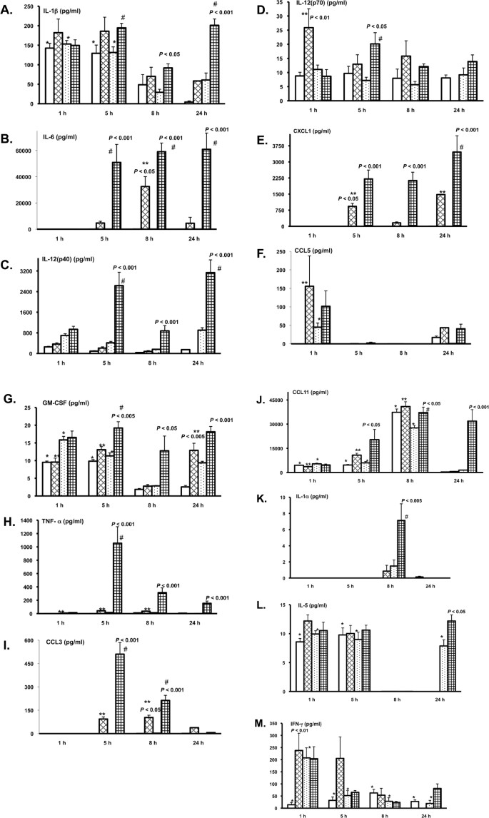
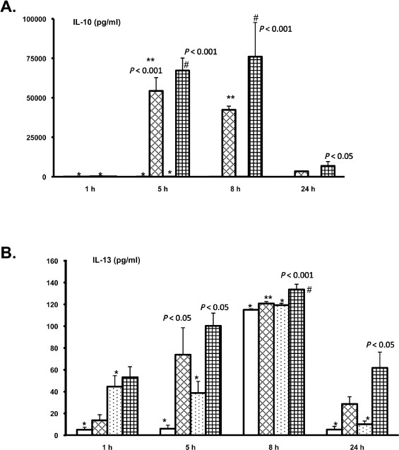
_PPTA_−/− and wild-type mice were killed at 1, 5, 8 and 24 h after sham or CLP surgery and 18 plasma cytokines were analyzed. Among all the cytokines tested (namely CCL11, GM-CSF, IFN-γ, IL-10, IL-12, IL-13, IL-17, IL-1α, IL-1β, IL-2, IL-3, IL-4, IL-5, IL-6, CXCL1, CCL3, CCL5, TNF-α), levels of both pro- (Figure 1A–M) and antiinflammatory (Figure 2A, B) cytokines were elevated significantly in the _PPTA_−/− septic mice compared with the wild-type mice. IL-2, IL-3, IL-4 and IL-17 levels were below the detection limit of the assay in all the samples. The measured basal cytokine levels for normal healthy _PPTA_−/− and wild-type mice were shown in Table 1.
Figure 1
Plasma proinflammatory cytokine profile in wild-type and PPTA−/− septic mice. (A) IL-1β; (B) IL-6; (C) IL-12(p40); (D) IL-12(p70); (E) CXCL1; (F) CCL5; (G) GM-CSF; (H) TNF-α; (I) CCL3; (J) CCL11; (K) IL-1α; (L) IL-5; (M) IFN-γ. Cytokine levels in plasma were measured 1, 5, 8 and 24 h after CLP or sham surgery in wild-type and _PPTA_−/− mice by multiplex immunoassay. Results were expressed as mean ± SEM (n = at least six mice per group). P values were shown for comparison with corresponding sham group. Symbols were used to denote significant differences between groups as a function of time. Key: balb/C sham, open bars; balb/C CLP, outlined diamond bars; _PPTA_−/− sham, dotted bars; _PPTA_−/− CLP, small grid bars. *P< 0.001 when compared with the corresponding normal value; **P< 0.05 when compared with the corresponding values of balb/C septic mice at different time points; #P < 0.05 when compared with the corresponding values of _PPTA_−/− septic mice at different time points; CLP, cecal ligation and puncture; GM-CSF, granulocyte-macrophage colony-stimulating factor; IL, interleukin; IFN-γ, interferon-γ; PPTA, preprotachykinin A; TNF-α, tumor necrosis factor-α.
Figure 2
Plasma antiinflammatory cytokine profile in wild-type and _PPTA_−/− septic mice. (A) IL-10; (B) IL-13. Cytokine levels in plasma were measured 1, 5, 8 and 24 h after CLP or sham surgery in wild-type and _PPTA_−/− mice by multiplex immunoassay. Results were expressed as mean ± SEM (n = at least six mice per group). P values were shown for comparison with corresponding sham group. Symbols were used to denote significant differences between groups as a function of time. Key: balb/C sham, open bars; balb/C CLP, outlined diamond bars; _PPTA_−/− sham, dotted bars; _PPTA_−/− CLP, small grid bars. *P < 0.001 when compared with the corresponding normal value; **P < 0.05 when compared with the corresponding values of balb/C septic mice at different time points; #P < 0.05 when compared with the corresponding values of _PPTA_−/− septic mice at different time points; CLP, cecal ligation and puncture; IL, interleukin; PPTA, preprotachykinin A.
Table 1 Basal levels of plasma cytokines for normal healthy _PPTA_−/− and wild-type mice (n = 6).
Cytokine Profile as a Function of Time for the Sham Groups
Mice subjected to sham surgery showed elevated levels of various cytokines at 1 and 5 h after the surgery (P < 0.05) (see Figures 1 and 2). IL-1β, GM-CSF, CCL11, IL-5, IFN-γ, IL-10 and IL-13 were increased in wild-type mice at the early time points studied (see Figures 1A, G, J, L, M, 2A, B respectively). Similarly, PPTA−/− mice showed elevated levels of IL-1β, CCL5, GM-CSF, CCL11, IL-5, IFN-γ, IL-10 and IL-13 (see Figures 1A, F, G, J, L, M, 2A, B respectively) at 1 and 5 h after sham surgery. However the levels were reduced in both _PPTA_−/− and wild-type sham groups at the later time points.
Cytokine Profile as a Function of Time for the Balb/C Septic Mice
Mice subjected to CLP have been reported to show elevated levels of proinflammatory cytokines such as IL-6, CXCL2 and TNF-α (24–27). Consistently, we found increased plasma IL-6, IL-12(p70), CXCL1, CCL5, GM-CSF, TNF-α, CCL3 and CCL11 levels (see Figure 1B, D-J respectively) in balb/C septic mice (P < 0.05). The elevated levels were apparent by 5 h after CLP for many of the cytokines and continued to remain high even at the 24-h time point (see Figure 1). However, CXCL1 and GM-CSF levels showed a reduction at 8-h time point (see Figure 1E, G).
Antiinflammatory cytokine, IL-10 levels were increased in wild-type mice only at 5 and 8 h after CLP (P < 0.001) (see Figure 2A). In addition, IL-13 levels showed significant increase only at 8 h after surgery (P < 0.05) (see Figure 2B).
Cytokine Profile as a Function of Time for the _PPTA_−/− Septic Mice
_PPTA_−/− septic mice also showed an increase in various cytokines over 24 h after induction of sepsis (P < 0.05). A significant elevation was observed for IL-1β, IL-6, IL-12(p40), IL-12(p70), CXCL1, GM-CSF, TNF-α, CCL3, CCL11, IL-1α, IL-5, IL-10 and IL-13 (see Figures 1A-E, G-L, 2A, B respectively). However, IL-1β and IL-5 levels were lowered at 8 h after CLP surgery (see Figure 1A, L).
Comparative Cytokine Profiles for the _PPTA_−/− and Wild-Type Septic Mice
Several sets of cytokines showed significantly different patterns across the _PPTA_−/− and the wild-type septic mice compared with their corresponding sham control groups.
Proinflammatory Cytokine Profiles
Plasma IL-1β levels were elevated more significantly in _PPTA_−/− septic mice during the later phase of sepsis (8 and 24 h) (P < 0.05 and P < 0.001 respectively) compared with the corresponding wild-type mice (Figure 1A). Levels of IL-6, an important proinflammatory cytokine in sepsis, were increased significantly in wild-type mice at 8 h after CLP (P < 0.05), but the increase was significantly higher in _PPTA_−/− septic mice at 5, 8 and 24 h after CLP (P < 0.001) and also when compared with the corresponding increase in wildtype group (see Figure 1B). Proinflammatory cytokine, IL-12(p70), is a heterodimer of IL-12(p40) and IL-12(p35) subunits connected by a disulphide bond that is essential for the biological activity (28). IL-12(p70) was significantly increased in wild-type mice only at 1 h after CLP (P < 0.01) but the difference was apparent in _PPTA_−/− septic mice at 5 h (P < 0.05) (see Figure 1D). Levels of IL-12(p40), a component of cytokines IL-12 and IL-23, were higher in _PPTA_−/− septic mice at 5, 8 and 24 h after CLP (P < 0.001) (see Figure 1C). CXCL1 and GM-CSF levels were also elevated significantly (P < 0.001 and P < 0.05 respectively) in _PPTA_−/− septic mice compared with the wild-type septic mice (see Figure 1E, G).
Systemic levels of TNF-α were increased significantly more in _PPTA_−/− mice 5 h after CLP (P < 0.001) and the increase was apparent up to 24 h post-CLP (P < 0.001) (Figure 1H). CCL3 protein levels in plasma were also elevated significantly in _PPTA_−/− mice at 5 and 8 h after CLP (P < 0.001) compared with the increase in wild-type mice at 8 h (P < 0.05), but this increase was reversed by 24 h post-CLP (see Figure 1I). In contrast, plasma CCL11 levels were elevated to a greater extent in _PPTA_−/− mice up to 24 h after CLP (see Figure 1J). IL-1α levels were found to be increased in _PPTA_−/− septic mice compared with the other groups only at 8 h after CLP (see Figure 1K). IL-5 plasma levels were not significantly different between the wild-type and _PPTA_−/− septic mice at any of the time points studied except at 24 h (P < 0.05) (see Figure 1L). Lastly, IFN-γ was found to be significantly increased in wild-type septic mice as early as 1 h (P < 0.01) and persisted up to 5 h after CLP, but the increase was not statistically significant in _PPTA_−/− septic mice (see Figure 1M).
Antiinflammatory Cytokine Profiles
Levels of IL-10 were increased after CLP in both _PPTA_−/− and wild-type mice, but the difference was significant in _PPTA_−/− mice at 5, 8 and 24 h (P < 0.001, P < 0.001 and P < 0.05 respectively) after the surgery (see Figure 2A). Similarly, plasma levels of another antiinflammatory cytokine, IL-13, were elevated in _PPTA_−/− and wild-type septic mice compared with the sham group, but the increase was more significant for the knockout mice especially at 5, 8 and 24 h (P < 0.05, P < 0.001 and P < 0.05 respectively) after the induction of sepsis (see Figure 2B).
Discussion
_PPTA_−/− mice are genetically modified animals that lack the neurokinin peptides SP and neurokinin A (NKA). In mammals, the PPTA gene encodes both SP and NKA. PPTA gene products have been reported earlier to be involved in neurogenic inflammation in various disease models (12). Immunoregulatory peptide SP is produced at various inflammation sites, in resident macrophages, circulating leukocytes and dendritic cells (29–31). Although SP and NKA are synthesized together, most studies have focused on the contribution of SP (32). SP is reported to increase postcapillary venule permeability, immune cell influx and glandular secretion in mammalian airways (33). It also induces the release of proinflammatory cytokines, lymphocyte proliferation and chemotaxis (34). Thus, deletion of SP could be beneficial in inflammatory conditions by potentially modulating cytokines and immune cells.
It is interesting to note that the PPTA gene deletion in mice contributed to a survival phenotype evidenced by a greater resilience to sepsis (12). However, the mechanism of tolerance and survival at elevated levels of systemic inflammatory cytokines has yet to be established. We have observed elevated levels of pulmonary cytokines in _PPTA_−/− mice subjected to polymicrobial sepsis (unpublished data). Although tissue-associated cytokine levels represent cytokine production more closely, systemic levels also provide a faster and reliable means of measurement, especially in clinical applications. Detectable plasma cytokines are likely to represent the excess of produced mediators which have not been contained within target tissues or organs. Using a bead-array based platform coupled with a flow-cytometric fluorescent based reader, we performed simultaneous measurement of 18 mouse cytokines using a very small volume (25 µL) of plasma per assay. Multiplexed bead-based arrays have been shown earlier to be especially useful for detection of analytes in precious small volume (27).
Many of the inflammatory cytokines studied were elevated at 1 h after surgery in the sham control group. Injury is known to trigger inflammation and coordinated cellular activities. It is possible that the surgical procedure as such initiates a transient proinflammatory response in sham group mice facilitating tissue regeneration. However, mice subjected to CLP showed significantly higher proinflammatory response and thus sham operated mice provide an efficient control to the surgical intervention involved in CLP.
Plasma cytokine time-point data showed that _PPTA_−/− mice subjected to CLP-induced sepsis exhibited elevated levels of both pro- and antiinflammatory cytokines. Indeed, early phase of lethal sepsis is reported to show overexpression of both pro- and antiinflammatory cytokines (14). Plasma concentrations of TNF-α, IL-1β, IL-6, IL-8, soluble cytokine receptors, cytokine receptor antagonists and counter-inflammatory cytokines are known to be elevated in human sepsis (35). We found significantly elevated levels of various proinflammatory cytokines such as TNF-α, CCL3, IL-1β, IL-6, CXCL1 and CCL11 in _PPTA_−/− mice compared with the wild-type mice, especially at later time points after induction sepsis. TNF-α, IL-1 and IL-6 coordinate the initiation of acute phase response in sepsis (36) that is triggered by the pathogen recognition and is important for survival in sepsis. CCL3, CCL6 and CXCL10 have been demonstrated to be protective in sepsis-induced injury and mortality in a murine CLP model (37–39). CCL22 also protected mice against CLP-induced death (40). In our previous study using _PPTA_−/− septic mice, only CCL2 and CXCL2 levels in lung and plasma were analyzed by ELISA (12). Although both the chemokines were elevated in _PPTA_−/− and wild-type septic mice, the increase was lower in the former group (12). These two chemokines were believed to act as chemoattractants to leukocytes and to play a role in tissue damage (12). We did not repeat these two chemokines in the present study, but the range of chemokines and cytokines studied showed a significant elevation up to 24 h after induction of sepsis. It is not clear yet as to why genetic deletion of SP, a product of PPTA gene, leads to significantly elevated cytokine levels, although it is possible that these proinflammatory cytokines are useful in countering the pathogenic invasion in the early phase of sepsis. A significant initial increase in IL-6 and subsequent reduction at a late stage has been reported to protect septic mice (41). Multiple mechanisms and mediators could be at play in this scenario which needs to be probed further. In addition, apart from SP, NKA also might play a role in sepsis and thus could become a potential lead.
Balance between proinflammatory and antiinflammatory mediators plays an important role in the pathophysiology of sepsis. Antiinflammatory cytokines such as IL-10 and IL-13 were also elevated significantly in _PPTA_−/− mice after sepsis. The increase was more significant in _PPTA_−/− septic mice compared with the corresponding wild-type mice. IL-10 levels detected in our assay were much higher compared with the values observed by others (42). It has been reported that antiinflammatory strategies applied early in patients with a hyperinflammatory immune response may prove to be lifesaving (43). Inhibition of IL-10 12 h after CLP has been shown to improve survival in mice (44). Depending on the time of intervention, IL-10 has been reported to be protective or deleterious in sepsis (45). _PPTA_−/− septic mice showed elevated levels of IL-10 at 5 and 8 h after sepsis and a subsequent reduction, both of which could have proved beneficial against mortality.
IL-12(p80), a homodimer of IL-12(p40) has been reported to be an antagonist of proinflammatory IL-12 receptor β1 (28). IL-12(p40) is released from various inflammatory cells in response to pathogenic or inflammatory signals (46). IL-12(p40) is reported to show both protective and pathogenic immune responses (28). Interestingly, we found significantly elevated levels of IL-12(p40) in _PPTA_−/− mice compared with the wildtype mice after the induction of sepsis. The increase corresponded with the elevation in proinflammatory cytokine IL-12(p70) in _PPTA_−/− septic mice. However, the significance of this effect is not very clear. The observed levels of IL-12(p40) were in agreement with the reported 50fold higher IL-12(p40) secretion compared with IL-12p70 in murine shock model (47). In addition, we also have seen elevated levels of another antiinflammatory cytokine, IL-1ra, in _PPTA_−/− mice compared with wild-type mice after sepsis (data not shown). IL-1ra plasma levels are reported to be elevated both in human volunteers injected with endotoxin as well as in patients with severe sepsis, although its function is not clear (48,49). Cytokine receptor antagonists are cytokine-like molecules binding to receptors but without signal transduction (35).
Although the specific role of antiinflammatory molecules in sepsis remains undefined, a complex interplay between cytokines and cytokine-neutralizing molecules is considered to govern the clinical presentation and outcome of sepsis (35). In patients with lethal septic shock, the level of secreted antiinflammatory molecules is believed to be insufficient to counter the overwhelming proinflammatory mediators (35). However, in _PPTA_−/− mice, we have uncovered that elevated levels of both the pro-and antiinflammatory mediators may act simultaneously and help resolve the infectious assault at the early stages of sepsis without excessively damaging the host tissue, and, thus, prolong the survival in these mice. Overall data indicates that multiple factors play protective roles in polymicrobial sepsis in _PPTA_−/− mice and render them resistant to microbial infection. The current time-dependent cytokine snapshot represents a rich source of information for further analysis and investigation.
Limited knowledge of the molecular mechanisms in sepsis has in the past led to the failure of various clinical trials of otherwise promising drug molecules from preclinical stages. Recently, improvements in methods of detecting genetic signatures of sepsis and biomarker identification more rapidly and cost effectively are beginning to provide added insight to both the research and clinical arenas (50). Finding a “magic bullet” is not more important than evaluating the complete immune response and inflammatory status and tailoring the treatment for individualized therapy in critically ill patients. Toward this end, our multiplexed approach of time-point analysis of cytokines, which are major mediators of sepsis, provides a relevant and valuable platform for further research and discovery, and a better diagnostic tool to profile septic patients clinically.
Disclosure
The authors declare that they have no competing interests as defined by Molecular Medicine, or other interests that might be perceived to influence the results and discussion reported in this paper.
References
- Matsuda M, Hattori Y. (2006) Systemic inflammatory response syndrome (SIRS): molecular pathophysiology and gene therapy. J. Pharmacol. Sci. 101:189–98.
Article CAS Google Scholar - Cavaillon JM, Annane D. (2006) Compartmentalization of the inflammatory response in sepsis and SIRS. J. Endotoxin Res. 12:151–70.
CAS PubMed Google Scholar - Ray CA, et al. (2005) Development, validation, and implementation of a multiplex immunoassay for the simultaneous determination of five cytokines in human serum. J. Pharm. Biomed. Anal. 36:1037–44.
Article CAS Google Scholar - de Jager W, Rijkers GT. (2006) Solid-phase and bead-based cytokine immunoassay: a comparison. Methods. 38:294–303.
Article Google Scholar - Ashare A, et al. (2005) Anti-inflammatory response is associated with mortality and severity of infection in sepsis. Am. J. Physiol. Lung Cell Mol. Physiol. 288:L633–40.
Article CAS Google Scholar - Reddy RC, Chen GH, Tekchandani PK, Standiford TJ. (2001) Sepsis-induced immunosuppression: from bad to worse. Immunol. Res. 24:273–87.
Article CAS Google Scholar - van der Poll T, Deventer SJ. (1999) Cytokines and anticytokines in the pathogenesis of sepsis. Infect. Dis. Clin. North Am. 13:413–26.
Article Google Scholar - Okazaki Y, Matsukawa A. (2009) Pathophysiology of sepsis and recent patents on the diagnosis, treatment and prophylaxis for sepsis. Recent Pat. Inflamm. Allergy Drug Discov. 3:26–32.
Article CAS Google Scholar - Marshall JC, et al. (2003). Measures, markers, and mediators: toward a staging system for clinical sepsis. A report of the Fifth Toronto Sepsis Roundtable, Toronto, Ontario, Canada, October 25–26, 2000. Crit. Care Med. 31:1560–7.
Article Google Scholar - Ramnath RD, et al. (2006) Inflammatory mediators in sepsis: Cytokines, chemokines, adhesion molecules and gases. J. Organ Dysfunction. 2:80–92.
Article Google Scholar - Ng SW, Zhang H, Hegde A, Bhatia M. (2008) Role of preprotachykinin-A gene products on multiple organ injury in LPS-induced endotoxemia. J. Leukoc. Biol. 83:288–95.
Article CAS Google Scholar - Puneet P, et al. (2006) Preprotachykinin-A gene products are key mediators of lung injury in polymicrobial sepsis. J. Immunol. 176:3813–20.
Article CAS Google Scholar - Zhang H, et al. (2007) Hydrogen sulfide upregulates substance P in polymicrobial sepsisassociated lung injury. J. Immunol. 179:4153–60.
Article CAS Google Scholar - Osuchowski MF, Welch K, Siddiqui J, Remick DG. (2006) Circulating cytokine/inhibitor profiles reshape the understanding of the SIRS/CARS continuum in sepsis and predict mortality. J. Immunol. 177:1967–74.
Article CAS Google Scholar - Kingsmore SF. (2006) Multiplexed protein measurement: technologies and applications of protein and antibody arrays. Nat. Rev. Drug Discov. 5:310–20.
Article CAS Google Scholar - Nolan JP, Mandy F. (2006) Multiplexed and microparticle-based analyses: quantitative tools for the large-scale analysis of biological systems. Cytometry A. 69:318–25.
Article Google Scholar - Vignali DA. (2000) Multiplexed particle-based flow cytometric assays. J. Immunol. Methods 243:243–55.
Article CAS Google Scholar - Dupont NC, Wang K, Wadhwa PD, Culhane JF, Nelson EL. (2005) Validation and comparison of Luminex multiplex cytokine analysis kits with ELISA: determinations of a panel of nine cytokines in clinical sample culture supernatants. J. Reprod. Immunol. 66:175–91.
Article CAS Google Scholar - Prabhakar U, Eirikis E, Davis HM. (2002) Simultaneous quantification of proinflammatory cytokines in human plasma using the LabMAP assay. J. Immunol. Methods. 260:207–18.
Article CAS Google Scholar - Linkov F, et al. (2007) Early detection of head and neck cancer: development of a novel screening tool using multiplexed immunobead-based biomarker profiling. Cancer Epidemiol. Biomarkers Prev. 16:102–7.
Article CAS Google Scholar - Ayala A, Herdon CD, Lehman DL, Ayala CA, Chaudry IH. (1996) Differential induction of apoptosis in lymphoid tissues during sepsis: variation in onset, frequency, and the nature of the mediators. Blood. 87:4261–75.
CAS PubMed Google Scholar - Zhou M, Chaudry IH, Wang P. (2001) The small intestine is an important source of adrenomedullin release during polymicrobial sepsis. Am. J. Physiol. Regul. Integr. Comp. Physiol. 281:R654–60.
Article CAS Google Scholar - Baker CC, Chaudry IH, Gaines HO, Baue AE. (1983) Evaluation of factors affecting mortality rate after sepsis in a murine cecal ligation and puncture model. Surgery. 94:331–5.
CAS PubMed Google Scholar - Zhang H, Zhi L, Moochhala S, Moore PK, Bhatia M. (2007) Hydrogen sulfide acts as an inflammatory mediator in cecal ligation and puncture-induced sepsis in mice by upregulating the production of cytokines and chemokines via NF-kappaB. Am. J. Physiol. Lung Cell Mol. Physiol. 292:L960–71.
Article CAS Google Scholar - Ertel W, et al. (1991) The complex pattern of cytokines in sepsis: Association between prostaglandins, cachectin, and interleukins. Ann. Surg. 214:141–8.
Article CAS Google Scholar - Salkowski CA, Detore G, Franks A, Falk MC, Vogel SN. (1998) Pulmonary and hepatic gene expression following cecal ligation and puncture: monophosphoryl lipid a prophylaxis attenuates sepsis-induced cytokine and chemokine expression and neutrophil infiltration. Infect. Immun. 66:3569–78.
CAS PubMed PubMed Central Google Scholar - Liu MY, et al. (2005) Multiplexed analysis of biomarkers related to obesity and the metabolic syndrome in human plasma, using the Luminex-100 System. Clin. Chem. 51:1102–9.
Article CAS Google Scholar - Cooper AM, Khader SA. (2007) IL-12p40: an inherently agonistic cytokine. Trends Immunol. 28:33–38.
Article CAS Google Scholar - Ho WZ, Lai JP, Zhu XH, Uvaydova M, Douglas SD. (1997) Human monocytes and macrophages express substance P and neurokinin-1 receptor. J. Immunol. 159:5654–60.
CAS PubMed Google Scholar - Lai JP, Douglas SD, Ho WZ. (1998) Human lymphocytes express substance P and its receptor. J. Neuroimmunol. 86:80–6.
Article CAS Google Scholar - O’Connor TM, et al. (2004) The role of substance P in inflammatory disease. J. Cell Physiol. 201:167–80.
Article Google Scholar - Cao YQ, et al. (1998) Primary afferent tachykinins are required to experience moderate to intense pain. Nature. 392:390–4.
Article CAS Google Scholar - Rizzo CA, Valentine AF, Egan RW, Kreutner W, Hey JA. (1999) NK(2)-receptor mediated contraction in monkey, guinea-pig and human airway smooth muscle. Neuropeptides. 33:27–34.
Article CAS Google Scholar - Groneberg DA, Quarcoo D, Frossard N, Fischer A. (2004) Neurogenic mechanisms in bronchial inflammatory diseases. Allergy. 59:1139–52.
Article CAS Google Scholar - Blackwell TS, Christman JW. (1996) Sepsis and cytokines: current status. Br. J. Anaesth. 77:110–7.
Article CAS Google Scholar - Sriskandan S, Altmann DM. (2008). The immunology of sepsis. J. Pathol. 214:211–23.
Article CAS Google Scholar - Ness TL, et al. (2004) CCR1 and CC chemokine ligand 5 interactions exacerbate innate immune responses during sepsis. J. Immunol. 173:6938–48.
Article CAS Google Scholar - Ness TL, Hogaboam CM, Strieter RM, Kunkel SL. (2003) Immunomodulatory role of CXCR2 during experimental septic peritonitis. J. Immunol. 171:3775–84.
Article CAS Google Scholar - Takahashi H, et al. (2002) An essential role of macrophage inflammatory protein 1alpha/CCL3 on the expression of host’s innate immunities against infectious complications. J. Leukocyte Biol. 72:1190–7.
CAS PubMed Google Scholar - Matsukawa A, et al. (2000) Pivotal role of the CC chemokine, macrophage-derived chemokine, in the innate immune response. J. Immunol. 164:5362–8.
Article CAS Google Scholar - Zhu S, et al. (2009) Spermine protects mice against lethal sepsis partly by attenuating surrogate inflammatory markers. Mol. Med. 15:275–82.
CAS PubMed PubMed Central Google Scholar - Torres MB, et al. (2005) IL-10 plasma levels are elevated after LPS injection in splenectomized A/J mice. J. Surg. Res. 129:101–6.
Article CAS Google Scholar - Hotchkiss RS, Karl IE. (2003). The pathophysiology and treatment of sepsis. N. Engl. J. Med. 348:138–50.
Article CAS Google Scholar - Song GY, Chung CS, Chaudry IH, Ayala A. (1999) What is the role of interleukin 10 in polymicrobial sepsis: anti-inflammatory agent or immunosuppressant? Surgery. 126:378–83.
Article CAS Google Scholar - Latifi SQ, O’Riordan MA, Levine AD. (2002) Interleukin-10 controls the onset of irreversible septic shock. Infect. Immun. 70:4441–6.
Article CAS Google Scholar - Trinchieri G, Pflanz S, Kastelein RA. (2003) The IL-12 family of heterodimeric cytokines: new players in the regulation of T cell responses. Immunity. 19:641–4.
Article CAS Google Scholar - Wysocka M, et al. (1995) Interleukin-12 is required for interferon-γ production and lethality in lipopolysaccharide-induced shock in mice. Eur. J. Immunol. 25:672–6.
Article CAS Google Scholar - Kuhns DB, Alvord WG, Gallin JI. (1995) Increased circulating cytokines, cytokine antagonists, and E-selectin after intravenous administration of endotoxin in humans. J. Infect. Dis. 171:145–52.
Article CAS Google Scholar - Gårdlund B, et al. (1995) Plasma levels of cytokines in primary septic shock in humans: correlation with disease severity. J. Infect. Dis. 172:296–301.
Article Google Scholar - Cinel I, Opal SM. (2009) Molecular biology of inflammation and sepsis: A primer. Crit. Care Med. 37:291–304.
Article CAS Google Scholar
Acknowledgements
This work was supported by research grants from NMRC (grant R-184-000-111-213) and JPP (grants R-184-000-144-123 and R-184-000-144-232). We thank Allan Basbaum (University of California, San Francisco, California, USA) for the gift of _PPTA_−/− mice. We also thank Shoon Mei Leng (Department of Pharmacology, National University of Singapore) for the technical assistance.
Author information
Author notes
- Madhav Bhatia
Present address: Department of Pathology, University of Otago, Christchurch, PO Box 4345, Christchurch, 8140, New Zealand - AH and MU contributed equally to this paper.
Authors and Affiliations
- Cardiovascular Biology Program, Department of Pharmacology, Yong Loo Lin School of Medicine, National University of Singapore, Singapore, Singapore
Akhil Hegde, Shabbir M. Moochhala & Madhav Bhatia - Defence Medical and Environmental Research Institute (DMERI), DSO National Laboratories, Singapore, Singapore
Mahesh Uttamchandani & Shabbir M. Moochhala
Authors
- Akhil Hegde
You can also search for this author inPubMed Google Scholar - Mahesh Uttamchandani
You can also search for this author inPubMed Google Scholar - Shabbir M. Moochhala
You can also search for this author inPubMed Google Scholar - Madhav Bhatia
You can also search for this author inPubMed Google Scholar
Corresponding author
Correspondence toMadhav Bhatia.
Rights and permissions
Open Access This article is published under license to BioMed Central Ltd. This is an Open Access article is distributed under the terms of the Creative Commons Attribution License (https://creativecommons.org/licenses/by/2.0 ), which permits unrestricted use, distribution, and reproduction in any medium, provided the original work is properly cited.
About this article
Cite this article
Hegde, A., Uttamchandani, M., Moochhala, S.M. et al. Plasma Cytokine Profiles in Preprotachykinin-A Knockout Mice Subjected to Polymicrobial Sepsis.Mol Med 16, 45–52 (2010). https://doi.org/10.2119/molmed.2009.00112
- Received: 24 September 2009
- Accepted: 28 October 2009
- Published: 29 October 2009
- Issue Date: January 2010
- DOI: https://doi.org/10.2119/molmed.2009.00112

