Lupus Nephritis: Enigmas, Conflicting Models and an Emerging Concept (original) (raw)
- Review Article
- Open access
- Published: 06 June 2013
Molecular Medicine volume 19, pages 161–169 (2013)Cite this article
- 972 Accesses
- 48 Citations
- Metrics details
Abstract
Autoantibodies to components of chromatin, which include double-stranded DNA (dsDNA), histones and nucleosomes, are central in the pathogenesis of lupus nephritis. How anti-chromatin autoantibodies exert their nephritogenic activity, however, is controversial. One model assumes that autoantibodies initiate inflammation when they cross-react with intrinsic glomerular structures such as components of membranes, matrices or exposed nonchromatin ligands released from cells. Another model suggests glomerular deposition of autoantibodies in complex with chromatin, thereby inducing classic immune complex-mediated tissue damage. Recent data suggest acquired error of renal chromatin degradation due to the loss of renal DNasel enzyme activity is an important contributing factor to the development of lupus nephritis in lupus-prone (NZBxNZW)F1 mice and in patients with lupus nephritis. Down-regulation of DNasel expression results in reduced chromatin fragmentation and in deposition of extracellular chromatin-IgG complexes in glomerular basement membranes in individuals who produce IgG anti-chromatin autoantibodies. The main focus of the present review is to discuss whether exposed chromatin fragments in glomeruli are targeted by potentially nephritogenic anti-dsDNA autoantibodies or if the nephritogenic activity of these autoantibodies is explained by cross-reaction with intrinsic glomerular constituents or if both models coexist in diseased kidneys. In addition, the role of silencing of the renal DNasel gene and the biological consequences of reduced chromatin fragmentation in nephritic kidneys are discussed.
ANTI-dsDNA Antibodies and Lupus Nephritis
Antibodies against DNA were described in 1957 by four independent research groups (1–4). Scientists at that time could not foresee that the discovery of antibodies to double-stranded DNA (dsDNA) would have an immense impact on our understanding of origin and regulation of autoimmunity in general, and more specifically on autoimmune-mediated inflammation.
Soon after their discovery, it was shown that anti-dsDNA autoantibodies were associated with lupus nephritis. This finding was supported by three facts: (i) DNA bound glomerular collagen (5,6); (ii) the nephritogenic antibodies were specific for DNA (7,8); and (iii) anti-dsDNA antibodies could be eluted from the nephritic kidneys (7),9–14; reviewed in [15]).
Despite several decades of research, there is no consensus on the basic mechanisms that promote lupus nephritis. Data on cross-reactivity of anti-dsDNA antibodies led to the interpretation that renal structures bound nephritogenic autoantibodies in vivo (reviewed in [15,16]; for details, see below). However, renal targets for anti-dsDNA antibodies can also be their homologous antigens (dsDNA or chromatin fragments) generated during apoptosis (reviewed in [15,17]). The release and accumulation of apoptotic chromatin fragments under normal physiological conditions is prevented by the rapid and silent clearance of apoptotic cells by macrophages. Pathological processes in systemic lupus erythematosus (SLE) that lead to accumulation and exposure of immunogenic chromatin fragments may include aberrant apoptosis, impaired clearance of apoptotic cells and reduced chromatin fragmentation (18–20).
Early mesangial nephritis is characterized by mesangial deposits of chromatin fragments in complex with antibodies to dsDNA, whereas advanced stages of lupus nephritis are characterized by deposition of immune complexes in both the mesangial matrix and the glomerular basement membrane (GBM) (21). Moreover, we have demonstrated that advanced stages of lupus nephritis are associated in time with an almost complete and selective silencing of the renal DNasel gene (22–24), the major endonuclease in the kidney (25), which is accompanied by a reduced chromatin fragmentation capacity in the nephritic kidneys (26).
The Paradox of Anti-dsDNA Antibodies: Are They Really Pathogenic?
Not all individuals with anti-dsDNA antibodies in their circulation develop nephritis, although anti-dsDNA antibodies are believed to be directly involved in the nephritic process (16). One model proposes that only those anti-dsDNA antibodies that cross-react with intrinsic glomerular antigens induce lupus nephritis, which could explain why not all patients with anti-dsDNA antibodies develop the disease. Another model states that the nephritogenic potential of anti-dsDNA/anti-chromatin antibodies is exerted because the antibodies target extracellular chromatin fragments in glomeruli (15). This result would explain that anti-dsDNA antibodies are pathogenic only in situations where chromatin is exposed in glomeruli.
Cross-reacting anti-dsDNA antibodies may react with, for example, α-actinin (27,28), extracellular matrix components (9,11,29), cell surface structures (30,31) and entactin (32). Until now, no results from prospective multicenter studies have been published that analyze in an unbiased way the impact of the described cross-reactive antibodies in the development of lupus nephritis. In one prospective clinical study in patients with lupus nephritis, a relationship was found between anti-dsDNA/anti-chromatin antibodies and renal parameters, which, for example, was not observed for anti-α-actinin antibodies (33). Lack of impact of anti-α-actinin antibodies on nephritis was also demonstrated in the (NZBxNZW)F1 (BW) mouse model for lupus nephritis, since antibodies eluted from diseased kidneys hardly bound α-actinin, whereas a large fraction of the antibodies bound dsDNA, histones and nucleosomes (10). Whether an anti-dsDNA/anti-chromatin antibody initiates and executes a pathogenic activity might therefore be determined by the availability of chromatin fragments within glomeruli and not by the potential cross-reactivity of these antibodies. In the absence of extracellular chromatin, the anti-dsDNA antibodies remain non-pathogenic epiphenomena, aside from their diagnostic potential for SLE.
What is the Evidence That Cross-Reaction of Anti-dsDNA Antibodies with Intrinsic Renal Antigens is a Sine Qua Non in the Process of Lupus Nephritis?
The cross-reaction model to explain the nephritogenic potential of anti-dsDNA antibodies shows some caveats. Dual specificity per se does not identify which one of the cross-reactive renal ligands actually are bound by these antibodies in vivo. Even proving that antibodies eluted from nephritic kidneys possess dual specificity, for example, by demonstrating that eluted anti-dsDNA antibodies cross-react with renal nonchromatin target structures such as a-actinin (27,28), laminin (29) or entactin (32), does not reveal which of the target structures these antibodies actually bind in vivo.
To determine the actual in vivo target structures for anti-dsDNA antibodies, different high-resolution analytical assays such as colocalization immune electron microscopy (IEM) techniques can be used (34,35). For example, colocalization IEM has the potential to reveal whether _in vivo_-bound IgG colocalizes or not with antibodies to dsDNA, histones and other chromatin-associated proteins added to the kidney sections in vitro. If a glomerular target antigen is a nonchromatin structure, but an intrinsic target structure, binding of anti-dsDNA/chromatin antibodies in vivo should be traced to the extracellular matrix, including the glomerular basement membrane (GBM), and not to deposited chromatin present as electron dense structures (EDSs) in the GBM (35). When we traced IgG bound in vivo to GBM-associated EDSs, we observed IgG binding confined to EDSs (35), which argue against in vivo binding of antibodies to, for example, laminin (29) or entactin (32). In addition, we could not demonstrate the presence of laminin and a-actinin within EDSs in murine (10,35) and human lupus nephritis (34). Thus, there seems to be no direct evidence that cross-reaction of anti-dsDNA antibodies with intrinsic renal antigens is important in the process of lupus nephritis.
What is the Probability that the Immune System Continuously Produces Cross-Reacting Anti-dsDNA Antibodies?
If anti-dsDNA antibodies are so central in the pathogenesis of lupus nephritis as we believe, and if cross-reaction of the antibodies with renal structures is a prerequisite for the pathogenic potential of the antibodies as others believe, then the immune system must be stimulated in a way that preserves the dual specificity of the antibodies over time.
Generation of antibody specificity is a result of several stochastic processes from random heavy-chain V-D-J and light-chain V-J gene segment recombination (combinatorial diversity), nontemplated nucleotide insertion (functional diversity), to heavy- and light-chain association and to somatic hypermutation (36,37) (for a scholarly overview, see relevant chapters by Murphy et al. [38]). These stochastic processes do not permit discrimination between self and nonself antigens, and generation of antibodies with specificity for dsDNA is therefore not a rare event (39–43). These anti-dsDNA antibodies are often observed to be cross-reacting or even polyspecific antibodies (44–48).
Many of the studies on the potential nephritogenic effect of cross-reactive antibody populations are performed by using monoclonal antibodies. In contrast to the dynamic affinity maturation as a consequence of continuous somatic mutations of antibodies during a humoral immune response (49,50), the specificity and affinity of monoclonal antibodies are frozen in time and therefore mostly retain their (dual) specificity that was present at the time of hybridoma cell generation. It may be argued whether the use of cross-reactive monoclonal anti-dsDNA antibodies to study their pathogenic potential is relevant to understand how anti-dsDNA antibodies bind in lupus nephritis. The biological meaning of somatic hypermutation is to increase affinity for the immunogen (in the case of SLE, components of chromatin) and to limit poly-specificity. Because in lupus nephritis both sets of possible target structures (that is, intrinsic glomerular structures and DNA/chromatin components) are present in the glomerulus (15,35), the following three considerations must be taken into account to clarify the role of cross-reactivity in lupus nephritis: (i) Is a given cross-reaction between DNA and a glomerular antigen retained during affinity maturation of the immune response? (ii) If cross-reaction is significant, and a given cross-reactive non-DNA glomerular antigen is supposed to bind the antibody, then are the affinities for both the DNA and the non-DNA antigen different? Theoretically, the affinity should be higher for the cross-reactive antigen than for DNA if binding to the cross-reactive antigen is the interaction that mediates the development of nephritis. (iii) Robust assays must be used to clarify the chemical nature and localization of glomerular structures targeted in vivo by nephritogenic anti-dsDNA antibodies. This step can be performed by electron microscopy and coherent studies of cross-reactive antibodies in sera and in renal eluates of diseased individuals.
Are Anti-dsDNA Antibodies Required for the Development of Lupus Nephritis?
Not all patients with lupus nephritis have detectable levels of anti-dsDNA antibodies in their circulation, indicating that lupus nephritis is not always linked to the presence of these antibodies. This has been experimentally demonstrated in murine models of lupus nephritis, where in fact the activity of T cells accounted for the kidney injury (51,52). Consistent with this latter observation, the production of anti-chromatin autoantibodies is not absolutely required for the development of lupus nephritis (53). Thus, the pathogenesis of lupus nephritis includes elements of antibody-independent processes.
Such observations may explain the results of the Lupus Nephritis Assessment with Rituximab (LUNAR) study (that is, an anti-B-cell therapy) (54). Although rituximab reduced CD19+ B cells and resulted in a relative improvement of serum levels of complement (C3 and C4) and reduced anti-dsDNA antibody levels, renal response rates were similar in the treatment and placebo groups. Thus, the nephritic process remained largely unaffected during the treatment period. This result may theoretically be explained in two ways: either reminiscent levels of anti-dsDNA antibodies were sufficient to maintain nephritis, or infiltrating autoimmune (possibly cytotoxic) T cells in the kidney may have been involved in a way that maintained the nephritic process, since T cells are not affected by rituximab. However, from the LUNAR study design, the role of T cells remains elusive, since renal biopsies were not analyzed at the end point. Nevertheless, although anti-dsDNA antibodies may not always be required in progressive lupus nephritis, most data so far indicate that anti-dsDNA antibodies are inflicted in the disease severity (8,55–57).
A Central Role for Chromatin in the Development of Lupus Nephritis
Our data in BW mice support the concept that the pathogenesis of murine lupus nephritis is characterized by two distinct stages. Late forms of membranoproliferative lupus nephritis evolve as a consequence of early mesangial lupus nephritis (24,58). In the next sections, we will outline the molecular and transcriptional mechanisms accounting for this two-stepped nephritic process.
Basic Observations in Early Murine Mesangial Lupus Nephritis
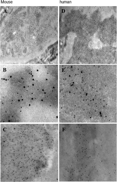
In several studies, we showed that chromatin fragments serve as central targets for nephritogenic anti-dsDNA/anti-nucleosome antibodies in all stages of lupus nephritis (35,59,60). The early phase of lupus nephritis correlated with deposition of chromatin fragment-IgG complexes in the mesangial matrix. By highresolution analytical approaches, such as IEM, colocalization IEM and colocalization terminal deoxynucleotidyl transferase dUTP nick end-labeling (TUNEL) IEM, IgG was never observed to bind in vivo directly to the GBM or to the mesangial matrix. But IgG did bind to TUNEL-positive EDSs associated with glomerular matrices and membranes (34,35) (Figure 1). This result was demonstrated in murine lupus nephritis (35) and was verified in patients with lupus nephritis (34).
Figure 1
Nephritogenic antibodies target chromatin exposed in glomeruli in the context of lupus nephritis as demonstrated by IEM. Nephritogenic antibodies bind chromatin fragments observed as EDSs in the GBM in murine (A-C) and human (D-F) lupus nephritis. _In vivo_-bound IgG antibodies are confined to EDSs present in the mesangial matrix and in the GBM in murine and human nephritis (A and D, respectively), as shown by IEM. In (A) and (D), the IgG molecules are stained with 5-nm gold particles. Their presence is confined to EDSs. In (B) and (E), the EDSs are shown to contain nicked DNA (traced by 10-nm gold particles) that colocalizes with _in vivo_-bound IgG (traced by 5-nm gold particles), as shown by a combination of IEM and the TUNEL assay (colocalization TUNEL IEM, (B) and (E) for murine and human nephritis, respectively). The colocalization TUNEL IEM was specific, since the assay performed in the absence of terminal deoxynucleotidyl transferase resulted in detection of _in vivo_-bound IgG (5 nm gold), but not in nicked DNA (absence of 10 nm gold, (C) and (F) for murine and human nephritis, respectively). These data show that targets for glomerular _in vivo_-bound IgG antibodies are observed as EDSs and contain DNA. The panels are previously unpublished images generated from the project linked to references (34) and (35) in the current article.
The link between production of anti-dsDNA antibodies and chromatin deposits in the mesangium indicates that the antibodies were directly responsible for this process (24), since we never observed chromatin in glomerular matrices and membranes in anti-dsDNA antibody-negative BW mice (22,24).
Progression of Murine Lupus Nephritis: A Possible Role for Renal Dnasei
At a certain time point, clinically silent mesangial nephritis progresses into endstage organ disease. This point was characterized by the deposition of chromatin fragment-IgG complexes in the GBM and by severe proteinuria. The progression was strongly associated with a loss of renal DNasel gene expression at both the transcriptional and the translational levels and with a severe loss of renal DNaseI enzyme activity as well. Silencing of the renal DNasel gene was demonstrated by Western blot, zymography, immunohistochemistry, immunofluorescence, quantitative polymerase chain reaction and unpublished Affymetrix microarray analyses (22–24,58). Because DNaseI is required for chromatin breakdown during apoptosis as well as necrosis (61,62), loss of this enzyme activity may lead to accumulation of apoptotic chromatin fragments in glomeruli. In line with this result, local accumulation of chromatin fragment-anti-dsDNA antibody complexes in GBM may be the core factor that imposes progressive renal inflammation in SLE (23,35,63).
The role of reduced DNasel in the pathogenesis of SLE has been discussed for decades. This discussion led to the opinion that DNasel could be a promising drug for substitutive therapy to prevent autoimmunity (64–66). Studies aimed at analyzing this option were performed in both BW mice (64,66) and human patients (65). However, this idea fell into oblivion for years because the treatment largely failed. Injection of DNasel in prenephritic BW mice delayed, but did not prevent, the disease in one study (64). In another study, administration of DNaseI in BW mice did not affect development or progression of lupus nephritis (66). Similarly, intravenous or subcutaneous administration of recombinant human DNaseI in patients with lupus nephritis class III–V did not affect kidney function or activity of the disease (65). These studies demonstrated indirectly that serum DNaseI had little or no influence on degradation of apoptotic chromatin in the kidney (61). Treatment failure may thus indicate that DNaseI in the extracellular compartments has a poor impact on chromatin deposited in the GBM.
Data from our laboratory as well as from other laboratories provide the basis for the following conclusions: (i) DNaseI is strongly expressed in healthy kidneys (67) (see also gene expression profiles from the BioGPS portal at https://doi.org/biogps.org) and account for 80% of total renal endonuclease activity (25); (ii) DNaseI expression is selectively reduced compared with other endonucleases expressed in nephritic kidneys (23); (iii) DNaseI expression is furthermore selectively silenced in affected kidneys and not in other organs and tissues that express DNaseI (23); (iv) silencing of the renal DNaseI gene is the result of an active regulation of gene expression and not caused by cell death, since other renal genes analyzed so far are not silenced but are expressed at normal levels (23,58); and (v) there is a highly significant inverse correlation between loss of DNaseI enzyme activity and progression of lupus nephritis, and with deposition of chromatin fragments in the GBM, while with normal or subnormal renal DNaseI gene expression, no deposits in GBM were observed (24,58). These conclusions do not, however, clarify if extracellular chromatin deposition secondary to loss of renal DNaseI is a direct or indirect consequence of local or systemic tissue damage and if the formation of anti-dsDNA antibodies in complex with chromatin is a secondary event that participates in development of nephritis. What the data demonstrate, however, is that chromatin-IgG complexes are involved in progression of lupus nephritis.
Biological Consequences of Loss of Dnasei Expression in Nephritic Kidneys
The biological consequences of renal DNasel shutdown and reduced chromatin fragmentation, beyond a classic immune complex disease, may also involve activation of cells of the innate immune system. Recently, we performed detailed analyses of renal expression of toll-like receptors (TLR) 7–9 and the necrosis-related Clec4e receptor in murine and human lupus nephritis. Furthermore, analyses were performed to determine if upregulation of matrix metalloproteases (MMPs) correlate with increased TLR7-9/Clec4e expression (68), since stimulation of TLR has been shown to upregulate certain MMPs in, for example, macrophages (69,70). For example, engagement of TLRs can upregulate the expression of proinflammatory cytokines (tumor necrosis factor [TNF]α and interferon [IFN]γ [71]) and interleukins (72,73) that directly induce expression of MMPs (71–73). Alternatively, incomplete clearance and degradation of apoptotic cells may transform them into secondary necrotic cell debris (63,74). This step may be significant, since necrotic cell debris contains SAP130, which serves as a ligand for the inflammation-related receptor Clec4e (75,76). Downstream signaling induced by SAP130-Clec4e receptor interaction also promotes production of proinflammatory cytokines (77) and a consequent upregulation of MMP expression. Increased MMP activity may (because of their gelatinase activity) disintegrate glomerular membranes (78). MMPs may therefore open membrane structures and facilitate deposition of chromatin fragment-IgG complexes in both the mesangial matrix and in the GBM.
We demonstrated that the expression of TLR7-9 and Clec4e was significantly upregulated in the BW mice at the same time when we observed the deposition of chromatin-IgG complexes in the GBM and the loss of renal DNaseI gene expression (68). We hypothesize that the presence of chromatin can be responsible for the increased TLR7-9/Clec4e expression. Thus, silencing of renal DNaseI expression may have immediate and harmful consequences linked to activation of the complement system by chromatin-IgG complexes (79). In addition, accumulated apoptotic chromatin fragments that are enriched in apoptosis-induced chromatin modifications (80–82) may affect cells of the innate immune system by triggering TLR7-9 and the Clec4e receptor. The apoptosis-induced chromatin modifications present in accumulated chromatin may be involved in sustained systemic autoimmune responses against chromatin (80–82).
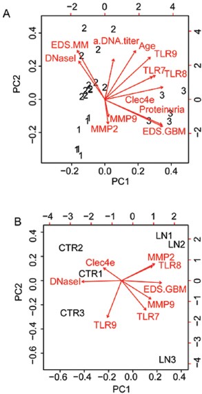
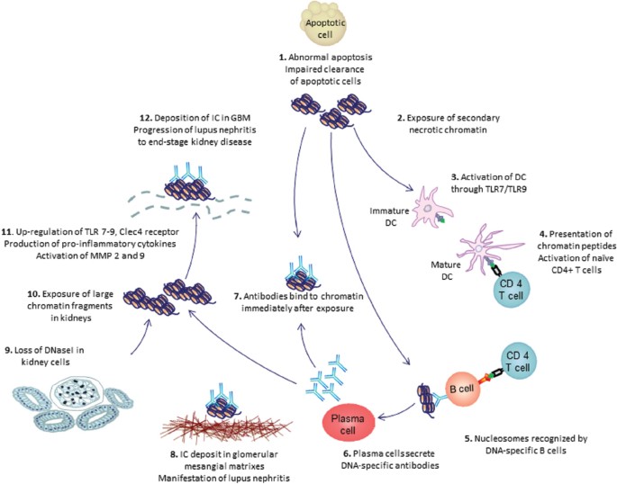
A principal component analysis (PCA) biplot of gene expression data from murine (Figure 2A) and human (Figure 2B) lupus nephritis demonstrated the importance of renal DNaseI gene shutdown for progression of the inflammatory organ disease. The data are taken from murine and human parameters published by Thiyagarajan et al. (68). These PCA biplots aim to display variances of the parameters. The angles between the various biplot axes (shown as arrows) indicate the correlations between the variables (shown as arrows). The angles between the arrows tell whether they are clustered and indicate that they correlate with each other. The different arrows point at the variables. The figure, therefore, shows which variables are clustered to indicate that they appear together. Similarly, the position of the samples of individual mice (Figure 2A, shown as the signs 1, 2 and 3 for group 1–3 mice) and of individual human controls and patients (Figure 2B) relative to the arrows, indicate which variable(s) have had the strongest impact on disease progression. In summary, the multiple effects of extracellular chromatin in lupus nephritis aggravate the autoinflammatory process that in the end will lead to end-stage renal disease, as outlined in Figure 3.
Figure 2
PCA of murine (A) and human (B) nephritic parameters (68). PCA biplots aim to optimally display variances and not correlations. The angles between the various biplot axes indicate the correlations between the parameters (shown as arrows). Similarly, the position of the samples of individual mice (shown as the signs 1, 2 and 3 for group 1–3 mice) relative to the arrows, indicates which variable(s) have had the largest effect on disease progression. The result of the biplots demonstrates that groups emerging from this analysis perfectly correlated with the groups of BW mice, as defined by Licht et al. (20), defined as prenephritic BW mice (group 1), BW mice with deposits of EDSs in the mesangial matrix (group 2) or with deposits in the GBM (group 3). In (B), a similar biplot was generated for the human data, where the three patients with low DNaseI expression levels are included. The most striking observation in these biplots is that the DNaseI vector points away from the individuals with severe lupus nephritis (to the left in the biplots), demonstrating that loss of DNaseI correlates inversely with disease progression, whereas MMP-2, MMP-9, TLRs and EDSs associated with GBM are clustered and points at the most severely diseased murine and human individuals, in harmony with statistical analyses demonstrated by Thiyagarajan et al. (68). This figure is modified from Thiyagarajan et al. (68).
Figure 3
The role of extracellular chromatin fragments and anti-dsDNA antibodies in progressive lupus nephritis. Retention of chromatin is assumed to start with reduced clearance of apoptotic cells (station 1). Secondary to this, chromatin may be exposed in tissues (station 2) and is assumed to activate dendritic cells (station 3). These cells present chromatin-derived peptides in the context of MHC class II molecules to peptide-specific CD4+ T cells (station 4). When primed, the peptide-specific T cells recirculate and bind the same chromatin-derived peptides presented in the context of MHC class II by chromatin-specific B-cells (here recognizing dsDNA in chromatin; station 5). As a consequence of cognate interaction of dsDNA-specific B cells and peptide-specific CD4+ T cells, the B cells transform into plasma cells that secrete IgG anti-dsDNA antibodies (station 6), which bind chromatin fragments (station 7). Immune complexes that consist of IgG antibodies and chromatin fragments bind in the glomerular mesangial matrix and initiate mesangial lupus nephritis (station 8). This early inflammation is followed by silencing of renal DnaseI in tubular and glomerular cells (station 9) and accumulation of undigested chromatin fragments (station 10). These fragments promote upregulation of TLRs, proinflammatory cytokines and matrix metalloproteases (station 11). Finally, IgG autoantibodies recognize and bind the chromatin fragments, and these immune complexes deposit in the GBMs and aggravate renal inflammation (station 12). In this sense, chromatin fragments and anti-chromatin (here: anti-dsDNA) antibodies are the partners that impose the classic murine and human lupus nephritis. Thus, anti-chromatin antibodies are pathogenic only in the context of exposed chromatin structures. This model does not exclude other processes that can initiate and maintain lupus nephritis, as discussed in the text.
Aberrant Renal DNaseI Gene Expression in Lupus Nephritis: Possible Regulatory Mechanisms
Silencing of the DNaseI gene in kidneys during progression of lupus nephritis may be controlled by different mechanisms. One possibility is a direct effect of early inflammation and secretion of proinflammatory cytokines in the context of early mesangial nephritis (studies in progress). By inspection of the DNaseI gene organization in the University of California, Santa Cruz, genome browser (https://doi.org/genome.ucsc.edu/index.html), we found an overlap of 59 nucleotides in the annotated transcript with a transcript from the convergently transcribed tumor necrosis factor receptor-associated protein 1 (Trap1) gene in the 3′-untranslated regions. This gene organization is peculiar and is likely to preclude coexpression of the two genes. Given the fact that transcription proceeds well beyond the 3′ end of the mature transcript (83,84), the overlap between the transcripts of the two genes is substantial, and recent data demonstrate that it is unlikely that the two genes can be transcribed simultaneously. This was directly demonstrated by Hobson et al. (85) when they analyzed transcription of convergent gene pairs. They used a combination of biochemical and genetic approaches and demonstrated that polymerases transcribing opposite and overlapping DNA strands cannot bypass each other. On the contrary, RNA polymerase II (RNAPII) molecules stop, and most importantly, do not dissociate from the DNA strands on head-to-head collision (85). This result suggests that opposing polymerases represent insurmountable blockades for each other (85). Finally, RNAPII induces chromatin modification, such as specific histone methylation and deacetylation, through the Set2/Rpd3S pathway (86,87). Such modifications will result in low to no expression of both genes.
The mutual suppression of one gene by expression of the other is called transcriptional interference (88). Recently, we showed that transcription of the DNaseI gene is inversely affected by Trap1 gene expression (89). More detailed analyses on regulation of DNaseI gene expression and the role of transcriptional interference with Trap1 gene expression are currently performed in our laboratory, involving studies of other regulators of DNaseI gene transcription such as miRNA (manuscript in preparation, KD Horvei, D Thiyagarajan, S Fismen, OP Rekvig, SD Johansen, H Nielsen) and the effect of DNA methylation.
Origin of Exposed Glomerular Chromatin
The processes that lead to exposure of renal chromatin are still an unresolved. Accelerated apoptosis or impaired clearance of apoptotic cells is thought to be involved (90–92). However, despite intensive investigations during the last decade, existing data on problems linked to disturbed apoptosis or clearance of apoptotic cells are not conclusive. Recent data expand our insight into these processes, since it was demonstrated that a loss-of-function variant in DNase1L3 causes a familial form of SLE with lupus nephritis (93). DNase1L3 is an apoptotic endonuclease predominantly expressed in macrophages (94) and dendritic cells (see https://doi.org/biogps.org) and therefore links an inactive apoptotic executer to a possible cause of lupus nephritis.
The cellular origin of chromatin fragments exposed in glomeruli in the context of lupus nephritis is difficult to assess. Accumulation of chromatin fragments in the GBM correlates in time with the loss of expression of the most important renal endonuclease (that is, DNaseI), and this result suggests a renal origin of the chromatin fragments. The fact that chromatin found in the GBM in human lupus nephritis can contain the polyomavirus BK large T antigen (95,96) also suggests a renal origin, since the BK virus possesses a selective tropism for tubular cells (97).
Another possibility is that that chromatin fragments reach glomeruli through the circulation, eventually in complex with IgG (40,63,98). This result explains the systemic character of tissue damage in SLE. Comparative studies of components of immune complexes and their localization in skin and glomerulus membranes and matrices demonstrated that they have similar structure (99,100). However, deposition of immune complexes in glomeruli did not predict deposition in skin. Examination of DNaseI and MMP expression in the skin in MRL-lpr/lpr mice demonstrated a stable activity of DNaseI and an increase in MMP-2 and MMP-9 enzyme activities during disease progression (100). These results indicate that circulating immune complexes can impose SLE manifestation in different organs, while progression of lupus nephritis may be organ-restricted because of the secondary mechanisms linked to silencing of the renal DNaseI gene.
Conclusion
There is comprehensive and coherent support for a model of lupus nephritis in which extracellular chromatin plays a direct role as a target structure for anti-dsDNA/anti-chromatin antibodies. Thus, a notable change in thinking entailed by recent studies is that chromatin fragments exposed in glomeruli are released from dying renal cells and that these fragments are not appropriately degraded during the programmed cell death process because of an acquired loss of the dominant renal endonuclease DNaseI. In this situation, accumulated chromatin fragments may be targeted by potentially nephritogenic anti-chromatin antibodies. In this sense, interaction of the antibodies and the chromatin targets is a homologous interaction. Therefore, the core nature of both murine and human lupus nephritis may point at exposure of chromatin in glomeruli and its complex formation with IgG as key events in disease pathogenesis and disease progression.
Disclosure
The authors declare that they have no competing interests as defined by Molecular Medicine, or other interests that might be perceived to influence the results and discussion reported in this paper.
References
- Ceppellini R, Polli E, Celada F. (1957) A DNA-reacting factor in serum of a patient with lupus erythematosus diffusus. Proc. Soc. Exp. Biol. Med. 96:572–4.
Article CAS PubMed Google Scholar - Robbins WC, Holman HR, Deicher H, Kungel HG. (1957) Complement fixation with cell nuclei and DNA in lupus erythematosus. Proc. Soc. Exp. Biol. Med. 96:575–9.
Article CAS PubMed Google Scholar - Seligmann M. (1957) Demonstration in the blood of patients with disseminated lupus erythematosus a substance determining a precipitation reaction with desoxyribonucleic acid [in French]. C. R. Hebd. Seances Acad. Sci. 245:243–5.
CAS PubMed Google Scholar - Miescher P, Strassle R. (1957) New serological methods for the detection of the L.E. factor. Vox Sang 2:283–7.
CAS PubMed Google Scholar - Izui S, Lambert PH, Fournie GJ, Turler H, Miescher PA. (1977) Features of systemic lupus erythematosus in mice injected with bacterial lipopolysaccharides: identificantion of circulating DNA and renal localization of DNA-anti-DNA complexes. J. Exp. Med. 145:1115–30.
Article CAS PubMed Google Scholar - Izui S, Lambert PH, Miescher PA. (1976) In vitro demonstration of a particular affinity of glomerular basement membrane and collagen for DNA: a possible basis for a local formation of DNA-anti-DNA complexes in systemic lupus erythematosus. J. Exp. Med. 144:428–43.
Article CAS PubMed Google Scholar - Khalil M, Spatz L, Diamond B. (1999) Anti-DNA antibodies. In Systemic Lupus Erythematosus. Lahita RG, Ed. Academic Press, San Diego, CA, pp. 197–217.
Google Scholar - Hahn BH. (1998) Antibodies to DNA. N. Engl. J. Med. 338:1359–68.
Article CAS PubMed Google Scholar - Xie C, Liang Z, Chang S, Mohan C. (2003) Use of a novel elution regimen reveals the dominance of polyreactive antinuclear autoantibodies in lupus kidneys. Arthritis Rheum. 48:2343–52.
Article CAS PubMed Google Scholar - Kalaaji M, Sturfelt G, Mjelle JE, Nossent H, Rekvig OP. (2006) Critical comparative analyses of anti-alpha-actinin and glomerulus-bound antibodies in human and murine lupus nephritis. Arthritis Rheum. 54:914–26.
Article CAS PubMed Google Scholar - Van Bruggen MC, Kramers C, Hylkema MN, Smeenk RJ, Berden JH. (1996) Significance of anti-nuclear and anti-extracellular matrix autoantibodies for albuminuria in murine lupus nephritis; a longitudinal study on plasma and glomerular eluates in MRL/l mice. Clin. Exp. Immunol. 105:132–9.
Article PubMed Google Scholar - Dang H, Harbeck RJ. (1984) The in vivo and in vitro glomerular deposition of isolated anti-double-stranded-DNA antibodies in NZB/W mice. Clin. Immunol. Immunopathol. 30:265–78.
Article CAS PubMed Google Scholar - Dang H, Harbeck RJ. (1982) A comparison of anti-DNA antibodies from serum and kidney eluates of NZB x NZW F1 mice. J. Clin. Lab. Immunol. 9:139–45.
CAS PubMed Google Scholar - Winfield JB, Faiferman I, Koffler D. (1977) Avidity of anti-DNA antibodies in serum and IgG glomerular eluates from patients with systemic lupus erythematosus: association of high avidity antinative DNA antibody with glomerulonephritis. J. Clin. Invest. 59:90–6.
Article CAS PubMed PubMed Central Google Scholar - Mortensen ES, Rekvig OP. (2009) Nephritogenic potential of anti-DNA antibodies against necrotic nucleosomes. J. Am. Soc. Nephrol. 20:696–704.
Article CAS PubMed Google Scholar - Jang YJ, Stollar BD. (2003) Anti-DNA antibodies: aspects of structure and pathogenicity. Cell. Mol. Life Sci. 60:309–20.
Article CAS PubMed Google Scholar - van der Vlag J, Berden JH. (2011) Lupus nephritis: role of antinucleosome autoantibodies. Semin. Nephrol. 31:376–89.
Article PubMed Google Scholar - Munoz LE, et al. (2009) Remnants of secondarily necrotic cells fuel inflammation in systemic lupus erythematosus. Arthritis Rheum. 60:1733–42.
Article CAS PubMed Google Scholar - Kruse K, et al. (2010) Inefficient clearance of dying cells in patients with SLE: anti-dsDNA autoantibodies, MFG-E8, HMGB-1 and other players. Apoptosis. 15:1098–113
Article CAS PubMed Google Scholar - Licht R, Dieker JW, Jacobs CW, Tax WJ, Berden JH. (2004) Decreased phagocytosis of apoptotic cells in diseased SLE mice. J. Autoimmun. 22:139–45.
Article CAS PubMed Google Scholar - Weening JJ, et al. (2004) The classification of glomerulonephritis in systemic lupus erythematosus revisited. J. Am. Soc. Nephrol. 15:241–50.
Article PubMed Google Scholar - Seredkina N, Zykova SN, Rekvig OP. (2009) Progression of murine lupus nephritis is linked to acquired renal Dnase1 deficiency and not to up-regulated apoptosis. Am. J. Pathol. 175:97–106.
Article CAS PubMed PubMed Central Google Scholar - Seredkina S, Rekvig OP. (2011) Acquired loss of renal nuclease activity is restricted to DNaseI and is an organ-selective feature in murine lupus nephritis. Am. J. Pathol. 179:1120–8.
Article CAS PubMed PubMed Central Google Scholar - Fenton K, et al. (2009) Anti-dsDNA antibodies promote initiation, and acquired loss of renal Dnase1 promotes progression of lupus nephritis in autoimmune (NZBxNZW)F1 mice. PLoS One. 4:e8474.
Article PubMed PubMed Central Google Scholar - Basnakian AG, et al. (2005) Cisplatin nephrotoxicity is mediated by deoxyribonuclease I. J. Am. Soc. Nephrol. 16:697–702.
Article CAS PubMed Google Scholar - Zykova SN, Seredkina N, Benjaminsen J, Rekvig OP. (2008) Reduced fragmentation of apoptotic chromatin is associated with nephritis in lupusprone (NZB × NZW)F(1) mice. Arthritis Rheum. 58:813–25.
Article CAS PubMed Google Scholar - Mostoslavsky G, et al. (2001) Lupus anti-DNA autoantibodies cross-react with a glomerular structural protein: a case for tissue injury by molecular mimicry. Eur. J. Immunol. 31:1221–7.
Article CAS PubMed Google Scholar - Zhao Z, et al. (2005) Cross-reactivity of human lupus anti-DNA antibodies with alpha-actinin and nephritogenic potential. Arthritis Rheum. 52:522–30.
Article CAS PubMed Google Scholar - Amital H, et al. (2005) Treatment with a laminin-derived peptide suppresses lupus nephritis. J. Immunol. 175:5516–23.
Article CAS PubMed Google Scholar - Raz E, Ben Bassat H, Davidi T, Shlomai Z, Eilat D. (1993) Cross-reactions of anti-DNA autoantibodies with cell surface proteins. Eur. J. Immunol. 23:383–90.
Article CAS PubMed Google Scholar - D’Andrea DM, Coupaye Gerard B, Kleyman TR, Foster MH, Madaio MP. (1996) Lupus autoantibodies interact directly with distinct glomerular and vascular cell surface antigens. Kidney Int. 49:1214–21.
Article PubMed Google Scholar - Krishnan MR, Wang C, Marion TN. (2012) Anti-DNA autoantibodies initiate experimental lupus nephritis by binding directly to the glomerular basement membrane in mice. Kidney Int. 82:184–92.
Article CAS PubMed PubMed Central Google Scholar - Manson JJ, et al. (2009) Relationship between anti-dsDNA, anti-nucleosome and anti-alpha-actinin antibodies and markers of renal disease in patients with lupus nephritis: a prospective longitudinal study. Arthritis Res. Ther. 11:R154.
Article PubMed PubMed Central Google Scholar - Kalaaji M, et al. (2007) Glomerular apoptotic nucleosomes are central target structures for nephritogenic antibodies in human SLE nephritis. Kidney Int. 71:664–72.
Article CAS PubMed Google Scholar - Kalaaji M, Mortensen E, Jorgensen L, Olsen R, Rekvig OP. (2006) Nephritogenic lupus antibodies recognize glomerular basement membrane-associated chromatin fragments released from apoptotic intraglomerular cells. Am. J. Pathol. 168:1779–92.
Article CAS PubMed PubMed Central Google Scholar - Schroeder K, Herrmann M, Winkler TH. (2013) The role of somatic hypermutation in the generation of pathogenic antibodies in SLE. Autoimmunity. 46:121–7.
Article CAS PubMed Google Scholar - Wellmann U, et al. (2005) The evolution of human anti-double-stranded DNA autoantibodies. Proc. Natl. Acad. Sci. U. S. A. 102:9258–63.
Article CAS PubMed PubMed Central Google Scholar - Murphy K. (2011) Janeway’s Immunobiology. Garland Science, New York. Chapter 4, Antigen Recognition by B-cell and T-cell Receptors; pp. 127–156; Chapter 5, The Generation of Lymphocyte Antigen Receptors; pp. 157–200; Chapter 10, The Humoral Immune Response; pp. 387–428.
Google Scholar - Starke C, et al. (2011) High frequency of autoantibody-secreting cells and long-lived plasma cells within inflamed kidneys of NZB/W F1 lupus mice. Eur. J. Immunol. 41:2107–12.
Article CAS PubMed Google Scholar - Radic M, Herrmann M, van der Vlag J, Rekvig OP. (2011) Regulatory and pathogenetic mechanisms of autoantibodies in SLE. Autoimmunity 44:349–56.
Article CAS PubMed Google Scholar - Xu H, Li H, Suri-Payer E, Hardy RR, Weigert M. (1998) Regulation of anti-DNA B cells in recombination-activating gene-deficient mice. J. Exp. Med. 188:1247–54.
Article CAS PubMed PubMed Central Google Scholar - Chen C, et al. (1994) Deletion and editing of B cells that express antibodies to DNA. J. Immunol. 152:1970–82.
Article CAS PubMed Google Scholar - Marion TN, Krishnan MR, Desai DD, Jou NT, Tillman DM. (1997) Monoclonal anti-DNA antibodies: structure, specificity, and biology. Methods. 11:3–11.
Article CAS PubMed Google Scholar - Carroll P, Stafford D, Schwartz RS, Stollar BD. (1985) Murine monoclonal anti-DNA autoantibodies bind to endogenous bacteria. J. Immunol. 135:1086–90.
Article CAS PubMed Google Scholar - Shoenfeld Y, et al. (1983) Polyspecificity of monoclonal lupus autoantibodies produced by humanhuman hybridomas. N. Engl. J. Med. 308:414–20.
Article CAS PubMed Google Scholar - Andrzejewski C Jr, Rauch J, Lafer E, Stollar BD, Schwartz RS. (1981) Antigen-binding diversity and idiotypic cross-reactions among hybridoma autoantibodies to DNA. J. Immunol. 126:226–31.
Article CAS PubMed Google Scholar - Lafer EM, et al. (1981) Polyspecific monoclonal lupus autoantibodies reactive with both polynucleotides and phospholipids. J. Exp. Med. 153:897–909.
Article CAS PubMed Google Scholar - van Bavel CC, Fenton KA, Rekvig OP, van der Vlag J, Berden JH. (2008) Glomerular targets of nephritogenic autoantibodies in systemic lupus erythematosus. Arthritis Rheum. 58:1892–9.
Article PubMed Google Scholar - Shlomchik M, et al. (1990) Anti-DNA antibodies from autoimmune mice arise by clonal expansion and somatic mutation. J. Exp. Med. 171:265–92.
Article CAS PubMed Google Scholar - Marion TN, Krishnan MR, Steeves MA, Desai DD. (2003) Affinity maturation and autoimmunity to DNA. Curr. Dir. Autoimmun. 6:123–53.
Article PubMed Google Scholar - Chan OT, Hannum LG, Haberman AM, Madaio MP, Shlomchik MJ. (1999) A novel mouse with B cells but lacking serum antibody reveals an antibody-independent role for B cells in murine lupus. J. Exp. Med. 189:1639–48.
Article CAS PubMed PubMed Central Google Scholar - Li S, Holdsworth SR, Tipping PG. (1997) Antibody independent crescentic glomerulonephritis in mu chain deficient mice. Kidney Int. 51:672–8.
Article CAS PubMed Google Scholar - Waters ST, et al. (2004) Breaking tolerance to double stranded DNA, nucleosome, and other nuclear antigens is not required for the pathogenesis of lupus glomerulonephritis. J. Exp. Med. 199:255–64.
Article CAS PubMed PubMed Central Google Scholar - Rovin BH, et al. (2012) Efficacy and safety of rituximab in patients with active proliferative lupus nephritis: the Lupus Nephritis Assessment with Rituximab study. Arthritis Rheum. 64:1215–26.
Article CAS PubMed Google Scholar - Ehrenstein MR, et al. (1995) Human IgG anti-DNA antibodies deposit in kidneys and induce proteinuria in SCID mice. Kidney Int. 48:705–11.
Article CAS PubMed Google Scholar - Ravirajan CT, et al. (1998) Genetic, structural and functional properties of an IgG DNA-binding monoclonal antibody from a lupus patient with nephritis. Eur. J. Immunol. 28:339–50.
Article CAS PubMed Google Scholar - Choi J, Kim ST, Craft J. (2012) The pathogenesis of systemic lupus erythematosus: an update. Curr. Opin. Immunol. 24:651–7.
Article CAS PubMed PubMed Central Google Scholar - Zykova SN, Tveita AA, Rekvig OP. (2010) Renal Dnase1 enzyme activity and protein expression is selectively shut down in murine and human membranoproliferative lupus nephritis. PLoS One. 10:5.
Google Scholar - Berden JH, Licht R, Van Bruggen MC, Tax WJ. (1999) Role of nucleosomes for induction and glomerular binding of autoantibodies in lupus nephritis. Curr. Opin. Nephrol. Hypertens. 8:299–306.
Article CAS PubMed Google Scholar - Van Bruggen MC, et al. (1997) Antigen specificity of anti-nuclear antibodies complexed to nucleosomes determines glomerular basement membrane binding in vivo. Eur. J. Immunol. 27:1564–9.
Article PubMed Google Scholar - Samejima K, Earnshaw WC. (2005) Trashing the genome: the role of nucleases during apoptosis. Nat. Rev. Mol. Cell. Biol. 6:677–88.
Article CAS PubMed Google Scholar - Kawane K, Nagata S. (2008) Nucleases in programmed cell death. Methods Enzymol. 442:271–87.
Article CAS PubMed Google Scholar - Berden JH, Grootscholten C, Jurgen WC, van der Vlag J. (2002) Lupus nephritis: a nucleosome waste disposal defect? J. Nephrol. 15 Suppl 6: S1–10.
CAS PubMed Google Scholar - Macanovic M, et al. (1996) The treatment of systemic lupus erythematosus (SLE) in NZB/W F1 hybrid mice: studies with recombinant murine DNase and with dexamethasone. Clin. Exp. Immunol. 106:243–52.
Article CAS PubMed PubMed Central Google Scholar - Davis JC Jr, et al. (1999) Recombinant human Dnase I (rhDNase) in patients with lupus nephritis. Lupus. 8:68–76.
Article PubMed Google Scholar - Verthelyi D, Dybdal N, Elias KA, Klinman DM. (1998) DNAse treatment does not improve the survival of lupus prone (NZB x NZW)F1 mice. Lupus. 7:223–30.
Article CAS PubMed Google Scholar - Napirei M, Ricken A, Eulitz D, Knoop H, Mannherz HG. (2004) Expression pattern of the deoxyribonuclease 1 gene: lessons from the Dnase1 knockout mouse. Biochem. J. 380:929–37.
Article CAS PubMed PubMed Central Google Scholar - Thiyagarajan D, et al. (2012) Silencing of renal DNaseI in murine lupus nephritis imposes exposure of large chromatin fragments and activation of toll like receptors and the Clec4e. PLoS One. 7:e34080.
Article CAS PubMed PubMed Central Google Scholar - Merrell MA, et al. (2006) Toll-like receptor 9 agonists promote cellular invasion by increasing matrix metalloproteinase activity. Mol. Cancer Res. 4:437–47.
Article CAS PubMed Google Scholar - Lim EJ, et al. (2006) Activation of toll-like receptor-9 induces matrix metalloproteinase-9 expression through Akt and tumor necrosis factor-alpha signaling. FEBS Lett. 580:4533–8.
Article CAS PubMed Google Scholar - Han YP, Tuan TL, Wu H, Hughes M, Garner WL. (2001) TNF-alpha stimulates activation of pro-MMP2 in human skin through NF-(kappa)B mediated induction of MT1-MMP. J. Cell Sci. 114:131–9.
Article CAS PubMed Google Scholar - Ben DD, Reznick AZ, Srouji S, Livne E. (2008) Exposure to pro-inflammatory cytokines upregulates MMP-9 synthesis by mesenchymal stem cells-derived osteoprogenitors. Histochem. Cell. Biol. 129:589–97.
Article Google Scholar - Triantafyllopoulou A, et al. (2010) Proliferative lesions and metalloproteinase activity in murine lupus nephritis mediated by type I interferons and macrophages. Proc. Natl. Acad. Sci. U. S. A. 107:3012–7.
Article CAS PubMed PubMed Central Google Scholar - Gaipl US, et al. (2006) Inefficient clearance of dying cells and autoreactivity. Curr. Top. Microbiol. Immunol. 305:161–76.
CAS PubMed Google Scholar - Brown GD. (2008) Sensing necrosis with Mincle. Nat. Immunol. 9:1099–100.
Article CAS PubMed Google Scholar - Yamasaki S, et al. (2008) Mincle is an ITAM-coupled activating receptor that senses damaged cells. Nat. Immunol. 9:1179–88.
Article CAS PubMed Google Scholar - Geijtenbeek TB, Gringhuis SI. (2009) Signalling through C-type lectin receptors: shaping immune responses. Nat. Rev. Immunol. 9:465–79.
Article CAS PubMed PubMed Central Google Scholar - Overall CM, Butler GS. (2007) Protease yoga: extreme flexibility of a matrix metalloproteinase. Structure. 15:1159–61.
Article CAS PubMed Google Scholar - Walport MJ. (2002) Complement and systemic lupus erythematosus. Arthritis Res. 4 Suppl 3: S279–93.
Article PubMed PubMed Central Google Scholar - van Bavel CC, et al. (2011) Apoptosis-induced histone H3 methylation is targeted by autoantibodies in systemic lupus erythematosus. Ann. Rheum. Dis. 70:201–7.
Article PubMed Google Scholar - Dieker JW, et al. (2007) Apoptosis-induced acetylation of histones is pathogenic in systemic lupus erythematosus. Arthritis Rheum. 56:1921–33.
Article CAS PubMed Google Scholar - Fransen JH, et al. (2009) Mouse dendritic cells matured by ingestion of apoptotic blebs induce T cells to produce interleukin-17. Arthritis Rheum. 60:2304–13.
Article CAS PubMed Google Scholar - Svejstrup JQ. (2013) RNA polymerase II transcript elongation. Biochim. Biophys. Acta. 1829:1.
Article CAS PubMed Google Scholar - Core LJ, Waterfall JJ, Lis JT. (2008) Nascent RNA sequencing reveals widespread pausing and divergent initiation at human promoters. Science. 322:1845–8.
Article CAS PubMed PubMed Central Google Scholar - Hobson DJ, Wei W, Steinmetz LM, Svejstrup JQ. (2012) RNA polymerase II collision interrupts convergent transcription. Mol. Cell. 48:365–74.
Article CAS PubMed PubMed Central Google Scholar - Kizer KO, et al. (2005) A novel domain in Set2 mediates RNA polymerase II interaction and couples histone H3 K36 methylation with transcript elongation. Mol. Cell. Biol. 25:3305–16.
Article CAS PubMed PubMed Central Google Scholar - Xiao T, et al. (2003) Phosphorylation of RNA polymerase II CTD regulates H3 methylation in yeast. Genes Dev. 17:654–63.
Article CAS PubMed PubMed Central Google Scholar - Shearwin KE, Callen BP, Egan JB. (2005) Transcriptional interference: a crash course. Trends Genet. 21:339–45.
Article CAS PubMed PubMed Central Google Scholar - Fismen S, et al. (2012) Impact of the tumor necrosis factor receptor-associated protein 1 (Trap1) on renal DNaseI shutdown and on progression of murine and human lupus nephritis. Am. J. Pathol. 182:688–700.
Article PubMed Google Scholar - Munoz LE, Lauber K, Schiller M, Manfredi AA, Herrmann M. (2010) The role of defective clearance of apoptotic cells in systemic autoimmunity. Nat. Rev. Rheumatol. 6:280–9.
Article PubMed Google Scholar - Dieker JW, van der Vlag J, Berden JH. (2004) Deranged removal of apoptotic cells: its role in the genesis of lupus. Nephrol. Dial. Transplant. 19:282–5.
Article PubMed Google Scholar - Kaplan MJ. (2004) Apoptosis in systemic lupus erythematosus. Clin. Immunol. 112:210–8.
Article CAS PubMed Google Scholar - Al-Mayouf SM, et al. (2011) Loss-of-function variant in DNASE1L3 causes a familial form of systemic lupus erythematosus. Nat. Genet. 43:1186–8.
Article CAS PubMed Google Scholar - Baron WF, et al. (1998) Cloning and characterization of an actin-resistant DNase I-like endonuclease secreted by macrophages. Gene. 215:291–301.
Article CAS PubMed Google Scholar - Bendiksen S, et al. (2008) Glomerular expression of large polyomavirus T antigen in binary tetoff regulated transgenic mice induces apoptosis, release of chromatin and initiates a lupus-like nephritis. Mol. Immunol. 45:728–39.
Article CAS PubMed Google Scholar - Fenton KA, Mjelle JE, Jakobsen S, Olsen R, Rekvig OP. (2008) Renal expression of polyomavirus large T antigen is associated with nephritis in human systemic lupus erythematosus. Mol. Immunol. 45:3117–24.
Article CAS PubMed Google Scholar - Fishman JA. (2002) BK virus nephropathy: polyomavirus adding insult to injury. N. Engl. J. Med. 347:527–30.
Article PubMed Google Scholar - Licht R, Van Bruggen MC, Oppers-Walgreen B, Rijke TP, Berden JH. (2001) Plasma levels of nucleosomes and nucleosome-autoantibody complexes in murine lupus: effects of disease progression and lipopolyssacharide administration. Arthritis Rheum. 44:1320–30.
Article CAS PubMed Google Scholar - Fismen S, et al. (2009) Circulating chromatin-anti-chromatin antibody complexes bind with high affinity to dermo-epidermal structures in murine and human lupus nephritis. Lupus. 18:597–607.
Article CAS PubMed Google Scholar - Hedberg A, Fismen S, Fenton KA, Mortensen ES, Rekvig OP. (2010) Deposition of chromatin-IgG complexes in skin of nephritic MRL-lpr/lpr mice is associated with increased local matrix metalloprotease activities. Exp. Dermatol. 19:e265–74.
Article PubMed Google Scholar
Acknowledgments
This study was supported by the Northern Norway Regional Health Authority Medical Research Program (grants SFP-100-04 and SFP-101-04), the Dutch Arthritis Association (grant 09-1-308) and the University of Tromsø as milieu support given to OP Rekvig.
Author information
Authors and Affiliations
- Molecular Pathology Research Group, Department of Medical Biology, Faculty of Health Sciences, University of Tromsø, N-9037, Tromsø, Norway
Natalya Seredkina, Elin Mortensen & Ole Petter Rekvig - Nephrology Research Laboratory, Department of Nephrology, Radboud University Nijmegen Medical Centre, Nijmegen, The Netherlands
Johan van der Vlag & Jo Berden
Authors
- Natalya Seredkina
You can also search for this author inPubMed Google Scholar - Johan van der Vlag
You can also search for this author inPubMed Google Scholar - Jo Berden
You can also search for this author inPubMed Google Scholar - Elin Mortensen
You can also search for this author inPubMed Google Scholar - Ole Petter Rekvig
You can also search for this author inPubMed Google Scholar
Corresponding author
Correspondence toOle Petter Rekvig.
Rights and permissions
Open Access This article is licensed under a Creative Commons Attribution-NonCommercial-NoDerivatives 4.0 International License, which permits any non-commercial use, sharing, distribution and reproduction in any medium or format, as long as you give appropriate credit to the original author(s) and the source, and provide a link to the Creative Commons license. You do not have permission under this license to share adapted material derived from this article or parts of it.
The images or other third party material in this article are included in the article’s Creative Commons license, unless indicated otherwise in a credit line to the material. If material is not included in the article’s Creative Commons license and your intended use is not permitted by statutory regulation or exceeds the permitted use, you will need to obtain permission directly from the copyright holder.
To view a copy of this license, visit (https://doi.org/creativecommons.org/licenses/by-nc-nd/4.0/)
About this article
Cite this article
Seredkina, N., van der Vlag, J., Berden, J. et al. Lupus Nephritis: Enigmas, Conflicting Models and an Emerging Concept.Mol Med 19, 161–169 (2013). https://doi.org/10.2119/molmed.2013.00010
- Received: 01 February 2013
- Accepted: 04 June 2013
- Published: 06 June 2013
- Issue Date: January 2013
- DOI: https://doi.org/10.2119/molmed.2013.00010


