Expression of sphingosine kinase gene in the interactions between human gastric carcinoma cell and vascular endothelial cell (original) (raw)
Gastric Cancer Open Access
Copyright ©The Author(s) 2002. Published by Baishideng Publishing Group Inc. All rights reserved.
World J Gastroenterol. Aug 15, 2002; 8(4): 602-607
Published online Aug 15, 2002. doi: 10.3748/wjg.v8.i4.602
Expression of sphingosine kinase gene in the interactions between human gastric carcinoma cell and vascular endothelial cell
Juan Ren, Department of Oncological Radiotherapy, First Hospital of Xi’an Jiaotong University, Xi’an 710061, Shaanxi Province, China
Lei Dong, Department of Gastroenterology, Second Hospital of Xi’an Jiaotong University, Xi’an 710004, Shaanxi Province, China
Cang-Bao Xu, Department of Pathophysiology, Lund University, Sweden
Bo-Rong Pan, Oncology Center, Xijing Hospital, Fourth Militry Medical University, Xi’an 710032, Shaanxi Province, China
ORCID number: $[AuthorORCIDs]
Author contributions: All authors contributed equally to the work.
Correspondence to: Dr. Juan Ren, Department of Oncological Radiotherapy, First Hospital, Xi’an Jiaotong University Xi’an 710061, Shaanxi Province, China. renjuan88@163.net
Telephone: +86-29-3058229 Fax: +86-29-4333028
Received: March 29, 2002
Revised: April 15, 2002
Accepted: April 20, 2002
Published online: August 15, 2002
Abstract
AIM: To study the interactions between human gastric carcinoma cell (HGCC) and human vascular endothelial cell (HVEC), and if the expression of sphingosine kinase (SPK) gene was involved in these interactions.
METHODS: The specific inhibitor to SPK, dimethyl sphingosine (DMS), was added acting on HGCC and HVEC, then the cell proliferation was measured by MTT. The conditioned mediums (CMs) of HGCC and HVEC were prepared. The CM of one kind of cell was added to the other kind of cell, and the cell proliferation was measured by MTT. After the action of CM, the cellular expression of SPK gene in mRNA level was detected with in situ hybridization (ISH).
RESULTS: DMS could almost completely inhibit the proliferation of HGCC and HVEC. The growth inhibitory rates could amount to 97.21%, 83.42%, respectively (P < 0.01). The CM of HGCC could stimulate the growth of HVEC (2.70 ± 0.01, P < 0.01) while the CM of HVEC could inhibit the growth of HGCC (52.97% ± 0.01%, P < 0.01). There was no significant change in the mRNA level of SPK gene in one kind of cell after the action of the CM of the other kind of cell.
CONCLUSION: SPK plays a key role in regulating the proliferation of HGCC and HVEC. There exist complicated interactions between HGCC and HVEC. HGCC can significantly stimulate the growth of HVEC while HVEC can significantly inhibit the growth of HGCC. The expression of SPK gene is not involved in the interactions.
Key Words: $[Keywords]
- Citation: Ren J, Dong L, Xu CB, Pan BR. Expression of sphingosine kinase gene in the interactions between human gastric carcinoma cell and vascular endothelial cell. World J Gastroenterol 2002; 8(4): 602-607
- URL: https://www.wjgnet.com/1007-9327/full/v8/i4/602.htm
- DOI: https://dx.doi.org/10.3748/wjg.v8.i4.602
INTRODUCTION
There exist many kinds of cells besides tumor cells in the solid neoplasm. These cells depend on each other and contribute together to the genesis, development, invasion and metastasis of tumor. The relations among all kinds of cells are very complicated. During the tumor angiogenesis and hematogenous metastasis, there exist complicated interactions between tumor cell (TC) and vascular endothelial cell (VEC)[1]. In the pre-angiogenesis, how does TC induce VEC to establish the tumor vascular system? On the other hand, the interactions between the two cells play a role in the tumor hematogenous metastasis. There exist some complicated mechanisms in these processes. The study of tumor angiogenesis mainly focuses on the interactions among the vascular component cells while the study of tumor metastasis mainly focuses on the interactions between TC and its surrounding stroma. Seldom does anyone notice the interactions between TC and VEC. To better understand some mechanisms in human gastric carcinoma angiogenesis and hematogenous metastasis, we selected human gastric carcinoma cell (HGCC) and human vascular endothelial cell (HVEC) to study the interactions between HGCC and HVEC and some mechanisms involved in these actions. Sphingosine kinase (SPK) is a newly found important kinase in regulating many biological functions of most kinds of cells. SPK can induce the synthesis of extracellular transmitter and intracellular second messanger, sphingosine-1-phosphate (SPP). To determine whether SPK took part in regulating the proliferation of HGCC and HVEC, the specific inhibitor to SPK, dimethyl sphingosine (DMS), was acted on the two cells[2]. The present study aims to probe into the interrelationships between HGCC and HVEC and if the expression of SPK gene was involved in these effects. The cell proliferation and the expression of SPK mRNA were measured after the action of the conditioned medium (CM) of the other kind of cell.
MATERIALS AND METHODS
Materials
Cell line HGCC line SGC7901 and HVEC line Eahy926 were employed.
Methods
Conditioned medium (CM) preparation, the ways of the actions of CM and DMS Cells in different confluent states were washed twice with PBS, then 3 mL culture medium was added to the cells. The medium what was taken as CM was collected after different periods. Cells were placed in serum free medium for growth arresting. After the growth of HGCC and HVEC was arrested for 24 h and 6 h respectively, the CM of the other kind of cell was added to the cell. The cell proliferation was measured by MTT after different culturing periods. The cell without the action of CM was taken as control. The action way of DMS was as same as the way of CM. The cell without the action of DMS was taken as control.
MTT (methyl tetrazolium colorimetry) 20 μL MTT solution (5 g/L) was added to 200 μL medium in each well of 96 well plate. Four hours later, the supernant was discarded, 150 μL DMSO was added in. After the crystal was dissolved completely, absorption spectrum (A) was measured at 490 nm in the enzyme linked immunosorbent assay meter. The inhibitory rate of cell proliferation = [1 - (the mean A of experimental group/The mean of control group)] × 100%
Detecting the expression of SPK gene After the action of CM of the other kind of cell, the cellular expression of SPK gene was detected by in situ hybridizition (ISH) for mRNA level. There has been no antibody to SPK available up to now, so that it is impossible to detect the level of SPK protein. Probe specific to SPK mRNA was designed with the software "rimer 3" The sequence of SPK probe is 5'-ATATACCAAGTAGGGGCATTCATACTC-3' Probe labeling and ISH were carried on according to the manual of the Dig Olignucleotide Tailing Kit and Dig Detection Kit (Boehringer Mannheim, Germany) respectively. PBS was substituted for anti-Dig-Ap as negative control. The sections were analyzed for A value in the image analysis apparatus.
Statistical analysis
t test was used to compare the means.
RESULTS
Effects of DMS on the proliferation of HGCC and HVEC
DMS could almost completely inhibit the proliferation of HGCC and HVEC. The growth inhibitory rates could amount to 97.21% and 83.42% respectively (b_P_ < 0.01 vs control). DMS could produce effects in 1 μmol/L and 3 h later. The inhibitory rate was related to the dose and action periods. Ten μmol/L DMS inhibited the proliferation of HGCC significantly, and the effect was increasing by leaps and bounds when the dose was from 3.5 μmol/L to 5 μmol/L or the action period was from 24 h to 48 h. Twenty-five μmol/L DMS inhibited the growth of HVEC significantly, and the effect was increasing by leaps and bounds when the dose was from 10 μmol/L to 15 μmol/L or the action period was from 48 h to 72 h (Table 1, Table 2).
Table 1 Dose-effect of DMS on HGCC and HVEC for 48 h (n = 8, ¯x ± s).
| DMS dose/(μmol/L) | Inhibitory rate of cell proliferation (%) | |
|---|---|---|
| HGCC | HVEC | |
| 0 (control) | 0 | 0 |
| 1 | 12.33b | 5.61b |
| 2 | 17.53b | 11.11b |
| 3.5 | 31.31b | 17.77b |
| 5 | 54.68b | 18.41b |
| 7.5 | 64.34b | 19.82b |
| 10 | 80.68b | 31.02b |
| 15 | 89.04b | 65.76b |
| 20 | 92.67b | 76.02b |
| 25 | 95.91b | 82.32b |
| 30 | 97.21b | 83.42b |
Table 2 Time-effect of 10 μmol/L DMS on HGCC and HVEC (n = 8, ¯x ± s).
| DMS action periods/h | Inhibitory rate of cell proliferation (%) | |
|---|---|---|
| HGCC | HVEC | |
| 0 (control) | 0 | 0 |
| 3 | 9.11b | 5.18b |
| 6 | 13.46b | 7.05b |
| 12 | 20.18b | 11.12b |
| 24 | 41.98b | 19.52b |
| 48 | 80.68b | 31.02b |
| 72 | 93.55b | 56.44b |
Effect of conditioned medium of gastric carcinoma cell on vascular endothelial cell
The conditioned medium of HGCC could stimulate the proliferation of HVEC (a_P_ < 0.05 vs no-CM action group) significantly. The stimulation effect was related to the different cell confluent states, different preparing periods, different volume fractions and different action periods (Table 3, Table 4).
Table 3 Different dose-time-effects of CMs of subconfluent and confluent HGCC on HVEC (n = 8, ¯x ± s).
| CM volume fraction _t/_h cell confluent state | A of CM group/A of control group | |||||||||
|---|---|---|---|---|---|---|---|---|---|---|
| 100%CM | 80%CM | 50%CM | 30%CM | 10%CM | ||||||
| subconfluent | confluent | subconfluent | confluent | subconfluent | confluent | subconfluent | confluent | subconfluent | confluent | |
| 24 | 2.38 ± 0.01a | 1.29 ± 0.00ab | 2.70 ± 0.01a | 1.55 ± 0.02ab | 2.21 ± 0.01a | 1.49 ± 0.02ab | 2.10 ± 0.03a | 1.44 ± 0.01ab | 1.92 ± 0.02a | 1.11 ± 0.01ab |
| 48 | 1.60 ± 0.01a | 1.22 ± 0.01ab | 1.54 ± 0.01a | 1.33 ± 0.02ab | 1.38 ± 0.02a | 1.23 ± 0.02ab | 1.37 ± 0.02a | 1.21 ± 0.01ab | 1.31 ± 0.01a | 1.15 ± 0.02ab |
| 72 | 0.89 ± 0.00 | 0.85 ± 0.00 | 0.99 ± 0.01 | 1.03 ± 0.01 | 1.07 ± 0.01a | 1.11 ± 0.01a | 1.27 ± 0.01a | 1.01 ± 0.00b | 1.01 ± 0.00 | 1.15 ± 0.01a |
Table 4 Different dose-time-effect of CMs of different preparing periods of confluent HGCC on HVEC (n = 8, ¯x ± s).
| CM volume fraction _t/_h Preparing periods | A of CM group/A of control group | |||||||||
|---|---|---|---|---|---|---|---|---|---|---|
| 100%CM | 80%CM | 50%CM | 30%CM | 10%CM | ||||||
| 24 hCM | 48 hCM | 24 hCM | 48 hCM | 24 hCM | 48 hCM | 24 hCM | 48 hCM | 24 hCM | 48 hCM | |
| 24 | 1.29 ± 0.01a | 1.38 ± 0.01ab | 1.55 ± 0.02a | 1.39 ± 0.01ab | 1.49 ± 0.01a | 1.38 ± 0.01ab | 1.44 ± 0.02a | 1.30 ± 0.01ab | 1.11 ± 0.01a | 1.00 ± 0.01b |
| 48 | 1.22 ± 0.01a | 1.11 ± 0.02ab | 1.33 ± 0.01a | 1.14 ± 0.02ab | 1.23 ± 0.01a | 1.03 ± 0.01b | 1.21 ± 0.01a | 1.03 ± 0.01b | 1.15 ± 0.01a | 1.02 ± 0.01b |
| 72 | 0.85 ± 0.01 | 0.78 ± 0.00 | 0.99 ± 0.01 | 0.92 ± 0.00 | 1.07 ± 0.02a | 1.14 ± 0.01ab | 1.27 ± 0.01a | 1.20 ± 0.01ab | 1.15 ± 0.01a | 1.16 ± 0.01a |
Effect of conditioned medium of vascular endothelial cell on gastric carcinoma cell
The conditioned medium of HVEC could inhibit the proliferation of HGCC significantly (b_P_ < 0.01 vs no-CM action group). The inhibitory effect was related to the different cell confluent states and different volume fractions (Table 5).
Table 5 Effects of CMs of subconflunt and confluent HVEC on HGCC for 48 h (n = 8, ¯x ± s).
| Volume fraction | Inhibitory rate of cell proliferation (%) | |
|---|---|---|
| CM of subconfluent HVEC | CM of confluent HVEC | |
| 100%CM | 52.97 ± 0.01a | 31.62 ± 0.02ab |
| 80%CM | 54.26 ± 0.01a | 30.46 ± 0.01ab |
| 50%CM | 23.46 ± 0.01a | 19.00 ± 0.01ab |
| 30%CM | 21.70 ± 0.00a | 2.13 ± 0.01b |
| 10%CM | 14.36 ± 0.00a | 1.61 ± 0.00b |
Expression of SPK gene in HVEC before and after the action of HGCC CM
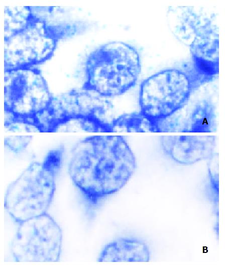
The mRNA level (A value of ISH) in HVEC before and after the action of HGCC CM were (0.265 ± 0.016), (0.264 ± 0.021) respectively. There was no significant difference between them. So, after the CM of HGCC acted on HVEC, the expression level of SPK gene in HVEC had no significant change (Figure 1).
Figure 1 Expression of SPK mRNA in HVEC after the action of the CM of HGCC. A: SPK mRNA in HVEC without the action of CM of HGCC (Control) (in situ hybridization); B: SPK mRNA in HVEC after the action of CM of HGCC (in situ hybridization).
Expression of SPK gene in HGCC before and after the action of HVEC CM
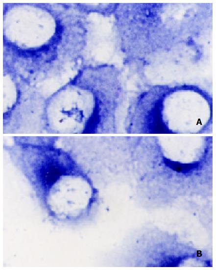
The mRNA level (A value of ISH) in HGCC before and after the action of HVEC CM were (0.244 ± 0.016), (0.243 ± 0.018) respectively. There was no significant difference between them. So, after the CM of HVEC acted on HGCC, the expression level of SPK gene in HGCC had not been significantly changed (Figure 2).
Figure 2 Expression of SPK mRNA in HGCC after the action of the CM of HVEC. A: SPK mRNA in HGCC without the action of CM of HVEC (Control) (in situ hybridization); B: SPK mRNA in HGCC after the action of CM of HVEC (in situ hybridization)
DISCUSSION
In the human solid neoplasm, there exist many kinds of cells besides the tumor cell, such as: interstitial cell, immunocyte and vascular cell. They depend on each other to contribute to the tumor genesis, development, invasion and metastasis. There exist complicated interactions among these cells. During tumor angiogenesis and tumor hematogenous metastasis, both vascular endothelial cell (VEC) and tumor cell (TC) contribute to finishing the pathology processes. The interactions between VEC and TC act in close coordination in establishing tumor vascular system and finishing hematogenous metastasis. There are very complicated interrelations between TC and VEC. In the pre-angiogenesis, how does TC induce VEC to establish the tumor vascular system? That is, how does TC influence the proliferation, degeneration, morphogenesis and functions of its neighboring VEC? Or, how does VEC influence these characters of its neighboring TC? These are all unclear. On the other hand, the interactions between two cells are involved in the tumor infiltration and hematogenous metastasis. Tumor vascular provides the passage for tumor infiltration and metastasis. The integrated vascular endothelium is the barrier to the tumor infiltration and metastasis. How do these two kinds of cells interact reciprocally to render tumor cell to adhere to and destroy the vascular endothelium to enter vascular lumen and damage it again to enter stroma? There exist some complicated mechanisms in this process. The study of tumor angiogenesis mainly focuses on the interactions among the vascular component cells while the study of tumor invasion and metastasis mainly focuses on the interactions between TC and surrounding stroma. Seldom has anyone noticed the interactions between TC and VEC. The relations between TC and VEC are not clear. Studies on this aspect are very few. Studies abroad always choose melanoma, glioblastoma, cephalo-cervical squamous carcinoma and hepatocellular carcinoma[3-18], but not gastric carcinoma as the target research yet. And the results are also controversial. We have not found anyone who studies gastric carcinoma yet. There are lots of studies on gastric carcinoma[19-29], but seldom in this aspect. To better understand some mechanisms in gastric carcinoma angiogenesis and hematogenous metastases, we select human gastric carcinoma cell (HGCC) and human vascular endothelial cell (HVEC) to study their interrelations and the mechanisms.
Results in this study showed that CMs of HGCC with different confluent states, different preparing periods, different volume fractions and different action periods could all stimulate the growth of HVEC significantly. The activity of CM of subconfluent cell was stronger than that of CM of confluent cell. The activity of CM preparing for 24 h was stronger than that of CM preparing for 48 h. After the nutrition exhaustion was replenished, the more of the volume fraction, the stronger of the activity. This is consistent with the results of some studies. Someone found CM of bladder carcinoma stimulated the growth of HVEC. Others found that cephalo-cervical squamous carcinoma cell stimulated the growth of HVEC through secreting FGF and VEGF. But there were other contrary viewpoints. Zhao found bladder carcinoma cell inhibited the growth of HVEC through a 10-16 bp fragment of tRNA. Albini found some kinds of TCs inhibited HVEC to form vascular through secreting IFN-γ. There was also a neutral objection: TC has little effect on the proliferation of HVEC. Some researchers found that although TC had no effect on the growth of HVEC, TC could change morphology of HVEC or its sensitivity to TNF-α. We think these different results are due to different cell types. Our results also showed that CM of HVEC could inhibit the growth of HGCC significantly. The activity of CM of subconfluent cell was stronger than that of CM of confluent cell. After the nutrition exhaustion was replenished, the more of the volume fraction, the stronger of the activity. We do not know what this means exactly. Maybe in the tumor angiogenesis, this inhibition could prevent HGCC to occupy the place of vascular or participate into the vascularition. Then the stimulation of vascular system to the growth of gastric carcinoma is not through the direct interactions between HGCC and HVEC, but through the establishment of the vascular system that passes nutrition to TC and excretes its metabolism waste. Whether HVEC secretes some growth-inhibiting factors to inhibit the proliferation of HGCC or not needs further study.
Sphingosine 1-phosphate (SPP) is a newly found important extracellular transmitter and intracellular second messanger. SPP regulates many biological functions of most kinds of cells through taking part in several signal transduction pathways which include MAPK/ERK (mitogen activated protein kinase/extracellular regulating kinase, MAPK/ERK) pathway, CAMP signal transduction pathway, Ca2+/CaM (Caldesmon, CaM) pathway and phospholipase D pathway. The functions of SPP include: regulating the proliferation, morphology, migration, adhesion and apoptosis of cells, maintaining the structure of the endothelium and epithelium, regulating vasculagenesis, regulating the cardiovascular function, regulating the intracellular level of Ca2+ and K+, regulating tumor metastasis and so on[29-39]. The synthesis of SPP depends on the activation of sphingosine kinase (SPK). The activation of SPK is closely related to the survival and function of cells[40-49]. Lots of factors can activate SPK such as PDGF (platelet derived growth factor, PDGF) and Fc receptors. The concentrations of SPK in the plasma and serum are 200 nM, 500 nM respectively. There are several phosphorylated sites and combination sites for Ca2+ and CaM in the SPK sequence. The sequence of SPK is highly conservative from yeast, protozoon to mammal. The activity of SPK can be detected in the cytoplasm and cell membrane. The functions of SPK are somewhat similar to the functions of PLC (phospholipase C, PLC). To define the role of SPK in regulating the proliferation of HGCC and HVEC, the specific inhibitor to SPK, DMS, was added to the two cells. Results showed that DMS could almost completely inhibit the proliferation of HGCC, HVEC and DMS could produce effects in 1 μmol/L and 3 h later. This illustrated that the regulation of DMS on the proliferation of two cells was prompt, sensitive and striking. Considering SPK was an important regulator to the growth of HGCC and HVEC, we studied the role of SPK in the interactions between these two cells. We found that the expression of SPK mRNA in one kind of cell with no significant change after the action of the CM of the other kind of cell. This showed that the expression of SPK gene didn't involve in the interactions between two cells. Maybe the regulation of one cell on the other cell is through some other factors or pathways. It needs further study to define the other mechanisms involved in the interrelationships between HGCC and HVEC.
Footnotes
Edited by Zhao P
References
| 1. | Ren J, Dong L, Xu CB, Pan BR, Li MZ. Interactions between the human gastric carcinoma cell and vascular endothelial cell. Shijie Huaren Xiaohua Zazhi. 2001;9:1254-1260. [PubMed] [DOI] [Cited in This Article: ] |
|---|
| 2. | Ren J, Dong L, Xu CB. Role of sphingosine-1-phosphate in regulating the proliferation of human gastric carcinoma cell and vascular endothelial cell. Xi’an Yike Daxue Xuebao. 2001;6:527-531. [PubMed] [DOI] [Cited in This Article: ] |
|---|
| 4. | Okamoto H, Ohigashi H, Nakamori S, Ishikawa O, Imaoka S, Mukai M, Kusama T, Fujii H, Matsumoto Y, Akedo H. Reciprocal functions of liver tumor cells and endothelial cells. Involvement of endothelial cell migration and tumor cell proliferation at a primary site in distant metastasis. Eur Surg Res. 2000;32:374-379. [PubMed] [DOI] [Cited in This Article: ] |
|---|
| 10. | Witt CJ, Gabel SP, Meisinger J, Werra G, Liu SW, Young MR. Interrelationship between protein phosphatase-2A and cytoskeletal architecture during the endothelial cell response to soluble products produced by human head and neck cancer. Otolaryngol Head Neck Surg. 2000;122:721-727. [PubMed] [DOI] [Cited in This Article: ] |
|---|
| 19. | Wu K, Zhao Y, Liu BH, Li Y, Liu F, Guo J, Yu WP. RRR-alpha-tocopheryl succinate inhibits human gastric cancer SGC-7901 cell growth by inducing apoptosis and DNA synthesis arrest. World J Gastroenterol. 2002;8:26-30. [PubMed] [DOI] [Cited in This Article: ] |
|---|
| 20. | Wang X, Lan M, Shi YQ, Lu J, Zhong YX, Wu HP, Zai HH, Ding J, Wu KC, Pan BR. Differential display of vincristine-resistance-related genes in gastric cancer SGC7901 cell. World J Gastroenterol. 2002;8:54-59. [PubMed] [DOI] [Cited in This Article: ] |
|---|
| 21. | Yao YL, Xu B, Song YG, Zhang WD. Overexpression of cyclin E in Mongolian gerbil with Helicobacter pylori-induced gastric precancerosis. World J Gastroenterol. 2002;8:60-63. [PubMed] [DOI] [Cited in This Article: ] |
|---|
| 22. | Yang SM, Fang DC, Luo YH, Lu R, Liu WW. Effect of antisense gene to hum an telomerase reverse transcriptase on telomerase activity and expression of apoptosis-associated gene. Shijie Huaren Xiaohua Zazhi. 2002;10:149-152. [PubMed] [DOI] [Cited in This Article: ] |
|---|
| 23. | Zheng ZH, Xun XJ, Qiu GR, Liu YX, Wang MX, Sun KL. E-cadherin gene mutation in precancerous condition、early and advanced stage of gastric cancer. Shijie Huaren Xiaohua Zazhi. 2002;10:153-156. [PubMed] [DOI] [Cited in This Article: ] |
|---|
| 24. | Wang W, Luo HS, Yu BP. Expression of human telomerase reverse transcriptase gene and c-myc protein in gastric carcinogenesis. Shijie Huaren Xiaohua Zazhi. 2002;10:258-261. [PubMed] [DOI] [Cited in This Article: ] |
|---|
| 25. | Li JY, Yu JP, Luo HS, Yu BP, Huang JA. Effects of nonsteroidal anti-inflammatory drugs on the proliferation and cyclooxygenase activity of gastric cancer cell line SGC7901. Shijie Huaren Xiaohua Zazhi. 2002;10:262-265. [PubMed] [DOI] [Cited in This Article: ] |
|---|
| 26. | Xue FB, Xu YY, Wan Y, Pan BR, Ren J, Fan DM. Association of H. pylori infection with gastric carcinoma: a Meta analysis. World J Gastroenterol. 2001;7:801-804. [PubMed] [DOI] [Cited in This Article: ] |
|---|
| 27. | Liu DH, Zhang XY, Fan DM, Huang YX, Zhang JS, Huang WQ, Zhang YQ, Huang QS, Ma WY, Chai YB. Expression of vascular endothelial growth factor and its role in oncogenesis of human gastric carcinoma. World J Gastroenterol. 2001;7:500-505. [PubMed] [DOI] [Cited in This Article: ] |
|---|
| 28. | Cai L, Yu SZ, Zhang ZF. Glutathione S-transferases M1, T1 genotypes and the risk of gastric cancer: a case-control study. World J Gastroenterol. 2001;7:506-509. [PubMed] [DOI] [Cited in This Article: ] |
|---|
| 29. | He XS, Su Q, Chen ZC, He XT, Long ZF, Ling H, Zhang LR. Expression, deletion [was deleton] and mutation of p16 gene in human gastric cancer. World J Gastroenterol. 2001;7:515-521. [PubMed] [DOI] [Cited in This Article: ] |
|---|
| 41. | Melendez AJ, Khaw AK. Dichotomy of Ca2 signals triggered by different phospholipid pathways in antigen stimulation of human mast cells. J Biol Chem. 2002;[epub ahead of print]. [PubMed] [DOI] [Cited in This Article: ] |
|---|
| 44. | Vann LR, Payne SG, Edsall LC, Twitty S, Spiegel S, Milstien S. Involvement of sphingosine kinase in TNF-alpha-stimulated tetrahydrobiopterin biosynthesis in C6 glioma cells. J Biol Chem. 2002;277:12649-12656. [PubMed] [DOI] [Cited in This Article: ] |
|---|

