Angiogenesis inhibited by drinking tea (original) (raw)
EGCG has been reported to inhibit urokinase, which may be used by some tumours for the invasion of neighbouring healthy tissues4. However, the effective concentration of EGCG seems to be too high for it to be physiologically relevant for tea drinkers5. The growth of almost all types of tumour is dependent on angiogenesis, and the enlargement and metastasis of tumours is disrupted when this is suppressed6.
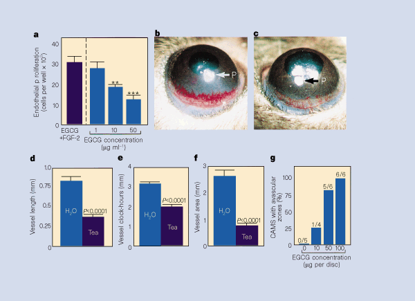
We thought that the inhibition of tumour growth by green tea might be mediated through the suppression of blood-vessel growth. To determine whether green tea could inhibit endothelial cell growth, we assayed EGCG on bovine capillary endothelial cells stimulated with the fibroblast growth factor FGF-2, as described7. We found that EGCG inhibited endothelial cell growth in a dose-dependent manner (Fig. 1a). The inhibition appeared to be specific to endothelial cells because non-endothelial cells, including murine T241 fibrosarcoma tumour cells, murine fibroblast cells, and rat smooth-muscle cells, were insensitive to EGCG treatment at the concentrations used.
Figure 1: Suppression of endothelial growth and angiogenesis by EGCG and green tea.
a, Results of a bovine capillary endothelial cell proliferation assay7. We seeded 10,000 cells per well in gelatinized 24-well plates and grew them in DME medium containing 5% bovine calf serum. EGCG samples in triplicates and FGF-2 at a final concentration of 1 ng ml−1 were added to each well. After 72 h incubation, cells were counted with a Coulter counter. Values are mean (±s.e.m.) number of cells from 3 determinations. **P <0.005, ***P <0.0001. **b-f,**Suppression of VEGF-stimulated vascularization by green tea in the mouse corneal model. Mice drank Chinese green-tea water extract9,10 starting at day 3 before VEGF implantation. Pellets containing 160 ng VEGF, sucrose, aluminium sulphate and hydron were implanted into corneal micropockets8. **b,**Cornea of a water-drinking mouse 5 days after implantation. **c,**Cornea of a green-tea-drinking mouse. **d-f,**Vessel length, clock-hours and vessel area. g, Results of the chorioallantoic membrane (CAM) assay8. Discs of methylcellulose containing EGCG dried on nylon mesh were implanted on the CAMs of individual embryos. Embryos and CAMs were examined by stereoscope after 48 h for the formation of avascular zones in the field of the implanted discs. At various doses, the number of CAMs with avascular zones over the total number of CAMs is indicated above each bar.
We examined the effect of EGCG on angiogenesis in the chick chorioallantoic membrane assay8. Over a concentration range of 1 to 100 µg per disc, EGCG inhibited new blood-vessel growth in a dose-dependent manner (Fig. 1g), as measured by the formation of avascular zones.
To investigate whether tea could suppress angiogenesis, we made green tea the sole drinking fluid for mice and examined the effect of oral consumption on the inhibition of corneal neovascularization stimulated by vascular endothelial growth factor (VEGF). The corneal model is a rigorous antiangiogenic assay, requiring systemic administration of a putative angiogenesis inhibitor to suppress neovascularization induced by 160 ng of VEGF in the cornea. The amount of green tea in the drinking water was 1.25% (4.69 mg ml-1) containing 708 µg ml-1 EGCG9,10. The concentration of EGCG in the plasma was previously reported to be in the range of 0.1-0.3 µM, which is similar to levels in humans after drinking two or three cups of tea11.
This tea preparation has been shown to significantly suppress the growth and progression of tumours in the lungs in animal models9. Compared with the control group that drank water alone (Fig. 1b), drinking tea significantly prevented VEGF-induced corneal neovascularization (Fig. 1c). Blood-vessel length (Fig. 1d), clock-hours of corneal neovascularization (the proportion of the circumference that is vascularized if the eye is viewed as a clock; Fig. 1e) and area of neovascularization (Fig. 1f) in seven corneas from four mice in the tea-drinking group were inhibited by approximately 55, 35 and 70%, respectively.
Our data indicate that EGCG suppresses endothelial cell growth in vitro and the formation of new blood vessels in chick chorioallantoic membrane. Drinking green tea significantly prevents corneal neovascularization induced by one of the most potent angiogenic factors, VEGF. Because the growth of all solid tumours is dependent on angiogenesis6, this finding may explain why drinking green tea prevents the growth of a variety of different types of tumour.
