Reactive syringofibroadenoma - Indian Journal of Dermatology, Venereology and Leprology (original) (raw)
Translate this page into:
Images in Clinical Practice
2020:86:6;681-682
doi: 10.4103/ijdvl.IJDVL_924_18
PMID: 31249214
Piyush Kumar1 , Anupam Das2 , PC Das3
1 Department of Dermatology, Katihar Medical College and Hospital, Katihar, Bihar, India
2 Department of Dermatology, KPC Medical College and Hospital, Kolkata, West Bengal, India
3 Consultant Dermatologist, Apurva Skin Centre, Katihar, Bihar, India
Correspondence Address:
Anupam Das
Building “Prerana,” 19 Phoolbagan, Kolkata - 700 086, West Bengal
India
How to cite this article:
Kumar P, Das A, Das P C. Reactive syringofibroadenoma. Indian J Dermatol Venereol Leprol 2020;86:681-682
Copyright: (C)2020 Indian Journal of Dermatology, Venereology, and Leprology
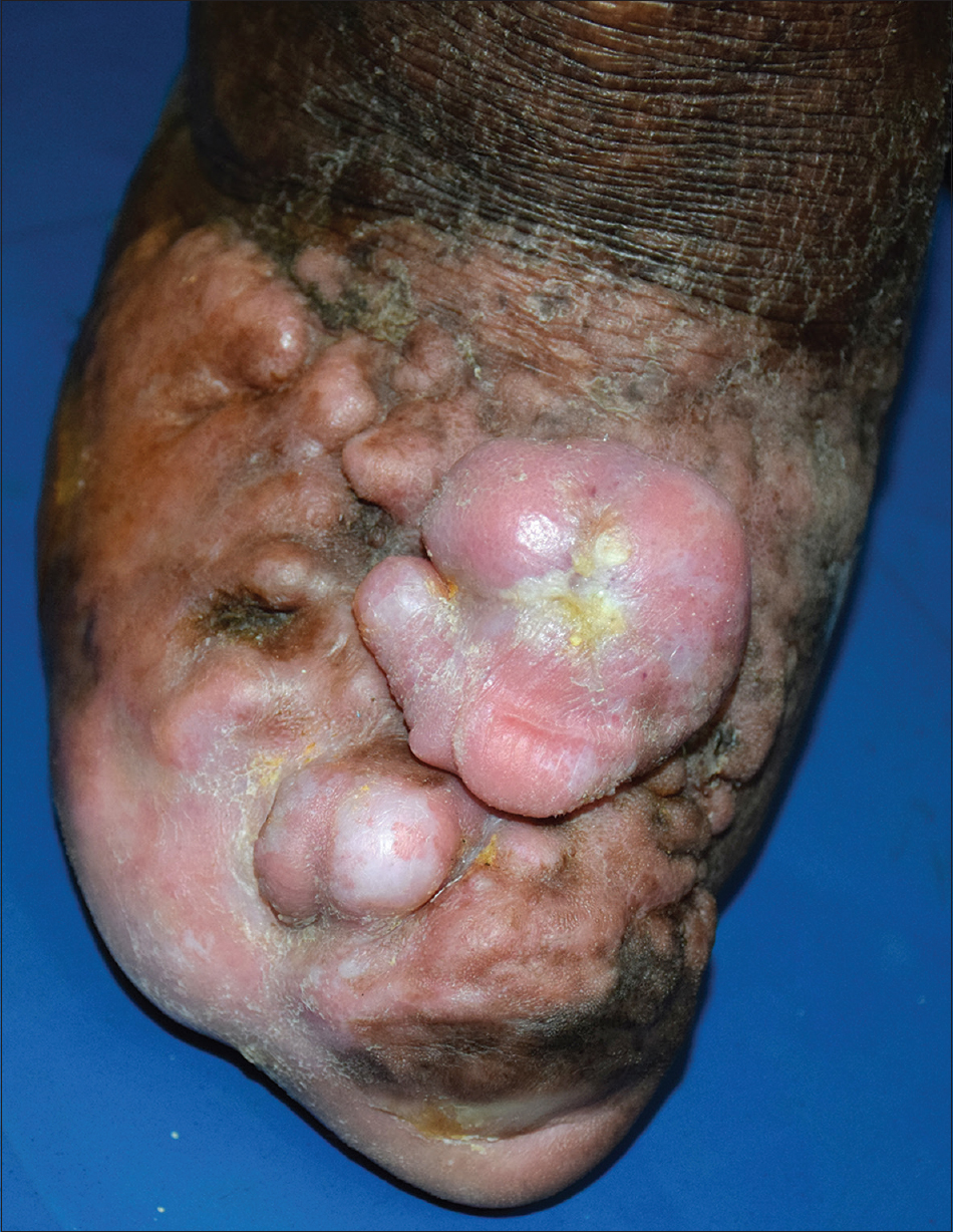
A 45-year-old diabetic and hypertensive man from Bihar presented with multiple, asymptomatic, elevated lesions on his right foot that developed over a period of 6 months. The lesions started developing gradually after auto-amputation of toes and have been increasing in size and thickness since then. He was treated with multi-drug therapy for Hansen's disease, 8 years before presenting to us. The further course was uneventful until he started developing auto-amputation of the toes of his right foot and he lost all of them over a period of 2 years. Cutaneous examination showed multiple, firm, slightly erythematous plaques and nodules having smooth surface, without any scaling or verrucosity, present over the distal aspect and dorsum of the right foot [Figure - 1]. Clinical differentials of dermatofibrosarcoma protuberans and hypertrophic scar were considered. Histology showed vertically oriented downward proliferation of epithelial cells, in the form of cords and strands, from undersurface of the epidermis. These epithelial downgrowths extended deep into the reticular dermis and appeared to be centered around the acrosyringium. A diagnosis of reactive syringofibroadenoma was made. In our case, most likely, micro-trauma had led to its development. The patient was referred to the general surgery department for debulking the lesions. This condition has been reported to occur on the pre-exisiting lesions of leprosy, lichen planus, bullous pemphigoid, burn scar, ileostomy stoma, venous stasis, nevus sebaceous, chronic diabetic foot ulcer, and so on. In such cases, addressing the underlying condition and proper foot care may prevent its development. The condition requires prompt intervention because untreated cases may progress towards eccrine syringofibrocarcinoma and squamous cell carcinoma. Therapeutic options include surgery, radiotherapy and carbon dioxide laser ablation.
 |
|---|
| Figure 1: Multiple plaques and nodules with smooth surface on the dorsum of right foot |
Declaration of patient consent
The authors certify that they have obtained all appropriate patient consent forms. In the form, the patient has given his consent for his images and other clinical information to be reported in the journal. The patient understands that name and initials will not be published and due efforts will be made to conceal identity, but anonymity cannot be guaranteed.
Financial support and sponsorship
Nil.
Conflicts of interest
There are no conflicts of interest.
Fulltext Views
3,416
PDF downloads
1,279