Alex KleinJan - Academia.edu (original) (raw)
Papers by Alex KleinJan

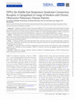
Enlarged alveolar airspaces in lungs of Fibulin-4 knockdown mice
<p>(A) Expression levels of Fibulin-4 in lungs isolated from newborn (n = 4, n = 4, n = 3) ... more <p>(A) Expression levels of Fibulin-4 in lungs isolated from newborn (n = 4, n = 4, n = 3) and adult (n = 4, n = 4, n = 4) Fibulin-4<sup>+/+</sup>, Fibulin-4<sup>+/R</sup> and Fibulin-4<sup>R/R</sup> mice relative to Fibulin-4<sup>+/+</sup> lungs (**p<0.01). (B) Mean peak inspiratory flow (PIF) and peak expiratory flow (PEF) values for Fibulin-4<sup>+/+</sup> (n = 4), Fibulin-4<sup>+/R</sup> (n = 4) and Fibulin-4<sup>R/R</sup> mice (observed for n = 4, but two animals died during the procedure) at 3-minute intervals. After a 9 minute adaptation period (the first three time intervals), PIF follows similar trends in Fibulin-4<sup>+/+</sup>, Fibulin-4<sup>+/R</sup> and Fibulin-4<sup>R/R</sup> mice, while Fibulin-4<sup>R/R</sup> mice show a decrease in PEF. (C) HE stained sections of formalin fixed lungs of male mice. Enlarged alveolar airspaces are observed in Fibulin-4<sup>+/R</sup> (middle, n = 3) and Fibulin-4<sup>R/R</sup> lungs (right, n = 3), with the latter being more pronounced, compared to Fibulin-4<sup>+/+</sup> (n = 3). Enlarged alveolar airspaces are already present in Fibulin-4<sup>R/R</sup> newborn lungs (n = 3), while lungs of Fibulin-4<sup>+/R</sup> littermates (n = 5) show no difference compared to Fibulin-4<sup>+/+</sup> lungs (n = 4). Scale bar 100 µm. Magnification 10x. (D) D<sub>2</sub> quantification (see methods and <a href="http://www.plosone.org/article/info:doi/10.1371/journal.pone.0106054#pone.0106054.s001" target="_blank">Figure S1</a> for further explanation) of the alveolar airspaces revealed a significant difference between adult Fibulin-4<sup>+/+</sup> and Fibulin-4<sup>+/R</sup> (*p<0.05) and between adult Fibulin-4<sup>+/+</sup> and Fibulin-4<sup>R/R</sup> lungs (**p<0.01) as well as between newborn Fibulin-4<sup>+/+</sup> and Fibulin-4<sup>R/R</sup> lungs (*p<0.05).</p

1Department of Pulmonary Medicine, Erasmus MC, ‘s-Gravendijkwal, Rotterdam, Netherlands. 2Cardiov... more 1Department of Pulmonary Medicine, Erasmus MC, ‘s-Gravendijkwal, Rotterdam, Netherlands. 2Cardiovascular and Pulmonary Branch, National Heart, Lung, and Blood Institute, NIH, Bethesda, Maryland USA. 3Department of Pulmonology, Franciscus Gasthuis, Rotterdam, Netherlands. 4Epirus Biopharmaceuticals Netherlands Yalelaan, Utrecht, Netherlands. 5VIB Center for Inflammation Research, Ghent University, Ghent, Belgium.

Frontiers in Pharmacology, 2020
Epithelial mast cells are generally present in the airways of patients with allergic asthma that ... more Epithelial mast cells are generally present in the airways of patients with allergic asthma that are inadequately controlled. Airway mast cells (MCs) are critically involved in allergic airway inflammation and contribute directly to the main symptoms of allergic patients. Phosphodiesterase 3 (PDE3) tailors signaling of cyclic adenosine monophosphate (cAMP) and cyclic guanosine monophosphate (cGMP), which are critical intracellular second messenger molecules in various signaling pathways. This paper investigates the pathophysiological role and disease-modifying effects of PDE3 in mouse bone marrow-derived MCs (bmMCs), human LAD2- and HMC1 mast cell lines, human blood basophils, and peripheral blood-derived primary human MCs (HuMCs). In a chronic house dust mite (HDM)-driven allergic airway inflammation mouse model, we observed that PDE3 deficiency or PDE3 inhibition (PDE3i) therapy reduced the numbers of epithelial MCs, when compared to control mice. Mouse bone marrow-derived MCs (bm...
Immune checkpoints skewing might be the cause of continuous immune activation in nasal polyps; new opportunities for therapies
Journal of Allergy and Clinical Immunology

Journal of Aerosol Medicine and Pulmonary Drug Delivery
Ferro et al. describe a patient with severe respiratory insufficiency caused by SARS-CoV-2 infect... more Ferro et al. describe a patient with severe respiratory insufficiency caused by SARS-CoV-2 infection, who improved greatly (without tracheal intubation) by administration of nebulized phosphodiesterase-3-(PDE3-) inhibitor enoximone. Using enoximone as a treatment for respiratory insufficiency due to SARS-CoV-2 infection was earlier described by Beute et al. (1) and suggested by Giorgi et al. (2) Beute et al. describe systemic delivery of PDE3, whereas Ferro used a nebulized administration form. Both are limited cases, but significant for the treatment of SARS-CoV-2-induced respiratory insufficiency, especially when linked to evidence of the efficiency of enoximone in asthma and status asthmaticus. A potent vasodilator, enoximone proved to be an equally potent bronchodilator, extremely suitable in respiratory diseases, even in low doses. (1,3,4) Ferro et al. used nebulized administration, choosing to minimize systemic exposure.

Journal of Allergy and Clinical Immunology

Mediators of Inflammation
Rationale. Sarcoidosis is a systemic inflammatory disorder characterized by the presence of granu... more Rationale. Sarcoidosis is a systemic inflammatory disorder characterized by the presence of granulomas in various organs, most commonly in the lungs. Although the ethology is unknown, sarcoidosis is thought to be mediated by T helper (Th)1 and Th17 lymphocytes. Chronic airway exposure to beryllium metal leads to chronic beryllium disease (CBD), which shares similarities with pulmonary sarcoidosis. Objective. To study airway pathophysiology and the role of dendritic cells (DCs) and IL-17 receptor (IL-17R) signals in a mouse model for CBD. Methods. Here, we present a CBD mouse model in which mice were exposed to beryllium during three weeks. We also exposed IL-17R-deficient mice and mice in which DCs were depleted. Results. Eight weeks after the initial beryllium exposure, an inflammatory response was detected in the lungs. Mice displayed inflammation of the lower airways that included focal dense infiltrates, granuloma-like foci, and tertiary lymphoid structure (TLS) containing T cel...

Basic Research in Cardiology
Pulmonary hypertension is common in heart failure with preserved ejection fraction (HFpEF). Here,... more Pulmonary hypertension is common in heart failure with preserved ejection fraction (HFpEF). Here, we tested the hypothesis that comorbidities [diabetes mellitus (DM, streptozotocin), hypercholesterolemia (HC, high-fat diet) and chronic kidney disease (CKD, renal microembolization)] directly impair pulmonary vasomotor control in a DM + HC + CKD swine model. 6 months after induction of DM + HC + CKD, pulmonary arterial pressure was similar in chronically instrumented female DM + HC + CKD (n = 19) and Healthy swine (n = 18). However, cardiac output was lower both at rest and during exercise, implying an elevated pulmonary vascular resistance (PVR) in DM + HC + CKD swine (153 ± 10 vs. 122 ± 9 mmHg∙L−1∙min∙kg). Phosphodiesterase 5 inhibition and endothelin receptor antagonism decreased PVR in DM + HC + CKD (− 12 ± 12 and − 22 ± 7 mmHg∙L−1∙min∙kg) but not in Healthy swine (− 1 ± 12 and 2 ± 14 mmHg∙L−1∙min∙kg), indicating increased vasoconstrictor influences of phosphodiesterase 5 and endo...

Journal of Clinical Investigation

Asthma Diagnosis and Management - Approach Based on Phenotype and Endotype
A recent status on asthmaticus multiple case report by Beute demonstrated the beneficial effects ... more A recent status on asthmaticus multiple case report by Beute demonstrated the beneficial effects of phosphodiesterase III (PDE3) and phosphodiesterase IV (PDE4) inhibition. This chapter reviews the possible underlying mechanisms, beside the known effect, for the beneficial effects of a mixed PDE3/4 inhibitor in allergic airway inflammation. Structural cells of the lung and immune system express PDE3 and 4. PDE3 and 4 inhibition have a number of consequences related to physical function and cytokine production. The most direct effect of PDE3 inhibition being relaxation of smooth muscle cells results in bronchodilation. However, PDE3 inhibition appears to go further than a mere inhibitory activity in bronchial smooth muscle. It also affects structural cells, and more importantly, it creates an improved barrier function in endothelial cells. PDE3 and 4 inhibition therefore strengthens the immune barrier; but in addition, it modifies the cells of the immune system itself, as these also express PDE3 and 4 activity, thus changing their function. All aspects of asthma-related pathophysiology seem to be affected by PDE3 and 4 inhibition. Clinical use of a mixed PDE3/4 inhibitor in respiratory diseases is currently limited to a few studies, including life-threatening asthma in which mixed PDE3/4 inhibition has a beneficial effect.
Targeting PDE3 Abrogates the Hallmarks of Asthma in Asthma Models
Clinical Problems

European Journal of Immunology
Influenza virus infection is an important cause of severe asthma exacerbations, but it remains un... more Influenza virus infection is an important cause of severe asthma exacerbations, but it remains unclear how a Th1-mediated antiviral response triggers a prototypical Th2 disease. We investigated CD4 + T cells and group 2 innate lymphoid cells (ILC2s) in influenza virus-infected mice. We found that ILC2s accumulated in the lung rapidly after influenza virus infection, but the induction of IL-5 and IL-13 secretion was delayed and concomitant with T cell activation. In an influenza-induced exacerbation of allergic airway inflammation model we noticed an initial reduction of ILC2 numbers and cytokine production in broncho-alveolar lavage compared to chronic house dust mite (HDM)-mediated airway inflammation alone. ILC2s phenotype was characterized by low T1/ST2, ICOS, KLRG1, and CD25 expression, resembling naïve ILC2s. The contribution of ILC2s to type 2 cytokine production in the early stage of the influenza-induced exacerbation was limited. In contrast, T cells showed increased IL-4 and IL-5 production when exposed to both HDM and influenza virus. Upon virus clearance, ILC2s regained an activated T1/ST2 high ICOS high KLRG1 high CD25 high phenotype paired with cytokine production and were major contributors to the type 2 cytokine milieu. Collectively, our data indicate that both T cells and ILC2s contribute to influenza-induced exacerbation of allergic airway inflammation, but with different kinetics.

The European respiratory journal, 2018
The lung-draining mediastinal lymph nodes (MLNs) are currently widely used to diagnose sarcoidosi... more The lung-draining mediastinal lymph nodes (MLNs) are currently widely used to diagnose sarcoidosis. We previously reported that T-helper (Th) 17.1 cells are responsible for the exaggerated interferon-γ production in sarcoidosis lungs. In this study, we aimed to investigate 1) whether Th17.1 cells are also increased in the MLNs of sarcoidosis patients and 2) whether frequencies of the Th17.1 cells at diagnosis may correlate with disease progression.MLN cells from treatment-naive pulmonary sarcoidosis patients (n=17) and healthy controls (n=22) and peripheral blood mononuclear cells (n=34) and bronchoalveolar lavage fluid (BALF) (n=36) from sarcoidosis patients were examined for CD4 T-cell subset proportions using flow cytometry.Higher proportions of Th17.1 cells were detected in sarcoidosis MLNs than in control MLNs. Higher Th17.1 cell proportions were found in sarcoidosis BALF compared with MLNs and peripheral blood. Furthermore, BALF Th17.1 cell proportions were significantly highe...

Journal of Allergy and Clinical Immunology
Background: The Notch signaling pathway has been implicated in the pathogenesis of allergic airwa... more Background: The Notch signaling pathway has been implicated in the pathogenesis of allergic airway inflammation. Targeting the active Notch transactivation complex by using the cellpermeable, hydrocarbon-stapled synthetic peptide stapled ahelical peptide derived from mastermind-like 1 (SAHM1) resulted in genome-wide suppression of Notch-activated genes in leukemic cells and other models. However, the efficacy of SAHM1 in allergic asthma models has remained unexplored. Objective: We aimed to investigate the therapeutic efficacy of SAHM1 in a house dust mite (HDM)-driven asthma model. Methods: Topical therapeutic intervention with SAHM1 or a control peptide was performed during sensitization, challenge, or both with HDM in mice. Airway inflammation was assessed by using multicolor flow cytometry, and bronchial hyperreactivity was studied. Additionally, SAHM1 therapy was investigated in mice with established allergic airway inflammation and in a model in which we neutralized IFN-g during HDM challenge to support the T H 2 response and exacerbate asthma. Results: SAHM1 treatment during the challenge phase led to a marked reduction of eosinophil and T cell numbers in bronchoalveolar lavage fluid compared with those in diluenttreated or control peptide-treated mice. Likewise, T-cell cytokine content and bronchial hyperreactivity were reduced. SAHM1 treatment dampened T H 2 inflammation during ongoing HDM challenge and enhanced recovery after established asthma. Additionally, in the presence of anti-IFN-g antibodies, SAHM1 downregulated expression of the key T H 2 transcription factor GATA3 and intracellular IL-4 in bronchoalveolar lavage fluid T cells, but expression of the T H 17 transcription factor retinoic acid-related orphan receptor gt or intracellular IL-17 was not affected. SAHM1 therapy also reduced serum IgE levels. Conclusions: Therapeutic intervention of Notch signaling by SAHM1 inhibits allergic airway inflammation in mice and is therefore an interesting new topical treatment opportunity in asthmatic patients.
Increased surface expression of NOTCH on memory T cells in peripheral blood from patients with asthma
Journal of Allergy and Clinical Immunology

JCI insight, Jan 25, 2018
Phosphodiesterase 3 (PDE3) and PDE4 regulate levels of cyclic AMP, which are critical in various ... more Phosphodiesterase 3 (PDE3) and PDE4 regulate levels of cyclic AMP, which are critical in various cell types involved in allergic airway inflammation. Although PDE4 inhibition attenuates allergic airway inflammation, reported side effects preclude its application as an antiasthma drug in humans. Case reports showed that enoximone, which is a smooth muscle relaxant that inhibits PDE3, is beneficial and lifesaving in status asthmaticus and is well tolerated. However, clinical observations also showed antiinflammatory effects of PDE3 inhibition. In this study, we investigated the role of PDE3 in a house dust mite-driven (HDM-driven) allergic airway inflammation (AAI) model that is characterized by T helper 2 cell activation, eosinophilia, and reduced mucosal barrier function. Compared with wild-type (WT) littermates, mice with a targeted deletion of the PDE3A or PDE3B gene showed significantly reduced HDM-driven AAI. Therapeutic intervention in WT mice showed that all hallmarks of HDM-d...

Frontiers in immunology, 2017
Group 2 innate lymphoid cells (ILC2) are implicated in allergic asthma as an early innate source ... more Group 2 innate lymphoid cells (ILC2) are implicated in allergic asthma as an early innate source of the type 2 cytokines IL-5 and IL-13. However, their induction in house dust mite (HDM)-mediated airway inflammation additionally requires T cell activation. It is currently unknown whether phenotypic differences exist between ILC2s that are activated in a T cell-dependent or T cell-independent fashion. Here, we compared ILC2s in IL-33- and HDM-driven airway inflammation. Using flow cytometry, we found that surface expression levels of various markers frequently used to identify ILC2s were dependent on their mode of activation, highly variable over time, and differed between tissue compartments, including bronchoalveolar lavage (BAL) fluid, lung, draining lymph nodes, and spleen. WhereasIL-33-activated BAL fluid ILC2s exhibited an almost uniform CD25CD127T1/ST2ICOSKLRG1phenotype, at a comparable time point after HDM exposure BAL fluid ILC2s had a very heterogeneous surface marker pheno...
Treatment of Vulvar Intraepithelial Neoplasia with Topical Imiquimod
Yearbook of Dermatology and Dermatologic Surgery
Alternatives to surgery are needed for the treatment of vulvar intraepithelial neoplasia. We inve... more Alternatives to surgery are needed for the treatment of vulvar intraepithelial neoplasia. We investigated the effectiveness of imiquimod 5% cream, a topical immune-response modulator, for the treatment of this condition. Fifty-two patients with grade 2 or 3 vulvar intraepithelial neoplasia were randomly assigned to receive either imiquimod or placebo, applied twice weekly for 16 weeks. The primary outcome was a reduction of more than 25% in lesion size at 20 weeks. Secondary outcomes were histologic regression, clearance of human papillomavirus (HPV) from the lesion, changes in immune cells in the epidermis and dermis of the vulva, relief of symptoms, improvement of quality of life, and durability of response. Reduction in lesion size was classified as complete response (elimination), strong partial response (76 to 99% reduction), weak partial response (26 to 75% reduction), or no response (&amp;amp;amp;amp;amp;amp;amp;amp;amp;amp;amp;amp;amp;amp;amp;amp;amp;amp;amp;amp;amp;amp;amp;amp;amp;amp;amp;amp;amp;amp;amp;amp;amp;amp;amp;amp;amp;amp;amp;amp;amp;amp;amp;amp;amp;amp;amp;amp;amp;amp;amp;amp;amp;amp;amp;amp;amp;amp;amp;lt; or =25% reduction). The follow-up period was 12 months. Lesion size was reduced by more than 25% at 20 weeks in 21 of the 26 patients (81%) treated with imiquimod and in none of those treated with placebo (P&amp;amp;amp;amp;amp;amp;amp;amp;amp;amp;amp;amp;amp;amp;amp;amp;amp;amp;amp;amp;amp;amp;amp;amp;amp;amp;amp;amp;amp;amp;amp;amp;amp;amp;amp;amp;amp;amp;amp;amp;amp;amp;amp;amp;amp;amp;amp;amp;amp;amp;amp;amp;amp;amp;amp;amp;amp;amp;amp;lt;0.001). Histologic regression was significantly greater in the imiquimod group than in the placebo group (P&amp;amp;amp;amp;amp;amp;amp;amp;amp;amp;amp;amp;amp;amp;amp;amp;amp;amp;amp;amp;amp;amp;amp;amp;amp;amp;amp;amp;amp;amp;amp;amp;amp;amp;amp;amp;amp;amp;amp;amp;amp;amp;amp;amp;amp;amp;amp;amp;amp;amp;amp;amp;amp;amp;amp;amp;amp;amp;amp;lt;0.001). At baseline, 50 patients (96%) tested positive for HPV DNA. HPV cleared from the lesion in 15 patients in the imiquimod group (58%), as compared with 2 in the placebo group (8%) (P&amp;amp;amp;amp;amp;amp;amp;amp;amp;amp;amp;amp;amp;amp;amp;amp;amp;amp;amp;amp;amp;amp;amp;amp;amp;amp;amp;amp;amp;amp;amp;amp;amp;amp;amp;amp;amp;amp;amp;amp;amp;amp;amp;amp;amp;amp;amp;amp;amp;amp;amp;amp;amp;amp;amp;amp;amp;amp;amp;lt;0.001). The number of immune epidermal cells increased significantly and the number of immune dermal cells decreased significantly with imiquimod as compared with placebo. Imiquimod reduced pruritus and pain at 20 weeks (P=0.008 and P=0.004, respectively) and at 12 months (P=0.04 and P=0.02, respectively). The lesion progressed to invasion (to a depth of &amp;amp;amp;amp;amp;amp;amp;amp;amp;amp;amp;amp;amp;amp;amp;amp;amp;amp;amp;amp;amp;amp;amp;amp;amp;amp;amp;amp;amp;amp;amp;amp;amp;amp;amp;amp;amp;amp;amp;amp;amp;amp;amp;amp;amp;amp;amp;amp;amp;amp;amp;amp;amp;amp;amp;amp;amp;amp;amp;lt;1 mm) in 3 of 49 patients (6%) followed for 12 months (2 in the placebo group and 1 in the imiquimod group). Nine patients, all treated with imiquimod, had a complete response at 20 weeks and remained free from disease at 12 months. Imiquimod is effective in the treatment of vulvar intraepithelial neoplasia. (Current Controlled Trials number, ISRCTN11290871 [controlled-trials.com].).
Clinical Infectious Diseases
Background. Middle East respiratory syndrome coronavirus (MERS-CoV) causes pneumonia with a relat... more Background. Middle East respiratory syndrome coronavirus (MERS-CoV) causes pneumonia with a relatively high case fatality rate in humans. Smokers and chronic obstructive pulmonary disease (COPD) patients have been reported to be more susceptible to MERS-CoV infection. Here, we determined the expression of MERS-CoV receptor, dipeptidyl peptidase IV (DPP4), in lung tissues of smokers without airflow limitation and COPD patients in comparison to nonsmoking individuals (never-smokers). Methods. DPP4 expression was measured in lung tissue of lung resection specimens of never-smokers, smokers without airflow limitation, COPD GOLD stage II patients and in lung explants of end-stage COPD patients. Both control subjects and COPD patients were well phenotyped and age-matched. The mRNA expression was determined using qRT-PCR and protein expression was quantified using immunohistochemistry. Results. In smokers and subjects with COPD, both DPP4 mRNA and protein expression were significantly higher compared to never-smokers. Additionally, we found that both DPP4 mRNA and protein expression were inversely correlated with lung function and diffusing capacity parameters. Conclusions. We provide evidence that DPP4 is upregulated in the lungs of smokers and COPD patients, which could partially explain why these individuals are more susceptible to MERS-CoV infection. These data also highlight a possible role of DPP4 in COPD pathogenesis. Keywords. Middle East respiratory syndrome coronavirus (MERS-CoV); dipeptidyl peptidase 4 (DPP4); smoking; chronic obstructive pulmonary disease (COPD). Middle East Respiratory Syndrome coronavirus (MERS-CoV) is a newly emerging pathogen that mainly causes pneumonia with a relatively high case-fatality rate [1, 2]. Since 2012, ~1900 laboratory-confirmed cases have been reported to the World Health Organization (WHO) [2]. The majority of cases occurred in familial or hospital-related clusters through human-to-human transmission [3-5]. The clinical course of MERS-CoV infection ranges from asymptomatic to acute respiratory distress syndrome with need for ventilatory support [3, 5-7]. To infect its host, MERS-CoV attaches to its receptor, an exopeptidase called dipeptidyl peptidase 4 (DPP4), also known as CD26 [8]. DPP4 is a type II transmembrane glycoprotein that is expressed in many cell types and organs in the body. It serves multiple functions among which post-translational cleavage of hormones and chemokines, T-cell activation, cell adhesion, and apoptosis [9-11]. In lungs, however, DPP4 is expressed at a minimum level [12], mainly in alveolar epithelial cells and endothelial cells, and to a lesser extent in bronchiolar epithelial cells, airway submucosal glands, alveolar macrophages, lymphocytes, and plasmacytoid dendritic cells [13-16]. Importantly, the alveolar epithelial cells are the main target for MERS-CoV [13, 17]. Several underlying comorbidities, including chronic lung diseases, have been reported to increase the risk of acquiring MERS-CoV infection [18]. Chronic obstructive pulmonary disease (COPD) is a highly prevalent chronic lung disease in older subjects and is currently the leading cause of death worldwide [19, 20]. The most common cause of COPD is chronic cigarette smoking. The inflammatory response to cigarette smoke results in an excessive release of chemokines and cytokines with a subsequent high influx of immune cells [20]. Because smoking has also been reported to increase susceptibility to MERS-CoV infection [18], we aimed to investigate the expression of the MERS-CoV receptor, DPP4, in a large well-phenotyped cohort of smokers, with and without airflow limitation, in comparison to age-matched individuals that never smoked (never-smokers).

Multidisciplinary Respiratory Medicine, 2017
Background: The intratracheal instillation of bleomycin in mice induces early damage to alveolar ... more Background: The intratracheal instillation of bleomycin in mice induces early damage to alveolar epithelial cells and development of inflammation followed by fibrotic tissue changes and represents the most widely used model of pulmonary fibrosis to investigate human IPF. Histopathology is the gold standard for assessing lung fibrosis in rodents, however it precludes repeated and longitudinal measurements of disease progression and does not provide information on spatial and temporal distribution of tissue damage. Here we investigated the use of the Micro-CT technique to allow the evaluation of disease onset and progression at different time-points in the mouse bleomycin model of lung fibrosis. Micro-CT was throughout coupled with histological analysis for the validation of the imaging results. Methods: In bleomycin-instilled and control mice, airways and lung morphology changes were assessed and reconstructed at baseline, 7, 14 and 21 days post-treatment based on Micro-CT images. Ashcroft score, percentage of collagen content and percentage of alveolar air area were detected on lung slides processed by histology and subsequently compared with Micro-CT parameters. Results: Extent (%) of fibrosis measured by Micro-CT correlated with Ashcroft score, the percentage of collagen content and the percentage of alveolar air area (r 2 = 0.91; 0.77; 0.94, respectively). Distal airway radius also correlated with the Ashcroft score, the collagen content and alveolar air area percentage (r 2 = 0.89; 0.78; 0.98, respectively). Conclusions: Micro-CT data were in good agreement with histological read-outs as micro-CT was able to quantify effectively and non-invasively disease progression longitudinally and to reduce the variability and number of animals used to assess the damage. This suggests that this technique is a powerful tool for understanding experimental pulmonary fibrosis and that its use could translate into a more efficient drug discovery process, also helping to fill the gap between preclinical setting and clinical practice.
Enlarged alveolar airspaces in lungs of Fibulin-4 knockdown mice
<p>(A) Expression levels of Fibulin-4 in lungs isolated from newborn (n = 4, n = 4, n = 3) ... more <p>(A) Expression levels of Fibulin-4 in lungs isolated from newborn (n = 4, n = 4, n = 3) and adult (n = 4, n = 4, n = 4) Fibulin-4<sup>+/+</sup>, Fibulin-4<sup>+/R</sup> and Fibulin-4<sup>R/R</sup> mice relative to Fibulin-4<sup>+/+</sup> lungs (**p<0.01). (B) Mean peak inspiratory flow (PIF) and peak expiratory flow (PEF) values for Fibulin-4<sup>+/+</sup> (n = 4), Fibulin-4<sup>+/R</sup> (n = 4) and Fibulin-4<sup>R/R</sup> mice (observed for n = 4, but two animals died during the procedure) at 3-minute intervals. After a 9 minute adaptation period (the first three time intervals), PIF follows similar trends in Fibulin-4<sup>+/+</sup>, Fibulin-4<sup>+/R</sup> and Fibulin-4<sup>R/R</sup> mice, while Fibulin-4<sup>R/R</sup> mice show a decrease in PEF. (C) HE stained sections of formalin fixed lungs of male mice. Enlarged alveolar airspaces are observed in Fibulin-4<sup>+/R</sup> (middle, n = 3) and Fibulin-4<sup>R/R</sup> lungs (right, n = 3), with the latter being more pronounced, compared to Fibulin-4<sup>+/+</sup> (n = 3). Enlarged alveolar airspaces are already present in Fibulin-4<sup>R/R</sup> newborn lungs (n = 3), while lungs of Fibulin-4<sup>+/R</sup> littermates (n = 5) show no difference compared to Fibulin-4<sup>+/+</sup> lungs (n = 4). Scale bar 100 µm. Magnification 10x. (D) D<sub>2</sub> quantification (see methods and <a href="http://www.plosone.org/article/info:doi/10.1371/journal.pone.0106054#pone.0106054.s001" target="_blank">Figure S1</a> for further explanation) of the alveolar airspaces revealed a significant difference between adult Fibulin-4<sup>+/+</sup> and Fibulin-4<sup>+/R</sup> (*p<0.05) and between adult Fibulin-4<sup>+/+</sup> and Fibulin-4<sup>R/R</sup> lungs (**p<0.01) as well as between newborn Fibulin-4<sup>+/+</sup> and Fibulin-4<sup>R/R</sup> lungs (*p<0.05).</p
1Department of Pulmonary Medicine, Erasmus MC, ‘s-Gravendijkwal, Rotterdam, Netherlands. 2Cardiov... more 1Department of Pulmonary Medicine, Erasmus MC, ‘s-Gravendijkwal, Rotterdam, Netherlands. 2Cardiovascular and Pulmonary Branch, National Heart, Lung, and Blood Institute, NIH, Bethesda, Maryland USA. 3Department of Pulmonology, Franciscus Gasthuis, Rotterdam, Netherlands. 4Epirus Biopharmaceuticals Netherlands Yalelaan, Utrecht, Netherlands. 5VIB Center for Inflammation Research, Ghent University, Ghent, Belgium.
Frontiers in Pharmacology, 2020
Epithelial mast cells are generally present in the airways of patients with allergic asthma that ... more Epithelial mast cells are generally present in the airways of patients with allergic asthma that are inadequately controlled. Airway mast cells (MCs) are critically involved in allergic airway inflammation and contribute directly to the main symptoms of allergic patients. Phosphodiesterase 3 (PDE3) tailors signaling of cyclic adenosine monophosphate (cAMP) and cyclic guanosine monophosphate (cGMP), which are critical intracellular second messenger molecules in various signaling pathways. This paper investigates the pathophysiological role and disease-modifying effects of PDE3 in mouse bone marrow-derived MCs (bmMCs), human LAD2- and HMC1 mast cell lines, human blood basophils, and peripheral blood-derived primary human MCs (HuMCs). In a chronic house dust mite (HDM)-driven allergic airway inflammation mouse model, we observed that PDE3 deficiency or PDE3 inhibition (PDE3i) therapy reduced the numbers of epithelial MCs, when compared to control mice. Mouse bone marrow-derived MCs (bm...
Immune checkpoints skewing might be the cause of continuous immune activation in nasal polyps; new opportunities for therapies
Journal of Allergy and Clinical Immunology
Journal of Aerosol Medicine and Pulmonary Drug Delivery
Ferro et al. describe a patient with severe respiratory insufficiency caused by SARS-CoV-2 infect... more Ferro et al. describe a patient with severe respiratory insufficiency caused by SARS-CoV-2 infection, who improved greatly (without tracheal intubation) by administration of nebulized phosphodiesterase-3-(PDE3-) inhibitor enoximone. Using enoximone as a treatment for respiratory insufficiency due to SARS-CoV-2 infection was earlier described by Beute et al. (1) and suggested by Giorgi et al. (2) Beute et al. describe systemic delivery of PDE3, whereas Ferro used a nebulized administration form. Both are limited cases, but significant for the treatment of SARS-CoV-2-induced respiratory insufficiency, especially when linked to evidence of the efficiency of enoximone in asthma and status asthmaticus. A potent vasodilator, enoximone proved to be an equally potent bronchodilator, extremely suitable in respiratory diseases, even in low doses. (1,3,4) Ferro et al. used nebulized administration, choosing to minimize systemic exposure.
Journal of Allergy and Clinical Immunology
Mediators of Inflammation
Rationale. Sarcoidosis is a systemic inflammatory disorder characterized by the presence of granu... more Rationale. Sarcoidosis is a systemic inflammatory disorder characterized by the presence of granulomas in various organs, most commonly in the lungs. Although the ethology is unknown, sarcoidosis is thought to be mediated by T helper (Th)1 and Th17 lymphocytes. Chronic airway exposure to beryllium metal leads to chronic beryllium disease (CBD), which shares similarities with pulmonary sarcoidosis. Objective. To study airway pathophysiology and the role of dendritic cells (DCs) and IL-17 receptor (IL-17R) signals in a mouse model for CBD. Methods. Here, we present a CBD mouse model in which mice were exposed to beryllium during three weeks. We also exposed IL-17R-deficient mice and mice in which DCs were depleted. Results. Eight weeks after the initial beryllium exposure, an inflammatory response was detected in the lungs. Mice displayed inflammation of the lower airways that included focal dense infiltrates, granuloma-like foci, and tertiary lymphoid structure (TLS) containing T cel...
Basic Research in Cardiology
Pulmonary hypertension is common in heart failure with preserved ejection fraction (HFpEF). Here,... more Pulmonary hypertension is common in heart failure with preserved ejection fraction (HFpEF). Here, we tested the hypothesis that comorbidities [diabetes mellitus (DM, streptozotocin), hypercholesterolemia (HC, high-fat diet) and chronic kidney disease (CKD, renal microembolization)] directly impair pulmonary vasomotor control in a DM + HC + CKD swine model. 6 months after induction of DM + HC + CKD, pulmonary arterial pressure was similar in chronically instrumented female DM + HC + CKD (n = 19) and Healthy swine (n = 18). However, cardiac output was lower both at rest and during exercise, implying an elevated pulmonary vascular resistance (PVR) in DM + HC + CKD swine (153 ± 10 vs. 122 ± 9 mmHg∙L−1∙min∙kg). Phosphodiesterase 5 inhibition and endothelin receptor antagonism decreased PVR in DM + HC + CKD (− 12 ± 12 and − 22 ± 7 mmHg∙L−1∙min∙kg) but not in Healthy swine (− 1 ± 12 and 2 ± 14 mmHg∙L−1∙min∙kg), indicating increased vasoconstrictor influences of phosphodiesterase 5 and endo...
Journal of Clinical Investigation
Asthma Diagnosis and Management - Approach Based on Phenotype and Endotype
A recent status on asthmaticus multiple case report by Beute demonstrated the beneficial effects ... more A recent status on asthmaticus multiple case report by Beute demonstrated the beneficial effects of phosphodiesterase III (PDE3) and phosphodiesterase IV (PDE4) inhibition. This chapter reviews the possible underlying mechanisms, beside the known effect, for the beneficial effects of a mixed PDE3/4 inhibitor in allergic airway inflammation. Structural cells of the lung and immune system express PDE3 and 4. PDE3 and 4 inhibition have a number of consequences related to physical function and cytokine production. The most direct effect of PDE3 inhibition being relaxation of smooth muscle cells results in bronchodilation. However, PDE3 inhibition appears to go further than a mere inhibitory activity in bronchial smooth muscle. It also affects structural cells, and more importantly, it creates an improved barrier function in endothelial cells. PDE3 and 4 inhibition therefore strengthens the immune barrier; but in addition, it modifies the cells of the immune system itself, as these also express PDE3 and 4 activity, thus changing their function. All aspects of asthma-related pathophysiology seem to be affected by PDE3 and 4 inhibition. Clinical use of a mixed PDE3/4 inhibitor in respiratory diseases is currently limited to a few studies, including life-threatening asthma in which mixed PDE3/4 inhibition has a beneficial effect.
Targeting PDE3 Abrogates the Hallmarks of Asthma in Asthma Models
Clinical Problems
European Journal of Immunology
Influenza virus infection is an important cause of severe asthma exacerbations, but it remains un... more Influenza virus infection is an important cause of severe asthma exacerbations, but it remains unclear how a Th1-mediated antiviral response triggers a prototypical Th2 disease. We investigated CD4 + T cells and group 2 innate lymphoid cells (ILC2s) in influenza virus-infected mice. We found that ILC2s accumulated in the lung rapidly after influenza virus infection, but the induction of IL-5 and IL-13 secretion was delayed and concomitant with T cell activation. In an influenza-induced exacerbation of allergic airway inflammation model we noticed an initial reduction of ILC2 numbers and cytokine production in broncho-alveolar lavage compared to chronic house dust mite (HDM)-mediated airway inflammation alone. ILC2s phenotype was characterized by low T1/ST2, ICOS, KLRG1, and CD25 expression, resembling naïve ILC2s. The contribution of ILC2s to type 2 cytokine production in the early stage of the influenza-induced exacerbation was limited. In contrast, T cells showed increased IL-4 and IL-5 production when exposed to both HDM and influenza virus. Upon virus clearance, ILC2s regained an activated T1/ST2 high ICOS high KLRG1 high CD25 high phenotype paired with cytokine production and were major contributors to the type 2 cytokine milieu. Collectively, our data indicate that both T cells and ILC2s contribute to influenza-induced exacerbation of allergic airway inflammation, but with different kinetics.
The European respiratory journal, 2018
The lung-draining mediastinal lymph nodes (MLNs) are currently widely used to diagnose sarcoidosi... more The lung-draining mediastinal lymph nodes (MLNs) are currently widely used to diagnose sarcoidosis. We previously reported that T-helper (Th) 17.1 cells are responsible for the exaggerated interferon-γ production in sarcoidosis lungs. In this study, we aimed to investigate 1) whether Th17.1 cells are also increased in the MLNs of sarcoidosis patients and 2) whether frequencies of the Th17.1 cells at diagnosis may correlate with disease progression.MLN cells from treatment-naive pulmonary sarcoidosis patients (n=17) and healthy controls (n=22) and peripheral blood mononuclear cells (n=34) and bronchoalveolar lavage fluid (BALF) (n=36) from sarcoidosis patients were examined for CD4 T-cell subset proportions using flow cytometry.Higher proportions of Th17.1 cells were detected in sarcoidosis MLNs than in control MLNs. Higher Th17.1 cell proportions were found in sarcoidosis BALF compared with MLNs and peripheral blood. Furthermore, BALF Th17.1 cell proportions were significantly highe...
Journal of Allergy and Clinical Immunology
Background: The Notch signaling pathway has been implicated in the pathogenesis of allergic airwa... more Background: The Notch signaling pathway has been implicated in the pathogenesis of allergic airway inflammation. Targeting the active Notch transactivation complex by using the cellpermeable, hydrocarbon-stapled synthetic peptide stapled ahelical peptide derived from mastermind-like 1 (SAHM1) resulted in genome-wide suppression of Notch-activated genes in leukemic cells and other models. However, the efficacy of SAHM1 in allergic asthma models has remained unexplored. Objective: We aimed to investigate the therapeutic efficacy of SAHM1 in a house dust mite (HDM)-driven asthma model. Methods: Topical therapeutic intervention with SAHM1 or a control peptide was performed during sensitization, challenge, or both with HDM in mice. Airway inflammation was assessed by using multicolor flow cytometry, and bronchial hyperreactivity was studied. Additionally, SAHM1 therapy was investigated in mice with established allergic airway inflammation and in a model in which we neutralized IFN-g during HDM challenge to support the T H 2 response and exacerbate asthma. Results: SAHM1 treatment during the challenge phase led to a marked reduction of eosinophil and T cell numbers in bronchoalveolar lavage fluid compared with those in diluenttreated or control peptide-treated mice. Likewise, T-cell cytokine content and bronchial hyperreactivity were reduced. SAHM1 treatment dampened T H 2 inflammation during ongoing HDM challenge and enhanced recovery after established asthma. Additionally, in the presence of anti-IFN-g antibodies, SAHM1 downregulated expression of the key T H 2 transcription factor GATA3 and intracellular IL-4 in bronchoalveolar lavage fluid T cells, but expression of the T H 17 transcription factor retinoic acid-related orphan receptor gt or intracellular IL-17 was not affected. SAHM1 therapy also reduced serum IgE levels. Conclusions: Therapeutic intervention of Notch signaling by SAHM1 inhibits allergic airway inflammation in mice and is therefore an interesting new topical treatment opportunity in asthmatic patients.
Increased surface expression of NOTCH on memory T cells in peripheral blood from patients with asthma
Journal of Allergy and Clinical Immunology
JCI insight, Jan 25, 2018
Phosphodiesterase 3 (PDE3) and PDE4 regulate levels of cyclic AMP, which are critical in various ... more Phosphodiesterase 3 (PDE3) and PDE4 regulate levels of cyclic AMP, which are critical in various cell types involved in allergic airway inflammation. Although PDE4 inhibition attenuates allergic airway inflammation, reported side effects preclude its application as an antiasthma drug in humans. Case reports showed that enoximone, which is a smooth muscle relaxant that inhibits PDE3, is beneficial and lifesaving in status asthmaticus and is well tolerated. However, clinical observations also showed antiinflammatory effects of PDE3 inhibition. In this study, we investigated the role of PDE3 in a house dust mite-driven (HDM-driven) allergic airway inflammation (AAI) model that is characterized by T helper 2 cell activation, eosinophilia, and reduced mucosal barrier function. Compared with wild-type (WT) littermates, mice with a targeted deletion of the PDE3A or PDE3B gene showed significantly reduced HDM-driven AAI. Therapeutic intervention in WT mice showed that all hallmarks of HDM-d...
Frontiers in immunology, 2017
Group 2 innate lymphoid cells (ILC2) are implicated in allergic asthma as an early innate source ... more Group 2 innate lymphoid cells (ILC2) are implicated in allergic asthma as an early innate source of the type 2 cytokines IL-5 and IL-13. However, their induction in house dust mite (HDM)-mediated airway inflammation additionally requires T cell activation. It is currently unknown whether phenotypic differences exist between ILC2s that are activated in a T cell-dependent or T cell-independent fashion. Here, we compared ILC2s in IL-33- and HDM-driven airway inflammation. Using flow cytometry, we found that surface expression levels of various markers frequently used to identify ILC2s were dependent on their mode of activation, highly variable over time, and differed between tissue compartments, including bronchoalveolar lavage (BAL) fluid, lung, draining lymph nodes, and spleen. WhereasIL-33-activated BAL fluid ILC2s exhibited an almost uniform CD25CD127T1/ST2ICOSKLRG1phenotype, at a comparable time point after HDM exposure BAL fluid ILC2s had a very heterogeneous surface marker pheno...
Treatment of Vulvar Intraepithelial Neoplasia with Topical Imiquimod
Yearbook of Dermatology and Dermatologic Surgery
Alternatives to surgery are needed for the treatment of vulvar intraepithelial neoplasia. We inve... more Alternatives to surgery are needed for the treatment of vulvar intraepithelial neoplasia. We investigated the effectiveness of imiquimod 5% cream, a topical immune-response modulator, for the treatment of this condition. Fifty-two patients with grade 2 or 3 vulvar intraepithelial neoplasia were randomly assigned to receive either imiquimod or placebo, applied twice weekly for 16 weeks. The primary outcome was a reduction of more than 25% in lesion size at 20 weeks. Secondary outcomes were histologic regression, clearance of human papillomavirus (HPV) from the lesion, changes in immune cells in the epidermis and dermis of the vulva, relief of symptoms, improvement of quality of life, and durability of response. Reduction in lesion size was classified as complete response (elimination), strong partial response (76 to 99% reduction), weak partial response (26 to 75% reduction), or no response (&amp;amp;amp;amp;amp;amp;amp;amp;amp;amp;amp;amp;amp;amp;amp;amp;amp;amp;amp;amp;amp;amp;amp;amp;amp;amp;amp;amp;amp;amp;amp;amp;amp;amp;amp;amp;amp;amp;amp;amp;amp;amp;amp;amp;amp;amp;amp;amp;amp;amp;amp;amp;amp;amp;amp;amp;amp;amp;amp;lt; or =25% reduction). The follow-up period was 12 months. Lesion size was reduced by more than 25% at 20 weeks in 21 of the 26 patients (81%) treated with imiquimod and in none of those treated with placebo (P&amp;amp;amp;amp;amp;amp;amp;amp;amp;amp;amp;amp;amp;amp;amp;amp;amp;amp;amp;amp;amp;amp;amp;amp;amp;amp;amp;amp;amp;amp;amp;amp;amp;amp;amp;amp;amp;amp;amp;amp;amp;amp;amp;amp;amp;amp;amp;amp;amp;amp;amp;amp;amp;amp;amp;amp;amp;amp;amp;lt;0.001). Histologic regression was significantly greater in the imiquimod group than in the placebo group (P&amp;amp;amp;amp;amp;amp;amp;amp;amp;amp;amp;amp;amp;amp;amp;amp;amp;amp;amp;amp;amp;amp;amp;amp;amp;amp;amp;amp;amp;amp;amp;amp;amp;amp;amp;amp;amp;amp;amp;amp;amp;amp;amp;amp;amp;amp;amp;amp;amp;amp;amp;amp;amp;amp;amp;amp;amp;amp;amp;lt;0.001). At baseline, 50 patients (96%) tested positive for HPV DNA. HPV cleared from the lesion in 15 patients in the imiquimod group (58%), as compared with 2 in the placebo group (8%) (P&amp;amp;amp;amp;amp;amp;amp;amp;amp;amp;amp;amp;amp;amp;amp;amp;amp;amp;amp;amp;amp;amp;amp;amp;amp;amp;amp;amp;amp;amp;amp;amp;amp;amp;amp;amp;amp;amp;amp;amp;amp;amp;amp;amp;amp;amp;amp;amp;amp;amp;amp;amp;amp;amp;amp;amp;amp;amp;amp;lt;0.001). The number of immune epidermal cells increased significantly and the number of immune dermal cells decreased significantly with imiquimod as compared with placebo. Imiquimod reduced pruritus and pain at 20 weeks (P=0.008 and P=0.004, respectively) and at 12 months (P=0.04 and P=0.02, respectively). The lesion progressed to invasion (to a depth of &amp;amp;amp;amp;amp;amp;amp;amp;amp;amp;amp;amp;amp;amp;amp;amp;amp;amp;amp;amp;amp;amp;amp;amp;amp;amp;amp;amp;amp;amp;amp;amp;amp;amp;amp;amp;amp;amp;amp;amp;amp;amp;amp;amp;amp;amp;amp;amp;amp;amp;amp;amp;amp;amp;amp;amp;amp;amp;amp;lt;1 mm) in 3 of 49 patients (6%) followed for 12 months (2 in the placebo group and 1 in the imiquimod group). Nine patients, all treated with imiquimod, had a complete response at 20 weeks and remained free from disease at 12 months. Imiquimod is effective in the treatment of vulvar intraepithelial neoplasia. (Current Controlled Trials number, ISRCTN11290871 [controlled-trials.com].).
Clinical Infectious Diseases
Background. Middle East respiratory syndrome coronavirus (MERS-CoV) causes pneumonia with a relat... more Background. Middle East respiratory syndrome coronavirus (MERS-CoV) causes pneumonia with a relatively high case fatality rate in humans. Smokers and chronic obstructive pulmonary disease (COPD) patients have been reported to be more susceptible to MERS-CoV infection. Here, we determined the expression of MERS-CoV receptor, dipeptidyl peptidase IV (DPP4), in lung tissues of smokers without airflow limitation and COPD patients in comparison to nonsmoking individuals (never-smokers). Methods. DPP4 expression was measured in lung tissue of lung resection specimens of never-smokers, smokers without airflow limitation, COPD GOLD stage II patients and in lung explants of end-stage COPD patients. Both control subjects and COPD patients were well phenotyped and age-matched. The mRNA expression was determined using qRT-PCR and protein expression was quantified using immunohistochemistry. Results. In smokers and subjects with COPD, both DPP4 mRNA and protein expression were significantly higher compared to never-smokers. Additionally, we found that both DPP4 mRNA and protein expression were inversely correlated with lung function and diffusing capacity parameters. Conclusions. We provide evidence that DPP4 is upregulated in the lungs of smokers and COPD patients, which could partially explain why these individuals are more susceptible to MERS-CoV infection. These data also highlight a possible role of DPP4 in COPD pathogenesis. Keywords. Middle East respiratory syndrome coronavirus (MERS-CoV); dipeptidyl peptidase 4 (DPP4); smoking; chronic obstructive pulmonary disease (COPD). Middle East Respiratory Syndrome coronavirus (MERS-CoV) is a newly emerging pathogen that mainly causes pneumonia with a relatively high case-fatality rate [1, 2]. Since 2012, ~1900 laboratory-confirmed cases have been reported to the World Health Organization (WHO) [2]. The majority of cases occurred in familial or hospital-related clusters through human-to-human transmission [3-5]. The clinical course of MERS-CoV infection ranges from asymptomatic to acute respiratory distress syndrome with need for ventilatory support [3, 5-7]. To infect its host, MERS-CoV attaches to its receptor, an exopeptidase called dipeptidyl peptidase 4 (DPP4), also known as CD26 [8]. DPP4 is a type II transmembrane glycoprotein that is expressed in many cell types and organs in the body. It serves multiple functions among which post-translational cleavage of hormones and chemokines, T-cell activation, cell adhesion, and apoptosis [9-11]. In lungs, however, DPP4 is expressed at a minimum level [12], mainly in alveolar epithelial cells and endothelial cells, and to a lesser extent in bronchiolar epithelial cells, airway submucosal glands, alveolar macrophages, lymphocytes, and plasmacytoid dendritic cells [13-16]. Importantly, the alveolar epithelial cells are the main target for MERS-CoV [13, 17]. Several underlying comorbidities, including chronic lung diseases, have been reported to increase the risk of acquiring MERS-CoV infection [18]. Chronic obstructive pulmonary disease (COPD) is a highly prevalent chronic lung disease in older subjects and is currently the leading cause of death worldwide [19, 20]. The most common cause of COPD is chronic cigarette smoking. The inflammatory response to cigarette smoke results in an excessive release of chemokines and cytokines with a subsequent high influx of immune cells [20]. Because smoking has also been reported to increase susceptibility to MERS-CoV infection [18], we aimed to investigate the expression of the MERS-CoV receptor, DPP4, in a large well-phenotyped cohort of smokers, with and without airflow limitation, in comparison to age-matched individuals that never smoked (never-smokers).
Multidisciplinary Respiratory Medicine, 2017
Background: The intratracheal instillation of bleomycin in mice induces early damage to alveolar ... more Background: The intratracheal instillation of bleomycin in mice induces early damage to alveolar epithelial cells and development of inflammation followed by fibrotic tissue changes and represents the most widely used model of pulmonary fibrosis to investigate human IPF. Histopathology is the gold standard for assessing lung fibrosis in rodents, however it precludes repeated and longitudinal measurements of disease progression and does not provide information on spatial and temporal distribution of tissue damage. Here we investigated the use of the Micro-CT technique to allow the evaluation of disease onset and progression at different time-points in the mouse bleomycin model of lung fibrosis. Micro-CT was throughout coupled with histological analysis for the validation of the imaging results. Methods: In bleomycin-instilled and control mice, airways and lung morphology changes were assessed and reconstructed at baseline, 7, 14 and 21 days post-treatment based on Micro-CT images. Ashcroft score, percentage of collagen content and percentage of alveolar air area were detected on lung slides processed by histology and subsequently compared with Micro-CT parameters. Results: Extent (%) of fibrosis measured by Micro-CT correlated with Ashcroft score, the percentage of collagen content and the percentage of alveolar air area (r 2 = 0.91; 0.77; 0.94, respectively). Distal airway radius also correlated with the Ashcroft score, the collagen content and alveolar air area percentage (r 2 = 0.89; 0.78; 0.98, respectively). Conclusions: Micro-CT data were in good agreement with histological read-outs as micro-CT was able to quantify effectively and non-invasively disease progression longitudinally and to reduce the variability and number of animals used to assess the damage. This suggests that this technique is a powerful tool for understanding experimental pulmonary fibrosis and that its use could translate into a more efficient drug discovery process, also helping to fill the gap between preclinical setting and clinical practice.