Firdaous Touarsa - Profile on Academia.edu (original) (raw)
Papers by Firdaous Touarsa

Iconographies supplémentaires de l'article : Hémorragie digestive haute sur gastropathie hypertensive portale révélatrice d’une rate baladeuse
Elsevier Masson, Aug 8, 2016
Iconography : Hémorragie digestive haute sur gastropathie hypertensive portale révélatrice d’une rate baladeuse
Elsevier Masson, Aug 8, 2016
Synovial chondrosarcoma, either primary or secondary, is very rare These two entities share a ver... more Synovial chondrosarcoma, either primary or secondary, is very rare These two entities share a very similar profile, not only epidemiologically and clinically, but also on the radiological and pathological level. Thus the distinction between synovial chondromatosis and synovial chondrosarcoma, is a real dilemma. However, this distinction has a major interest in view of the evolutionary and therapeutic modalities are very different. in this work, we report a case of synovial chondrosarcoma, a woman of 62 years old , whose diagnosis was confirmed by histological examination. We will then discuss the contribution of individual authors, and all scientific testing, to establish specific criteria and objectives, that suggest the aggressiveness of chondrosarcoma, hidden behind the benignity of synovial chondromatosis.
La Presse Médicale, 2016
Hémorragie digestive haute sur gastropathie hypertensive portale révélatrice d'une rate baladeuse... more Hémorragie digestive haute sur gastropathie hypertensive portale révélatrice d'une rate baladeuse tome xx > n8x > xx 2016 Pour citer cet article : Touarsa F, et al. Hémorragie digestive haute sur gastropathie hypertensive portale révélatrice d'une rate baladeuse. Presse Med. (2016),
Syndrome confusionnel chez un sujet HIV positif
Feuillets de Radiologie, 2016
Austin Journal of Radiology
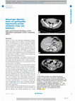
Progressive supranuclear palsy is one of the causes of atypical parkinsonian syndrome. We present... more Progressive supranuclear palsy is one of the causes of atypical parkinsonian syndrome. We present the case of an 83-year-old patient with typical symptomatology suggesting progressive supranuclear palsy with characteristic imaging.
Central neurocytoma—positive and differential diagnosis: An example through a case report
SAGE open medical case reports, 2023

Intracochlear schwannoma: Imaging diagnosis
Journal of Otology
A rare case of Fahr disease revealed by an epileptic seizure
Radiology Case Reports
Hypotension intracrânienne: apport de l'irm cérébrale
Journal of Neuroradiology
Malformations congénitales de l'oreille interne: ce que le radiologue doit savoir
Journal of Neuroradiology
Neurosyphilis en déguisement à l'irm
Journal of Neuroradiology
Apport de l'irm dans les agénésies calleuses: à propos d'une série de 17 cas
Journal of Neuroradiology
Syndrome de la dent couronnée cause rare de cervicalgies chroniques: à propos d'un cas
Journal of Neuroradiology
Osteomyelite du crane: une complication severe et un challenge diagnostique
Journal of Neuroradiology
Fulminant Susac syndrome—a rare cause of coma: The history of the fatal course in a young man
SAGE Open Medical Case Reports
Susac syndrome is a rare microangiopathy of indeterminate etiology, presumably autoimmune, charac... more Susac syndrome is a rare microangiopathy of indeterminate etiology, presumably autoimmune, characterized by a triad of encephalopathy, sensorineural hearing loss, and branch retinal artery occlusions occurring predominantly in women. The onset and progression patterns are multiple, mainly of three modes. Fulminant evolution is exceptional, rarely reported across literature. We report through this case a Susac syndrome in a young man in whom evolution was fatal. Magnetic resonance imaging is essential to raise the diagnosis and for follow-up, with almost pathognomonic findings, all the more useful as the clinical triad is usually incomplete and as the encephalopathy is the most limiting of the symptoms.
Apport de l'IRM dans les cholestéatomes opérés (résidu ? récidive ? Ou autres ?)
Journal of Neuroradiology
Spontaneous clival meningocele
SAGE Open Medical Case Reports
The occipital bone is an uncommon location for meningoceles protrusion. This condition occurs gen... more The occipital bone is an uncommon location for meningoceles protrusion. This condition occurs generally after a severe traumatism or surgical procedure. However, in some rare cases, the herniation can happen spontaneously. Nontraumatic clival meningoceles present an extremely rare entity and correspond to a herniating pachymeningeal collection containing cerebrospinal fluid through a zone of fragility in the clivus. Clinical presentation ranges from simple headache or rhinorrhea to severe complications such as recurrent bacterial meningitis or nerve compression. Computed tomography provides an analysis of the bone and magnetic resonance imaging provides a superior contrast resolution, helping to distinguish among the various types of clival lesions. We report the case of a young woman with a long history of idiopathic intracranial hypertension, who presented with a worsening headache. Magnetic resonance imaging confirmed a clival meningocele without other complications and the patie...
Incidental optic nerve sheath arachnoid cyst: A rare case finding with literature review
SAGE Open Medical Case Reports
Arachnoid cysts are the most common benign cystic abnormalities formed due to congenital splittin... more Arachnoid cysts are the most common benign cystic abnormalities formed due to congenital splitting of the arachnoid layer. They comprise 1% of intracranial masses, and the orbital location is even more rarely reported in history especially in the pediatric population. They might be discovered as an asymptomatic finding on imaging performed for a concomitant condition or, in most reported cases, as a result of ophthalmic impairment. They can be isolated or associated with gliomas, neurofibromas, empty sella syndrome, and frontotemporal porencephalic cysts. Computed tomography scan shows a non-enhancing liquid cystic lesion, and magnetic resonance imaging remains the best assessment tool confirming the similarity of the fluid to cerebrospinal fluid and evaluating the optic nerves. Herein, we report the case of an incidental discovery of an intraorbital arachnoid cyst on magnetic resonance imaging in a 53-year-old woman with a history of epilepsy. No treatment was performed as the cyst...