John Fassett - Profile on Academia.edu (original) (raw)
Papers by John Fassett

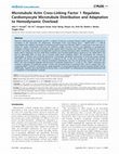
MACF1 depletion alters microtubule organization in neonatal rat cardiomyocytes
<p>Neonatal rat ventricular cardiomyocytes(NRVMs) transfected with MACF1 RNAi or non-target... more <p>Neonatal rat ventricular cardiomyocytes(NRVMs) transfected with MACF1 RNAi or non-targeting control RNAi were immunostained for tubulin (red). Hoescht stain (blue) was used to identify nuclei.</p
MACF1 deletion exacerbates pressure overload induced hypertrophy
<p>3 weeks after control or TAC conditions, WT or MACF1 KO hearts were collected and used t... more <p>3 weeks after control or TAC conditions, WT or MACF1 KO hearts were collected and used to measure left ventricle(LV) weight to body weight ratio (Figure 2A), Left atria (LA) to body weight ratio (Figure 2B), Right ventricle (RV) weight to body weight ratio (Figure 2C) and right atria weight to body weight ratio (Figure 2D). Some hearts(n = 5 per condition) were used for measuring cardiomyocyte cross-sectional area (Figure 2E), or for Sirius red staining for fibrosis (Figure 2F) Graphs represent average tissue weights from 8 WT and 8 MACF1 KO mice under control conditions, and 19 WT and 12 MACF1 KO mice under TAC conditions. Cardiomyocyte cross sectional area was averaged from 5 mice (∼300 cells per heart) from each condition (* indicates p<.05 as compared to control conditions, # indicates p<.05 as compared to WT under same conditions).</p
Maladaptive response to pressure overload is associated with redistribution of microtubules into distinct subcellular fractions
<p>The relationship between LV ejection fraction and the levels of Tubulin, as well as β-ac... more <p>The relationship between LV ejection fraction and the levels of Tubulin, as well as β-actin, and Desmin, in different subcellular fractions (Figure 5A) was examined by linear regression analysis (Figure 5B–5G). The data was collected from 18 WT mice and 6 MACF1 KO mice exposed to TAC. Relative values of the protein levels were determined as fold increase over non-banded controls from the same experiment. Samples with a red x are MACF1 KO.</p
MACF1 KO exacerbates pathological features of hypertrophy in response to TAC
<p>At 2 and 3 weeks after sham or TAC surgery, echocardiography was used to measure end sys... more <p>At 2 and 3 weeks after sham or TAC surgery, echocardiography was used to measure end systolic diameter (Figure 3A), end diastolic diameter (Figure 3B) percent ejection fraction (Figure 3C) and posterior wall thickness at end systole (Figure 3D) and end diastole (Figure 3E). After 3 weeks TAC, tissue was collected and lung weight to body weight ratio was used to indicate pulmonary congestion (Figure 3F) Expression of stress responsive proteins atrial natrurietic protein (ANP) (Figure 3G), Serca2A (Figure 3H) β-Myosin heavy chain (Figure 3I) were measured by western blot. α-MHC was used as a loading control (Figure 3J) (* indicates p<.05 as compared to control conditions, # indicates p<.05 as compared to WT under same conditions. For LV function measurements, WT control (n = 8), MACF1 KO control (n = 8), WT TAC (n = 16), KO TAC (n = 11)).</p
MACF1 expression is increased in response to pressure overload
<p>RT-PCR was performed to amplify between exons 10 and 14 of MACF1. A 269 b.p. band corres... more <p>RT-PCR was performed to amplify between exons 10 and 14 of MACF1. A 269 b.p. band corresponding to splicing between exon 10 and 14 (indicating loss of exons 11–13) is observed only in MACF1 KO mice, whereas WT mice express only the predicted size of 645 b.p. (Figure 1A) Western blot analysis of triton soluble (Figure 1B and 1C) and insoluble fractions (Figure 1B and 1D) of ventricular lysates of WT and MACF1 KO mice under control or after 3 weeks of TAC conditions (total of 7 weeks after tamoxifen treatment) demonstrates MACF1 expression increases in response to TAC, and is effectively silenced in MACF1 KO hearts (graphs represent averages of 3 WT or KO control mice, and 9 WT and KO TAC mice). Troponin I and vinculin were used as loading controls. (* indicates p<.05 as compared to control conditions, # indicates p<.05 as compared to WT under same conditions).</p
Abstract 1582: Adenosine A3 Receptor Deficiency Exerts Unanticipated Protective Effects on Pressure Overloaded-Induced Left Ventricular Hypertrophy and Dysfunction in Mice
Circulation, Oct 28, 2008
Endogenous adenosine can protect the overloaded heart against the development of hypertrophy and ... more Endogenous adenosine can protect the overloaded heart against the development of hypertrophy and heart failure, but the contribution of A 1 receptors (A 1 R) and A 3 receptors(A 3 R) is not known. To test the hypothesis A 1 R and A 3 R can protect the heart against systolic overload, we exposed A 3 R gene deficient (A 3 R KO) mice and A 1 R KO mice to transverse aortic constriction (TAC). Contrary to our hypothesis, A 3 R KO attenuated 5 weeks TAC-induced left ventricular (LV) hypertrophy (ratio of ventricular mass/body weight increased to 7.6 ±0.3 mg/g in wild type (Wt) mice as compared with 6.3±0.4 mg/g in KO), fibrosis and dysfunction (LV ejection fraction decreased to 43±2.5% and 55±4.2% in Wt and KO mice, respectively). A 3 R KO also attenuated the TAC-induced increases of myocardial ANP and the oxidative stress markers 3-nitrotyrosine(3-NT ) and 4-hydroxynonenal. In addition, A 3 R KO significantly attenuated TAC-induced activation of multiple MAP kinase pathways, and the activation of Akt-GSK signaling pathway. In contrast, A 1 R-KO increased TAC-induced mortality, but did not alter ventricular hypertrophy or dysfunction compared to Wt mice. In mice in which extracellular adenosine production was impaired by CD73 KO, TAC caused greater hypertrophy and dysfunction, and increased myocardial 3-NT, indicates that extracellular adenosine protects heart against TAC-induced ventricular oxidative stress and hypertrophy. In neonatal rat cardiomyocytes induced to hypertrophy with phenylephrine, the adenosine analogue 2-chloroadenosine (CADO) reduced cell area, protein synthesis, ANP and 3-NT. Antagonism of A3R significantly potentiated the anti-hypertrophic effects of CADO. Our data demonstrated that extracellular adenosine exerts protective effects on the overloaded heart, but A 3 R act counter to the protective effect of adenosine. The data suggest that selective attenuation of A 3 R activity might be a novel approach to attenuate pressure overload-induced myocardial oxidative stress, LV hypertrophy and dysfunction. This research has received full or partial funding support from the American Heart Association, AHA Midwest Affiliate (Illinois, Indiana, Iowa, Kansas, Michigan, Minnesota, Missouri, Nebraska, North Dakota, South Dakota & Wisconsin).
Abstract 16017: Metformin Protects the Heart from Systolic Overload-Induced Heart Failure Through an Ampka2 Independent Pathway
Circulation, Nov 22, 2011
Hu X global-DDAH1.1540.full
Abstract 1578: Endogenous Adenosine Protects the Heart from Severe Systolic Overload Induced Ventricular Hypertrophy and Congestive Heart Failure
Circulation, Oct 31, 2006
Adenosine regulates myocardial blood flow and protects the heart from acute ischemia-reperfusion ... more Adenosine regulates myocardial blood flow and protects the heart from acute ischemia-reperfusion induced damage. However, the effect of endogenous adenosine on chronic systolic overload induced left ventricular hypertrophy (LVH) and congestive heart failure (CHF) is unknown. CD73 uses AMP as substrate to generate adenosine, and CD73-deficiency attenuates extracellular adenosine production. To determine the effect of endogenous adenosine on LVH and CHF, the degree of LV hypertrophy, dilation and dysfunction in response to severe transverse aortic constriction (TAC) was determined in CD73-deficient (CD73-KO) mice and their wild type littermates. CD73-KO mice develop and grow normally without obvious cardiovascular defects or dysfunction. Although there was no difference in heart weight, lung weight, or LV function between CD73-KO mice and wild type littermates (WTL) under unstressed conditions, CD73-KO mice demonstrated more severe myocardial hypertrophy, LV dilation and LV dysfunction in response to systol...
A novel role for adenosine kinase (ADK) in cardiac autophagy
Intrinsic Activity, 2018
PLOS ONE, Sep 26, 2013
Aberrant cardiomyocyte microtubule growth is a feature of pressure overload induced cardiac hyper... more Aberrant cardiomyocyte microtubule growth is a feature of pressure overload induced cardiac hypertrophy believed to contribute to left ventricular (LV) dysfunction. Microtubule Actin Cross-linking Factor 1 (MACF1/Acf7) is a 600 kd spectraplakin that stabilizes and guides microtubule growth along actin filaments. MACF1 is expressed in the heart, but its impact on cardiac microtubules, and how this influences cardiac structure, function, and adaptation to hemodynamic overload is unknown. Here we used inducible cardiac-specific MACF1 knockout mice (MACF1 KO) to determine the impact of MACF1 on cardiac microtubules and adaptation to pressure overload (transverse aortic constriction (TAC).In adult mouse hearts, MACF1 expression was low under basal conditions, but increased significantly in response to TAC. While MACF1 KO had no observable effect on heart size or function under basal conditions, MACF1 KO exacerbated TAC induced LV hypertrophy, LV dilation and contractile dysfunction. Interestingly, subcellular fractionation of ventricular lysates revealed that MACF1 KO altered microtubule distribution in response to TAC, so that more tubulin was associated with the cell membrane fraction. Moreover, TAC induced microtubule redistribution into this cell membrane fraction in both WT and MACF1 KO mice correlated strikingly with the level of contractile dysfunction (r 2 = 0.786, p,.001). MACF1 disruption also resulted in reduction of membrane caveolin 3 levels, and increased levels of membrane PKCa and b1 integrin after TAC, suggesting MACF1 function is important for spatial regulation of several physiologically relevant signaling proteins during hypertrophy. Together, these data identify for the first time, a role for MACF1 in cardiomyocyte microtubule distribution and in adaptation to hemodynamic overload.
Adenosine kinase (ADK) inhibition with ABT-702 induces ADK protein degradation and a distinct form of sustained cardioprotection
European Journal of Pharmacology, May 1, 2022

Molecular Pharmacology, 2018
According to current views, oxidation of aldehyde dehydrogenase-2 (ALDH2) during glyceryltrinitra... more According to current views, oxidation of aldehyde dehydrogenase-2 (ALDH2) during glyceryltrinitrate (GTN) biotransformation is essentially involved in vascular nitrate tolerance and explains the dependence of this reaction on added thiols. Using a novel fluorescent intracellular nitric oxide (NO) probe expressed in vascular smooth muscle cells (VSMCs), we observed ALDH2-catalyzed formation of NO from GTN in the presence of exogenously added dithiothreitol (DTT), whereas only a short burst of NO, corresponding to a single turnover of ALDH2, occurred in the absence of DTT. This short burst of NO associated with oxidation of the reactive C302 residue in the active site was followed by formation of low-nanomolar NO, even without added DTT, indicating slow recovery of ALDH2 activity by an endogenous reductant. In addition to the thiol-reversible oxidation of ALDH2, thiol-refractive inactivation was observed, particularly under high-turnover conditions. Organ bath experiments with rat aortas showed that relaxation by GTN lasted longer than that caused by the NO donor diethylamine/NONOate, in line with the long-lasting nanomolar NO generation from GTN observed in VSMCs. Our results suggest that an endogenous reductant with low efficiency allows sustained generation of GTN-derived NO in the lownanomolar range that is sufficient for vascular relaxation. On a longer time scale, mechanism-based, thiol-refractive irreversible inactivation of ALDH2, and possibly depletion of the endogenous reductant, will render blood vessels tolerant to GTN. Accordingly, full reactivation of oxidized ALDH2 may not occur in vivo and may not be necessary to explain GTN-induced vasodilation. This work was supported by the Fonds zur Förderung der Wissenschaftlichen Forschung in Austria [Grant P24946].

Basic research in cardiology, May 1, 2017
Inflammatory responses play an important role in the development of left ventricular (LV) hypertr... more Inflammatory responses play an important role in the development of left ventricular (LV) hypertrophy and dysfunction. Recent studies demonstrated that increased T-cell infiltration and T-cell activation contribute to LV hypertrophy and dysfunction. Dendritic cells (DCs) are professional antigen-presenting cells that orchestrate immune responses, especially by modulating T-cell function. In this study, we investigated the role of bone marrow-derived CD11c(+) DCs in transverse aortic constriction (TAC)-induced LV fibrosis and hypertrophy in mice. We observed that TAC increased the number of CD11c(+) cells and the percentage of CD11c(+) MHCII(+) (major histocompatibility complex class II molecule positive) DCs in the LV, spleen and peripheral blood in mice. Using bone marrow chimeras and an inducible CD11c(+) DC ablation model, we found that depletion of bone marrow-derived CD11c(+) DCs significantly attenuated LV fibrosis and hypertrophy in mice exposed to 24 weeks of moderate TAC. C...
Induction of hepatocyte proliferation in vitro by inhibition of cell cycle inhibitors
Regulation of Hepatocyte Cell Cycle Progression and Differentiation by Type I Collagen Structure
Current Topics in Developmental Biology, 2005
Cell behavior is strongly influenced by the extracellular matrix (ECM) to which cells adhere. Bot... more Cell behavior is strongly influenced by the extracellular matrix (ECM) to which cells adhere. Both chemical determinants within ECM molecules and mechanical properties of the ECM network regulate cellular response, including proliferation, differentiation, and apoptosis. Type I collagen is the most abundant ECM protein in the body with a complex structure that can be altered in vivo by proteolysis, cross-linking, and other processes. Because of collagen&amp;amp;amp;amp;amp;amp;amp;amp;amp;amp;amp;amp;amp;amp;amp;amp;amp;amp;amp;#39;s complex and dynamic nature, it is important to define the changes in cell response to different collagen structures and its underlying mechanisms. This chapter reviews current knowledge of potential mechanisms by which type I collagen affects cell behavior, and it presents data that elucidate specific intracellular signaling pathways by which changes in type I collagen structure differentially regulate hepatocyte cell cycle progression and differentiation. A network of polymerized fibrillar type I collagen (collagen gel) induces a highly differentiated but growth-arrested phenotype in primary hepatocytes, whereas a film of monomeric collagen adsorbed to a rigid dish promotes cell cycle progression and dedifferentiation. Studies presented here demonstrate that protein kinase A (PKA) activity is significantly elevated in hepatocytes on type I collagen gel relative to collagen film, and inhibition of this elevated PKA activity can promote hepatocyte cell cycle progression on collagen gel. Additional studies are presented that examine changes in hepatocyte cell cycle progression and differentiation in response to increased rigidity of polymerized collagen gel by fiber cross-linking. Potential mechanisms underlying these cellular responses and their implications are discussed.
Cardiomyocyte Microtubule Distribution and Adaptation to Hemodynamic Overload
Aberrant cardiomyocyte microtubule growth is a feature of pressure overload induced cardiac hyper... more Aberrant cardiomyocyte microtubule growth is a feature of pressure overload induced cardiac hypertrophy believed to contribute to left ventricular (LV) dysfunction. Microtubule Actin Cross-linking Factor 1 (MACF1/Acf7) is a 600 kd spectraplakin that stabilizes and guides microtubule growth along actin filaments. MACF1 is expressed in the heart, but its impact on cardiac microtubules, and how this influences cardiac structure, function, and adaptation to hemodynamic overload is unknown. Here we used inducible cardiac-specific MACF1 knockout mice (MACF1 KO) to determine the impact of MACF1 on cardiac microtubules and adaptation to pressure overload (transverse aortic constriction (TAC).In adult mouse hearts, MACF1 expression was low under basal conditions, but increased significantly in response to TAC. While MACF1 KO had no observable effect on heart size or function under basal conditions, MACF1 KO exacerbated TAC induced LV hypertrophy, LV dilation and contractile dysfunction. Inte...
Abstract 1412: Critical Role Of Extracellular Sod For Protecting The Heart Against Myocardial Hypertrophy And Dysfunction After Myocardial Infarction
Circulation, Oct 16, 2007
<jats:p> Extracellular SOD ( <jats:bold>SOD3</jats:bold> ) contributes only a s... more <jats:p> Extracellular SOD ( <jats:bold>SOD3</jats:bold> ) contributes only a small fraction to total SOD activity in the normal heart, but is strategically located to scavenge free radicals in the extracellular compartment. SOD3 expression is decreased in the failing heart, but whether SOD3 can abrogate oxidative stress or modify left ventricular (LV) remodeling following myocardial infarction (MI) is unclear. To examine this question, we studied LV remodeling in SOD3 KO mice and wild type mice following MI. Under unstressed conditions, SOD3 KO had no effect on myocardial total SOD activity, SOD1 or SOD2 protein content, or myocardial nitrotyrosine or superoxide anion production, and caused no change in LV ejection fraction. However, 4 weeks or 8 weeks after MI, SOD3 KO mice developed more LV hypertrophy (8 weeks after MI, ventricular mass increased 1.64-fold in KO mice as compared to 1.35-fold in wild type mice, p<0.01) and had a greater reduction of LV ejection fraction (8 weeks after MI, LV ejection fraction was 35±2.4% in wild type mice as compared to 30±2.0% in KO mice, p<0.01). As compared with wild type mice, SOD3 KO mice had significantly greater increases of myocardial nitrotyrosine and superoxide anion production, a significantly greater decrease of ANP in the peri-infarct zone, and a significant more decrease of SERCA2a in both the peri-infarct and remote zones. In addition, MI caused greater activation of mitogen-activated protein kinase (MAPK) signaling pathways in SOD3 KO mice, as demonstrated by significantly greater increases of p-p38 <jats:sup>Thr180/</jats:sup> <jats:sub>Tyr182</jats:sub> , p-Erk <jats:sup>Thr202/Tyr204</jats:sup> and p-JNK <jats:sup>Thr183/Tyr185</jats:sup> in SOD3 KO mice 8 weeks after MI. The finding that SOD3 KO had no effect on myocardial total SOD activity, but significantly exacerbated MI induced LV remodeling implies that the specific extracellular location of SOD3 is more important than its contribution to overall SOD activity in protecting the heart against contractile dysfunction following myocardial infarct. </jats:p>

This manuscnpt has been reproduced from the microfilm master. UMI films the text directly from th... more This manuscnpt has been reproduced from the microfilm master. UMI films the text directly from the original or copy submitted. Thus, some thesis and dissertation copies are in typewriter tace, while others may be from any type of computer printer. The quality of this reproduction is dependent upon the quality of the copy submitted. Broken or indistinct print, colored or poor quality illustrations and photographs, print bleedthrough, substandard margins, and improper alignment can adversely affiect reproduction. In the unlikely event that the author dkJ not send UMI a complete manuscript and there are missing pages, these will be noted. Also, if unauthorized copyright material had to be removed, a note will indicate the deletk)n. Oversize materials (e.g., maps, drawings, charts) are reproduced by sectioning the original, beginning at the upper left-hand comer and continuing from left to right in equal sections with small overiaps. Photographs included in the original manuscript have been reproduced xerographically in this copy. Higher quality 6' x 9' black and wtiite photographic prints are available for any photographs or illustrations appearing in this copy for an additional charge. Contact UMI directly to order.
Adenosine kinase mediates adenosine attenuation of cardiomyocyte microtubule cytoskeletal densification
Intrinsic Activity, 2015