Teruo Nishida - Academia.edu (original) (raw)
Papers by Teruo Nishida

Investigative Ophthalmology & Visual Science, Apr 1, 2004
PURPOSE. Insulin-like growth factors (IGFs) and either substance P (SP) or an SP-derived peptide ... more PURPOSE. Insulin-like growth factors (IGFs) and either substance P (SP) or an SP-derived peptide (FGLM-amide) synergistically facilitate corneal epithelial wound healing in vitro and in vivo. The mechanism of this synergism and the clinical potential of these agents were further investigated by determination of the relevant functional domain of IGFs. METHODS. The effects of IGF-derived peptides on corneal epithelial cell migration were evaluated with the rabbit cornea in an organ culture system. Corneal epithelial wound closure in vivo was also evaluated in rabbits after epithelial debridement with n-heptanol. RESULTS. In the presence of FGLM-amide, peptides corresponding to the C domain of IGF-1 or-2 significantly promoted corneal epithelial migration in vitro to an extent similar to that apparent with the full-length molecules. In contrast, peptides corresponding to the D domain of these growth factors had no such effect. Mutation of serine-34 in the C domain of IGF-1 to alanine abolished the synergistic effect with FGLM-amide on corneal epithelial migration. The C peptide of proinsulin did not affect corneal epithelial migration in the absence or presence of FGLM-amide. The administration of eye drops containing both the C-domain peptide of IGF-1 and FGLM-amide significantly promoted corneal epithelial wound closure in vivo. CONCLUSIONS. The C domain of IGF-1 or-2, for which no biological function has previously been identified, is essential for the synergistic effect of these growth factors with SP on corneal epithelial migration.

Expert Opinion on Investigational Drugs, 1995
Hyaluronan (hyaluronic acid) is a high molecular weight glycosaminoglycan, distributed ubiquitous... more Hyaluronan (hyaluronic acid) is a high molecular weight glycosaminoglycan, distributed ubiquitously in the extracellular spaces in the body, and especially in the soft connective tissues. Early research into hyaluronan focused primarily upon its physicochemical properties, such as its viscosity and elasticity. Based upon these properties, hyaluronan has been applied clinically as a viscosurgical tool in ophthalmology and in otology. During the last decade, hyaluronan has been shown to be involved in a range of cell behaviours, such as cell movement, growth and differentiation. In addition, hyaluronan receptors have been isolated from a variety of cell surfaces. Hence, hyaluronan is no longer considered solely to be a constituent of the extracellular space, but, in addition, to have a role in modulating cellular functions. The roles of hyaluronan in such functions as wound healing and cell migration, locomotion and motility have been investigated. The epithelial surface of the body is one of the most important barrier systems to protect and maintain the internal environment. The epithelium possesses an active wound healing system to repair defects, and, in general, has a tremendous capacity to heal rapidly after injury. However, disturbance or delay to normal wound healing processes results in undesirable outcomes, such as persistent defects or recurrent erosions. In order to treat these epithelial disorders clinically, it is important to understand the mechanisms of epithelial wound healing. In this article, we review the recent developments in the use of hyaluronan in wound healing, and, particularly, the mechanisms of action of hyaluronan during corneal epithelial wound healing.

Experimental Eye Research, 1991
Hyaluronan (hyaluronic acid), well-known for its viscoelastic properties, is also recognized as a... more Hyaluronan (hyaluronic acid), well-known for its viscoelastic properties, is also recognized as a biological signal to cells. Using organ cultures of the rabbit cornea, we investigated the effects of hyaluronan on the migration of cornea1 epithelium. The addition of hyaluronan to the culture medium increased the length of the path of the cornea1 epithelial layer in a dose-dependent fashion. Other glycosaminoglycans (chondroitin, chondroitin sulphate. keratan sulphate and heparan sulphate) were also tried, but only hyaluronan exhibited a stimulatory effect on cornea1 epithelial migration. The effects of hyaluronan and fibronectin or epidermal growth factor (EGF) were additive: the addition of antisera against fibronectin or against EGF did not alter the stimulatory effect of hyaluronan. These results demonstrate that hyaluronan stimulates cornea1 epithelial migration by mechanism(s) different from those of fibronectin and EGF.

Effects of antimicrobials on corneal epithelial migration
Current Eye Research, 1993
The slowed healing rates observed by some investigators may be caused by vehicles or preservative... more The slowed healing rates observed by some investigators may be caused by vehicles or preservatives in the antimicrobials preparations tested. To determine whether antimicrobials directly inhibit corneal epithelial wound healing, we cultured blocks of the rabbit cornea in media containing various concentrations of antibiotics or antimicrobials (at 1, 10, or 100 micrograms/ml); after 24 hours, we measured the distance of epithelium that had migrated down the side of each block. The higher concentrations of fluoroquinolones (ofloxacin; 74 +/- 5.8% of control at 100 micrograms/ml, p < 0.05, ciprofloxacin; 4.4 +/- 1.5% of control at 100 micrograms/ml, p < 0.01, or norfloxacin; 71 +/- 7.0% at 10 mu g/ml, p < 0.01, and 1.5 +/- 0.4% of control at 100 mu g/ml, p < 0.01) and the highest concentrations of peptides (polymyxin B; 64 +/- 3.0% of control at 100 micrograms/ml, p < 0.01, or colistin; 67 +/- 5.7% of control at 100 micrograms/ml, p < 0.01) or fosfomycin (79 +/- 6.2% of control at 100 micrograms/ml, p < 0.05) had an inhibitory effect on corneal epithelial migration. Among aminoglycosides tested, sisomicin (85 +/- 10.0% of control, not significant), dibekacin (76 +/- 11.6% of control, p < 0.05) and streptomycin (77 +/- 9.4% of control, not significant) were inhibitory at 100 micrograms/ml, but tobramycin had no effect. Penicillins (aspoxicillin, sulbenicillin or ampicillin), cephalosporins (cefmenoxime or cefminox), oxytetracycline, erythromycin and chloramphenicol did not affect epithelial migration at all. These results demonstrate that some antimicrobials are inhibitory at high concentrations, but penicillins, cephalosporins, oxytetracycline, erythromycin or chloramphenicol has no inhibitory effect on corneal epithelial migration.

Journal of Leukocyte Biology, 2003
Activated corneal fbroblasts and infiltrated leukocytes are thought to contribute to corneal ulce... more Activated corneal fbroblasts and infiltrated leukocytes are thought to contribute to corneal ulceration. The potential roles of neutrophil-fibroblast and cell-matrix interactions in the degradation of stromal collagen associated with corneal ulceration have now been investigated with the use of three-dimensional cultures of rabbit cells in collagen gels. Degradation of collagen fibrils during culture was measured by spectrophotometric determination of released hydroxyproline. Whereas corneal fibroblasts alone degraded collagen fibrils to a small extent, neutrophils did not. However, the addition of neutrophils or neutrophil–conditioned medium (CM) to cultures of corneal fibroblasts resulted in a marked increase in the amount of collagen degraded by the fibroblasts. The effect of CM from neutrophils cultured in collagen gels on collagen degradation by corneal fibroblasts was greater than that of medium conditioned by neutrophils in monolayer culture. Immunoblot as well as reverse tra...
Extracellular matrix and growth factors in corneal wound healing
Current Opinion in Ophthalmology, 1995
Healing of corneal wounds is a complex process involving epithelial, keratocyte, and endothelial ... more Healing of corneal wounds is a complex process involving epithelial, keratocyte, and endothelial interactions that are affected by their associations with wound bed matrix and by cytokine availability and activation. The spectrum of possible cellular-matrix-growth factor interactions is indeed great and growing. Several of the significant contributions made during the past year include development of an organotypic organ culture model of the cornea that allows in vitro assembly of the epithelial extracellular matrix-anchoring complex, demonstration of epithelial synthesis of Bowman's layer collagens, demonstration of transforming growth factor-beta 2's inhibition of stromal cell collagenase synthesis, and demonstration of the paracrine pathway of keratinocyte growth factor action in the cornea.

Current Opinion in Ophthalmology, 2009
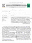
The cornea is one of the most heavily innervated and most sensitive tissues in the body. The dens... more The cornea is one of the most heavily innervated and most sensitive tissues in the body. The density of nerve endings in the cornea is thus about 300-400 times greater than that in the skin. Most of the sensory nerves in the cornea are derived from the ciliary nerves of the ophthalmic branch of the trigeminal nerve. These long ciliary nerves constitute the perilimbal nerve ring. Nerve fibers penetrate the cornea in the deep peripheral stroma radially and then course anteriorly, forming a terminal subepithelial plexus. The nerve fibers lose their myelination within a short distance of their point of entry into the
![Modified from [3]. Table 1 Causes of neurotrophic keratopathy](https://figures.academia-assets.com/41826486/table_001.jpg) ](
](
The Effects of Dexamethasone on the Na,K-ATPase Activity and Pump Function of Corneal Endothelial Cells
Current Eye Research, 2009
The Na(+)- and K(+)-dependent ATPase (Na,K-ATPase) expressed in the basolateral membrane of corne... more The Na(+)- and K(+)-dependent ATPase (Na,K-ATPase) expressed in the basolateral membrane of corneal endothelial cells plays an important role in the pump function of the corneal endothelium. We investigated the possible role of dexamethasone in the regulation of Na,K-ATPase activity and pump function in corneal endothelial cells. Confluent monolayers of mouse corneal endothelial cells were exposed to dexamethasone. ATPase activity of the cells was evaluated by spectrophotometric measurement of phosphate released from ATP with the use of ammonium molybdate, with Na,K-ATPase activity being defined as the portion of total ATPase activity sensitive to ouabain. Pump function of the cells was measured with the use of an Ussing chamber, with the pump function attributable to Na,K-ATPase activity being defined as the portion of the total short-circuit current sensitive to ouabain. Western blot analysis was examined to measure the expression of the Na,K-ATPase alpha(1)-subunit. Dexamethasone (1 or 10 microM) increased the Na,K-ATPase activity and pump function of the cultured cells. These effects of dexamethasone were blocked by cycloheximide, a protein synthesis inhibitor. Western blot analysis also indicated that dexamethasone increased the expression of the Na,K-ATPase alpha(1)-subunit, whereas it decreased the expression of the phospho-Na,K-ATPase alpha(1)-subunit. Our results suggest that dexamethasone stimulates Na,K-ATPase activity in mouse corneal endothelial cells. The effect of dexamethasone activation in these cells is mediated by Na,K-ATPase synthesis and increase in an enzymatic activity by dephosphorylation of Na,K-ATPase alpha(1)-subunits.
Substance P in Human Tears
Cornea, 2003
To determine the levels and biochemical characteristics of substance P-like immunoreactivity (SPL... more To determine the levels and biochemical characteristics of substance P-like immunoreactivity (SPLI) in human tears and ascertain whether substance P (SP) concentrations in tears reflect the condition of the ocular surface. Unstimulated tears were collected with a micropipette. Tear samples were partially purified using C-18 cartridges. Levels of SPLI in purified samples were measured using an enzyme immunoassay (EIA). For biochemical characterization of SPLI, tear extracts were fractionated using high-performance liquid chromatography (HPLC); each fraction was then subjected to EIA. To determine the catabolism of SP in tears, synthetic SP was incubated in medium containing pooled tears and then analyzed using HPLC. The concentration of SPLI in normal human tears was 306.0 +/- 96.5 pg/mL (mean +/- SD, range 148-555 pg/mL). Levels of SPLI did not vary significantly by age or gender. Concentrations of SPLI in tears from eyes with unilateral corneal hypesthesia were lower than those in tears from contralateral healthy eyes. Diclofenac sodium eye drops reduced concentrations of prostaglandin E2 and SPLI in tears. Analysis using HPLC indicated that five different substances contributed to SPLI in tears and that SP was broken down into several fragments, including SP(8-11), by enzymes present in tears. Substance P is a normal component of human tears. Levels of SPLI in tears might reflect the denervated status of the ocular surface. Substance P is catabolized by degradative enzymes in tears to maintain the ocular surface by exerting the trophic effects of SP while avoiding undesirable effects.
Open Clinical Study of Eye Drops Containing the Fibronectin-Derived Peptide PHSRN for Treatment of Persistent Corneal Epithelial Defects
Cornea, 2012
We have previously shown that the fibronectin-derived peptide PHSRN (Pro-His-Ser-Arg-Asn) promote... more We have previously shown that the fibronectin-derived peptide PHSRN (Pro-His-Ser-Arg-Asn) promotes corneal epithelial wound healing in vivo. We have now examined the clinical efficacy of eye drops containing the PHSRN peptide for treatment of persistent epithelial defects (PEDs) of the cornea. Seven patients (5 men and 2 women; mean age±SD, 78.3±9.4 years) with PEDs were treated by administration of eye drops containing PHSRN. The duration of the PEDs before treatment was 7.4±5.5 weeks. The eye drops were administered as 1 drop per eye 4 times a day. Epithelial defects were observed with a slit-lamp microscope and photographed during the treatment course. Epithelial defects in 5 of the 7 affected eyes (71%) responded to PHSRN treatment as manifested by complete epithelial resurfacing within the 4-week period after treatment initiation. The mean±SD time required for complete epithelial resurfacing in the 5 responding subjects was 15.8±3.4 days. No adverse effects of treatment were observed in any of the subjects. Eye drops containing the fibronectin-derived PHSRN peptide are clinically efficacious for the treatment of PEDs.
Spreading of Cultured Corneal Epithelial Cells on Fibronectin and Other Extracellular Matrices
Cornea, 1990
Previous investigations demonstrated that fibronectin is an essential adhesive glycoprotein for t... more Previous investigations demonstrated that fibronectin is an essential adhesive glycoprotein for the corneal epithelial cells. Fibronectin mediates attachment and spreading of the corneal epithelial cells. However, it seems to be important to know the changes in cellular receptor activity in order to understand the interactions of corneal epithelial cells and underlying extracellular matrix. In this investigation, we studied the effects of various culture times and conditions on the receptor activity of rabbit corneal epithelial cells for fibronectin. The cultured cells were placed on tissue culture plates that were coated with various concentrations of four extracellular matrices: fibronectin, laminin and collagen types I and IV. Freshly isolated corneal epithelial cells without culture did not spread on fibronectin and laminin; they spread only on collagen types I and IV. When the epithelial cells were cultured for 12 h or more, they spread on fibronectin. However, the spreading of the cells on collagen types I and IV was the same regardless of the culture period (up to 20 h). Only a small number of epithelial cells spread on laminin at the highest concentration examined after culture for 12 h or more. Thus, the corneal epithelial cells responded differently to fibronectin, collagen types I and IV, and laminin. Perhaps the receptor for fibronectin appears when epithelial cells are cultured, but the receptor(s) for collagen types I and IV are always present on the cell surface of corneal epithelial cells.
Evaluation of povidone-iodine as a disinfectant solution for contact lenses: Antimicrobial activity and cytotoxicity for corneal epithelial cells
Contact Lens and Anterior Eye, 2006
Povidone-iodine (PVP-I) possesses broad-spectrum antimicrobial activity and is used clinically as... more Povidone-iodine (PVP-I) possesses broad-spectrum antimicrobial activity and is used clinically as a disinfectant. We evaluated the disinfectant properties and safety of PVP-I for use as a contact lens solution. The concentrations of PVP-I required to reduce the number of Staphylococcus aureus or Candida albicans by 3 log units were lower than were those of hydrogen peroxide, polyhexamethylene biguanide (PHMB), and benzalkonium chloride (BAK). The cytotoxicity of PVP-I for cultured human corneal epithelial (HCE) cells was less than that of the other three agents. The safety margin for PVP-I was thus greatest among the tested compounds. PVP-I appears suited for use as a contact lens disinfectant.

Clinical Ophthalmology, 2009
Purpose: To investigate whether the GLC3A locus harboring the CYP1B1 gene is associated with norm... more Purpose: To investigate whether the GLC3A locus harboring the CYP1B1 gene is associated with normal tension glaucoma (NTG) in Japanese patients. Materials and Methods: One hundred forty-two Japanese patients with NTG and 101 Japanese healthy controls were recruited. Patients exhibiting a comparatively early onset were selected as this suggests that genetic factors may show stronger involvement. Genotyping and assessment of allelic diversity was performed on 13 highly polymorphic microsatellite markers in and around the GLC3A locus. Results: There were decreased frequencies of the 444 allele of D2S0416i and the 258 allele of D2S0425i in cases compared to controls (P = 0.022 and P = 0.034, respectively). However, this statistical signifi cance disappeared when corrected (Pc Ͼ 0.05). We did not fi nd any signifi cant association between the remaining 11 microsatellite markers, including D2S177, which may be associated with CYP1B1, and NTG (P Ͼ 0.05). Conclusions: Our study showed no association between the GLCA3 locus and NTG, suggesting that the CYP1B1 gene, which is reportedly involved in a range of glaucoma phenotypes, may not be an associated factor in the pathogenesis of NTG.
Quantitative evaluation of corneal epithelial oedema by confocal microscopy
Clinical & Experimental Ophthalmology, 2009
To develop a novel quantitative index for evaluation of corneal epithelial oedema, the pixel inte... more To develop a novel quantitative index for evaluation of corneal epithelial oedema, the pixel intensity of confocal microscopic images was measured derived from the basal cell layer (BCL) of the corneal epithelium in normal eyes, eyes before and after cataract surgery, and eyes affected by bullous keratopathy. Five eyes of five normal volunteers, 14 eyes of 11 cataract patients and 12 eyes of 12 bullous keratopathy patients were examined by confocal microscopy. The cataract patients underwent cataract surgery, and they were examined by confocal microscopy, corneal pachymetry, and anterior fluorometry both before and at various times after surgery. The pixel intensity of BCL images obtained by confocal microscopy was measured and expressed as the BCL index. The coefficient of variation for repeated (five times) measurement of the BCL index in each of the five normal eyes was 3.4%. The BCL index was 54.8 5.3 (mean SD) before surgery, increased significantly to 65.2 10.0 on the day after surgery, and gradually decreased thereafter in the cataract patients. The time-course of the BCL index coincided well with that of corneal thickness and anterior fluorometry value. The BCL index in eyes affected by bullous keratopathy was significantly increased at 95.0 6.4. The BCL index was increased after cataract surgery and in eyes affected by bullous keratopathy, conditions associated with corneal epithelial oedema. This quantitative measure obtained by confocal microscopy may prove useful in the clinical evaluation of corneal epithelial oedema.

British Journal of Ophthalmology, 2005
Open clinical study of eye-drops containing tetrapeptides derived from substance P and insulin-like growth factor-1 for treatment of persistent corneal epithelial defects associated with neurotrophic keratopathy
British Journal of Ophthalmology, 2008
Loss of corneal sensation results in the development of persistent corneal epithelial defects. Th... more Loss of corneal sensation results in the development of persistent corneal epithelial defects. The combination of a substance P-derived peptide (FGLM-amide) and an insulin-like growth factor-1 (IGF-1)-derived peptide (SSSR) stimulates rabbit corneal epithelial migration in vitro and rabbit corneal epithelial wound closure in vivo. The clinical efficacy of eye-drops containing FGLM-amide and SSSR for the treatment of persistent corneal epithelial defects in individuals with neurotrophic keratopathy was examined in a prospective open study. Twenty-five consecutive patients (26 eyes) with persistent corneal epithelial defects associated with neurotrophic keratopathy were treated by administration of eye-drops containing FGLM-amide and SSSR. The course of epithelial healing was monitored by slit-lamp examination. Epithelial defects resurfaced completely in 19 of the 26 eyes (73%) within 4 weeks after treatment initiation. Complete resurfacing of epithelial defects was apparent in 18 of 22 (82%) or in one of four (25%) eyes without or with limbal stem cell deficiency, respectively. No adverse effects of treatment were observed in any subject. Eye-drops containing FGLM-amide and SSSR induced the rapid resurfacing of persistent epithelial defects in stem cell-positive individuals with neurotrophic keratopathy.

British Journal of Ophthalmology, 2008
Purpose: This study was performed to separately assess the aqueous flow applied with hyaluronic a... more Purpose: This study was performed to separately assess the aqueous flow applied with hyaluronic acid, and the behaviour of hyaluronic acid itself on the ocular surface. Methods: Two different fluorescent dyes, fluorescein sodium dissolved in 0.1% hyaluronic acid (HA) solution and 0.1% fluorescein conjugated with hyaluronic acid (F-HA) dissolved in saline, were used. A volume of 20 ml of tested solution was applied to the eye of 10 healthy volunteers. Fluorescein sodium dissolved in saline served as a control. The fluorescent intensity of the precorneal tear film was measured at the central cornea every minute for 10 min. The turnover rate was calculated using the equation that plots fluorescent intensity against time in a semilog plot and expressed as %/min. Results: Turnover rates of topically applied 0.1% F-HA, 0.1% HA and saline were 8.1 (SD 3.6)%/min, 21.6 (2.8)%/ min, and 31.0 (3.7)%/min, respectively. The turnover rate of F-HA was significantly lower than those of HA and saline (p = 0.00012 and p = 0.00000022, respectively; Mann-Whitney test). The turnover rate of HA was significantly lower than that of saline (p = 0.00001; Mann-Whitney test). Conclusion: Our results indicate that the bulk aqueous flow applied with HA and the turnover of HA itself are different. HA molecules may adhere to the ocular surface by surface-chemical and/or biochemical properties. The long retention time of HA on the ocular surface may explain the mechanism in which hyaluronic acid has been shown to enhance tear film stability for a few hours. -Published by bjo.bmj.com Downloaded from 2008 92: 108-111 Br J Ophthalmol H Mochizuki, M Yamada, S Hato, et al. applied hyaluronic acid precorneal residence time of topically Fluorophotometric measurement of the http://bjo.bmj.com/content/92/1/108.full.html
Up-regulation of HSP70 by the fibronectin-derived peptide PHSRN in human corneal epithelial cells
Biochemical and Biophysical Research Communications, 2008
HSP70 is a member of the heat shock protein family and is induced by various types of cellular st... more HSP70 is a member of the heat shock protein family and is induced by various types of cellular stress. We examined whether HSP70 might play a role in the healing of corneal epithelial wounds. Given that the PHSRN peptide, which corresponds to the second cell-binding domain of fibronectin, promotes corneal epithelial migration, we investigated the effect of this peptide on HSP70 expression in cultured human corneal epithelial cells. Reverse transcription-polymerase chain reaction and immunoblot analyses revealed that PHSRN increased the amounts of HSP70 mRNA and protein in these cells in a concentration- and time-dependent manner, whereas a control peptide (NRSHP) had no such effects. Furthermore, the PHSRN-induced up-regulation of HSP70 was blocked by SB203580, an inhibitor of p38 mitogen-activated protein kinase (MAPK), but it was not affected by PD98059 or SP600125, inhibitors of signaling by the MAPKs ERK and JNK, respectively. Our results suggest that induction of HSP70 expression may contribute to the promotion of corneal epithelial migration by PHSRN and hence to corneal epithelial wound healing.
Biochemical and Biophysical Research Communications, 2008
Semaphorins are a family of glycoproteins that play an important role in repulsive axon guidance ... more Semaphorins are a family of glycoproteins that play an important role in repulsive axon guidance during embryogenesis. We have now investigated the effect of corneal epithelial cells on the expression of Sema3A in corneal fibroblasts with the use of a coculture system in which the two cell types are separated by a collagen vitrigel membrane. Reverse transcription-polymerase chain reaction and immunoblot analyses revealed that the presence of immortalized human corneal epithelial (HCE) cells increased the expression of Sema3A in human corneal fibroblasts at both the mRNA and protein levels. This effect of HCE cells was mimicked by recombinant human epidermal growth factor (EGF) in a concentrationand time-dependent manner. An inhibitor of the tyrosine kinase activity of the EGF receptor, PD153035, blocked the EGF-induced up-regulation of both Sema3A mRNA and protein in corneal fibroblasts. Depletion of EGF in HCE cells by RNA interference largely abolished the effect of these cells on Sema3A expression in corneal fibroblasts. These findings indicate that EGF released from corneal epithelial cells up-regulates the expression of Sema3A in corneal fibroblasts. This effect of EGF may play an important role in maintenance of corneal structure and repair of corneal damage.

Biochemical and Biophysical Research Communications, 2010
Interactions between epithelial cells and fibroblasts play important roles in tissue homeostasis.... more Interactions between epithelial cells and fibroblasts play important roles in tissue homeostasis. Semaphorins are a family of glycoproteins that contribute to axon guidance during development. With the use of a coculture system in which human corneal fibroblasts and epithelial cells are cultured on opposite sides of a collagen membrane, we have examined the effects of overexpression of semaphorin 3A in corneal fibroblasts on the expression of junctional proteins in corneal epithelial cells. Reverse transcriptionpolymerase chain reaction as well as immunoblot and immunofluorescence analyses revealed that the overexpression of semaphorin 3A in corneal fibroblasts increased the expression of E-cadherin and N-cadherin in corneal epithelial cells at both the mRNA and protein levels. These effects of semaphorin 3A were dependent on the presence of extracellular Ca 2+ . Our findings indicate that semaphorin 3A released from corneal fibroblasts may play an important role in the regulation of intercellular communication between corneal epithelial cells as well as in the maintenance of corneal structure and function.
Investigative Ophthalmology & Visual Science, Apr 1, 2004
PURPOSE. Insulin-like growth factors (IGFs) and either substance P (SP) or an SP-derived peptide ... more PURPOSE. Insulin-like growth factors (IGFs) and either substance P (SP) or an SP-derived peptide (FGLM-amide) synergistically facilitate corneal epithelial wound healing in vitro and in vivo. The mechanism of this synergism and the clinical potential of these agents were further investigated by determination of the relevant functional domain of IGFs. METHODS. The effects of IGF-derived peptides on corneal epithelial cell migration were evaluated with the rabbit cornea in an organ culture system. Corneal epithelial wound closure in vivo was also evaluated in rabbits after epithelial debridement with n-heptanol. RESULTS. In the presence of FGLM-amide, peptides corresponding to the C domain of IGF-1 or-2 significantly promoted corneal epithelial migration in vitro to an extent similar to that apparent with the full-length molecules. In contrast, peptides corresponding to the D domain of these growth factors had no such effect. Mutation of serine-34 in the C domain of IGF-1 to alanine abolished the synergistic effect with FGLM-amide on corneal epithelial migration. The C peptide of proinsulin did not affect corneal epithelial migration in the absence or presence of FGLM-amide. The administration of eye drops containing both the C-domain peptide of IGF-1 and FGLM-amide significantly promoted corneal epithelial wound closure in vivo. CONCLUSIONS. The C domain of IGF-1 or-2, for which no biological function has previously been identified, is essential for the synergistic effect of these growth factors with SP on corneal epithelial migration.
Expert Opinion on Investigational Drugs, 1995
Hyaluronan (hyaluronic acid) is a high molecular weight glycosaminoglycan, distributed ubiquitous... more Hyaluronan (hyaluronic acid) is a high molecular weight glycosaminoglycan, distributed ubiquitously in the extracellular spaces in the body, and especially in the soft connective tissues. Early research into hyaluronan focused primarily upon its physicochemical properties, such as its viscosity and elasticity. Based upon these properties, hyaluronan has been applied clinically as a viscosurgical tool in ophthalmology and in otology. During the last decade, hyaluronan has been shown to be involved in a range of cell behaviours, such as cell movement, growth and differentiation. In addition, hyaluronan receptors have been isolated from a variety of cell surfaces. Hence, hyaluronan is no longer considered solely to be a constituent of the extracellular space, but, in addition, to have a role in modulating cellular functions. The roles of hyaluronan in such functions as wound healing and cell migration, locomotion and motility have been investigated. The epithelial surface of the body is one of the most important barrier systems to protect and maintain the internal environment. The epithelium possesses an active wound healing system to repair defects, and, in general, has a tremendous capacity to heal rapidly after injury. However, disturbance or delay to normal wound healing processes results in undesirable outcomes, such as persistent defects or recurrent erosions. In order to treat these epithelial disorders clinically, it is important to understand the mechanisms of epithelial wound healing. In this article, we review the recent developments in the use of hyaluronan in wound healing, and, particularly, the mechanisms of action of hyaluronan during corneal epithelial wound healing.
Experimental Eye Research, 1991
Hyaluronan (hyaluronic acid), well-known for its viscoelastic properties, is also recognized as a... more Hyaluronan (hyaluronic acid), well-known for its viscoelastic properties, is also recognized as a biological signal to cells. Using organ cultures of the rabbit cornea, we investigated the effects of hyaluronan on the migration of cornea1 epithelium. The addition of hyaluronan to the culture medium increased the length of the path of the cornea1 epithelial layer in a dose-dependent fashion. Other glycosaminoglycans (chondroitin, chondroitin sulphate. keratan sulphate and heparan sulphate) were also tried, but only hyaluronan exhibited a stimulatory effect on cornea1 epithelial migration. The effects of hyaluronan and fibronectin or epidermal growth factor (EGF) were additive: the addition of antisera against fibronectin or against EGF did not alter the stimulatory effect of hyaluronan. These results demonstrate that hyaluronan stimulates cornea1 epithelial migration by mechanism(s) different from those of fibronectin and EGF.
Effects of antimicrobials on corneal epithelial migration
Current Eye Research, 1993
The slowed healing rates observed by some investigators may be caused by vehicles or preservative... more The slowed healing rates observed by some investigators may be caused by vehicles or preservatives in the antimicrobials preparations tested. To determine whether antimicrobials directly inhibit corneal epithelial wound healing, we cultured blocks of the rabbit cornea in media containing various concentrations of antibiotics or antimicrobials (at 1, 10, or 100 micrograms/ml); after 24 hours, we measured the distance of epithelium that had migrated down the side of each block. The higher concentrations of fluoroquinolones (ofloxacin; 74 +/- 5.8% of control at 100 micrograms/ml, p < 0.05, ciprofloxacin; 4.4 +/- 1.5% of control at 100 micrograms/ml, p < 0.01, or norfloxacin; 71 +/- 7.0% at 10 mu g/ml, p < 0.01, and 1.5 +/- 0.4% of control at 100 mu g/ml, p < 0.01) and the highest concentrations of peptides (polymyxin B; 64 +/- 3.0% of control at 100 micrograms/ml, p < 0.01, or colistin; 67 +/- 5.7% of control at 100 micrograms/ml, p < 0.01) or fosfomycin (79 +/- 6.2% of control at 100 micrograms/ml, p < 0.05) had an inhibitory effect on corneal epithelial migration. Among aminoglycosides tested, sisomicin (85 +/- 10.0% of control, not significant), dibekacin (76 +/- 11.6% of control, p < 0.05) and streptomycin (77 +/- 9.4% of control, not significant) were inhibitory at 100 micrograms/ml, but tobramycin had no effect. Penicillins (aspoxicillin, sulbenicillin or ampicillin), cephalosporins (cefmenoxime or cefminox), oxytetracycline, erythromycin and chloramphenicol did not affect epithelial migration at all. These results demonstrate that some antimicrobials are inhibitory at high concentrations, but penicillins, cephalosporins, oxytetracycline, erythromycin or chloramphenicol has no inhibitory effect on corneal epithelial migration.
Journal of Leukocyte Biology, 2003
Activated corneal fbroblasts and infiltrated leukocytes are thought to contribute to corneal ulce... more Activated corneal fbroblasts and infiltrated leukocytes are thought to contribute to corneal ulceration. The potential roles of neutrophil-fibroblast and cell-matrix interactions in the degradation of stromal collagen associated with corneal ulceration have now been investigated with the use of three-dimensional cultures of rabbit cells in collagen gels. Degradation of collagen fibrils during culture was measured by spectrophotometric determination of released hydroxyproline. Whereas corneal fibroblasts alone degraded collagen fibrils to a small extent, neutrophils did not. However, the addition of neutrophils or neutrophil–conditioned medium (CM) to cultures of corneal fibroblasts resulted in a marked increase in the amount of collagen degraded by the fibroblasts. The effect of CM from neutrophils cultured in collagen gels on collagen degradation by corneal fibroblasts was greater than that of medium conditioned by neutrophils in monolayer culture. Immunoblot as well as reverse tra...
Extracellular matrix and growth factors in corneal wound healing
Current Opinion in Ophthalmology, 1995
Healing of corneal wounds is a complex process involving epithelial, keratocyte, and endothelial ... more Healing of corneal wounds is a complex process involving epithelial, keratocyte, and endothelial interactions that are affected by their associations with wound bed matrix and by cytokine availability and activation. The spectrum of possible cellular-matrix-growth factor interactions is indeed great and growing. Several of the significant contributions made during the past year include development of an organotypic organ culture model of the cornea that allows in vitro assembly of the epithelial extracellular matrix-anchoring complex, demonstration of epithelial synthesis of Bowman's layer collagens, demonstration of transforming growth factor-beta 2's inhibition of stromal cell collagenase synthesis, and demonstration of the paracrine pathway of keratinocyte growth factor action in the cornea.
Current Opinion in Ophthalmology, 2009
The cornea is one of the most heavily innervated and most sensitive tissues in the body. The dens... more The cornea is one of the most heavily innervated and most sensitive tissues in the body. The density of nerve endings in the cornea is thus about 300-400 times greater than that in the skin. Most of the sensory nerves in the cornea are derived from the ciliary nerves of the ophthalmic branch of the trigeminal nerve. These long ciliary nerves constitute the perilimbal nerve ring. Nerve fibers penetrate the cornea in the deep peripheral stroma radially and then course anteriorly, forming a terminal subepithelial plexus. The nerve fibers lose their myelination within a short distance of their point of entry into the
The Effects of Dexamethasone on the Na,K-ATPase Activity and Pump Function of Corneal Endothelial Cells
Current Eye Research, 2009
The Na(+)- and K(+)-dependent ATPase (Na,K-ATPase) expressed in the basolateral membrane of corne... more The Na(+)- and K(+)-dependent ATPase (Na,K-ATPase) expressed in the basolateral membrane of corneal endothelial cells plays an important role in the pump function of the corneal endothelium. We investigated the possible role of dexamethasone in the regulation of Na,K-ATPase activity and pump function in corneal endothelial cells. Confluent monolayers of mouse corneal endothelial cells were exposed to dexamethasone. ATPase activity of the cells was evaluated by spectrophotometric measurement of phosphate released from ATP with the use of ammonium molybdate, with Na,K-ATPase activity being defined as the portion of total ATPase activity sensitive to ouabain. Pump function of the cells was measured with the use of an Ussing chamber, with the pump function attributable to Na,K-ATPase activity being defined as the portion of the total short-circuit current sensitive to ouabain. Western blot analysis was examined to measure the expression of the Na,K-ATPase alpha(1)-subunit. Dexamethasone (1 or 10 microM) increased the Na,K-ATPase activity and pump function of the cultured cells. These effects of dexamethasone were blocked by cycloheximide, a protein synthesis inhibitor. Western blot analysis also indicated that dexamethasone increased the expression of the Na,K-ATPase alpha(1)-subunit, whereas it decreased the expression of the phospho-Na,K-ATPase alpha(1)-subunit. Our results suggest that dexamethasone stimulates Na,K-ATPase activity in mouse corneal endothelial cells. The effect of dexamethasone activation in these cells is mediated by Na,K-ATPase synthesis and increase in an enzymatic activity by dephosphorylation of Na,K-ATPase alpha(1)-subunits.
Substance P in Human Tears
Cornea, 2003
To determine the levels and biochemical characteristics of substance P-like immunoreactivity (SPL... more To determine the levels and biochemical characteristics of substance P-like immunoreactivity (SPLI) in human tears and ascertain whether substance P (SP) concentrations in tears reflect the condition of the ocular surface. Unstimulated tears were collected with a micropipette. Tear samples were partially purified using C-18 cartridges. Levels of SPLI in purified samples were measured using an enzyme immunoassay (EIA). For biochemical characterization of SPLI, tear extracts were fractionated using high-performance liquid chromatography (HPLC); each fraction was then subjected to EIA. To determine the catabolism of SP in tears, synthetic SP was incubated in medium containing pooled tears and then analyzed using HPLC. The concentration of SPLI in normal human tears was 306.0 +/- 96.5 pg/mL (mean +/- SD, range 148-555 pg/mL). Levels of SPLI did not vary significantly by age or gender. Concentrations of SPLI in tears from eyes with unilateral corneal hypesthesia were lower than those in tears from contralateral healthy eyes. Diclofenac sodium eye drops reduced concentrations of prostaglandin E2 and SPLI in tears. Analysis using HPLC indicated that five different substances contributed to SPLI in tears and that SP was broken down into several fragments, including SP(8-11), by enzymes present in tears. Substance P is a normal component of human tears. Levels of SPLI in tears might reflect the denervated status of the ocular surface. Substance P is catabolized by degradative enzymes in tears to maintain the ocular surface by exerting the trophic effects of SP while avoiding undesirable effects.
Open Clinical Study of Eye Drops Containing the Fibronectin-Derived Peptide PHSRN for Treatment of Persistent Corneal Epithelial Defects
Cornea, 2012
We have previously shown that the fibronectin-derived peptide PHSRN (Pro-His-Ser-Arg-Asn) promote... more We have previously shown that the fibronectin-derived peptide PHSRN (Pro-His-Ser-Arg-Asn) promotes corneal epithelial wound healing in vivo. We have now examined the clinical efficacy of eye drops containing the PHSRN peptide for treatment of persistent epithelial defects (PEDs) of the cornea. Seven patients (5 men and 2 women; mean age±SD, 78.3±9.4 years) with PEDs were treated by administration of eye drops containing PHSRN. The duration of the PEDs before treatment was 7.4±5.5 weeks. The eye drops were administered as 1 drop per eye 4 times a day. Epithelial defects were observed with a slit-lamp microscope and photographed during the treatment course. Epithelial defects in 5 of the 7 affected eyes (71%) responded to PHSRN treatment as manifested by complete epithelial resurfacing within the 4-week period after treatment initiation. The mean±SD time required for complete epithelial resurfacing in the 5 responding subjects was 15.8±3.4 days. No adverse effects of treatment were observed in any of the subjects. Eye drops containing the fibronectin-derived PHSRN peptide are clinically efficacious for the treatment of PEDs.
Spreading of Cultured Corneal Epithelial Cells on Fibronectin and Other Extracellular Matrices
Cornea, 1990
Previous investigations demonstrated that fibronectin is an essential adhesive glycoprotein for t... more Previous investigations demonstrated that fibronectin is an essential adhesive glycoprotein for the corneal epithelial cells. Fibronectin mediates attachment and spreading of the corneal epithelial cells. However, it seems to be important to know the changes in cellular receptor activity in order to understand the interactions of corneal epithelial cells and underlying extracellular matrix. In this investigation, we studied the effects of various culture times and conditions on the receptor activity of rabbit corneal epithelial cells for fibronectin. The cultured cells were placed on tissue culture plates that were coated with various concentrations of four extracellular matrices: fibronectin, laminin and collagen types I and IV. Freshly isolated corneal epithelial cells without culture did not spread on fibronectin and laminin; they spread only on collagen types I and IV. When the epithelial cells were cultured for 12 h or more, they spread on fibronectin. However, the spreading of the cells on collagen types I and IV was the same regardless of the culture period (up to 20 h). Only a small number of epithelial cells spread on laminin at the highest concentration examined after culture for 12 h or more. Thus, the corneal epithelial cells responded differently to fibronectin, collagen types I and IV, and laminin. Perhaps the receptor for fibronectin appears when epithelial cells are cultured, but the receptor(s) for collagen types I and IV are always present on the cell surface of corneal epithelial cells.
Evaluation of povidone-iodine as a disinfectant solution for contact lenses: Antimicrobial activity and cytotoxicity for corneal epithelial cells
Contact Lens and Anterior Eye, 2006
Povidone-iodine (PVP-I) possesses broad-spectrum antimicrobial activity and is used clinically as... more Povidone-iodine (PVP-I) possesses broad-spectrum antimicrobial activity and is used clinically as a disinfectant. We evaluated the disinfectant properties and safety of PVP-I for use as a contact lens solution. The concentrations of PVP-I required to reduce the number of Staphylococcus aureus or Candida albicans by 3 log units were lower than were those of hydrogen peroxide, polyhexamethylene biguanide (PHMB), and benzalkonium chloride (BAK). The cytotoxicity of PVP-I for cultured human corneal epithelial (HCE) cells was less than that of the other three agents. The safety margin for PVP-I was thus greatest among the tested compounds. PVP-I appears suited for use as a contact lens disinfectant.
Clinical Ophthalmology, 2009
Purpose: To investigate whether the GLC3A locus harboring the CYP1B1 gene is associated with norm... more Purpose: To investigate whether the GLC3A locus harboring the CYP1B1 gene is associated with normal tension glaucoma (NTG) in Japanese patients. Materials and Methods: One hundred forty-two Japanese patients with NTG and 101 Japanese healthy controls were recruited. Patients exhibiting a comparatively early onset were selected as this suggests that genetic factors may show stronger involvement. Genotyping and assessment of allelic diversity was performed on 13 highly polymorphic microsatellite markers in and around the GLC3A locus. Results: There were decreased frequencies of the 444 allele of D2S0416i and the 258 allele of D2S0425i in cases compared to controls (P = 0.022 and P = 0.034, respectively). However, this statistical signifi cance disappeared when corrected (Pc Ͼ 0.05). We did not fi nd any signifi cant association between the remaining 11 microsatellite markers, including D2S177, which may be associated with CYP1B1, and NTG (P Ͼ 0.05). Conclusions: Our study showed no association between the GLCA3 locus and NTG, suggesting that the CYP1B1 gene, which is reportedly involved in a range of glaucoma phenotypes, may not be an associated factor in the pathogenesis of NTG.
Quantitative evaluation of corneal epithelial oedema by confocal microscopy
Clinical & Experimental Ophthalmology, 2009
To develop a novel quantitative index for evaluation of corneal epithelial oedema, the pixel inte... more To develop a novel quantitative index for evaluation of corneal epithelial oedema, the pixel intensity of confocal microscopic images was measured derived from the basal cell layer (BCL) of the corneal epithelium in normal eyes, eyes before and after cataract surgery, and eyes affected by bullous keratopathy. Five eyes of five normal volunteers, 14 eyes of 11 cataract patients and 12 eyes of 12 bullous keratopathy patients were examined by confocal microscopy. The cataract patients underwent cataract surgery, and they were examined by confocal microscopy, corneal pachymetry, and anterior fluorometry both before and at various times after surgery. The pixel intensity of BCL images obtained by confocal microscopy was measured and expressed as the BCL index. The coefficient of variation for repeated (five times) measurement of the BCL index in each of the five normal eyes was 3.4%. The BCL index was 54.8 5.3 (mean SD) before surgery, increased significantly to 65.2 10.0 on the day after surgery, and gradually decreased thereafter in the cataract patients. The time-course of the BCL index coincided well with that of corneal thickness and anterior fluorometry value. The BCL index in eyes affected by bullous keratopathy was significantly increased at 95.0 6.4. The BCL index was increased after cataract surgery and in eyes affected by bullous keratopathy, conditions associated with corneal epithelial oedema. This quantitative measure obtained by confocal microscopy may prove useful in the clinical evaluation of corneal epithelial oedema.
British Journal of Ophthalmology, 2005
Open clinical study of eye-drops containing tetrapeptides derived from substance P and insulin-like growth factor-1 for treatment of persistent corneal epithelial defects associated with neurotrophic keratopathy
British Journal of Ophthalmology, 2008
Loss of corneal sensation results in the development of persistent corneal epithelial defects. Th... more Loss of corneal sensation results in the development of persistent corneal epithelial defects. The combination of a substance P-derived peptide (FGLM-amide) and an insulin-like growth factor-1 (IGF-1)-derived peptide (SSSR) stimulates rabbit corneal epithelial migration in vitro and rabbit corneal epithelial wound closure in vivo. The clinical efficacy of eye-drops containing FGLM-amide and SSSR for the treatment of persistent corneal epithelial defects in individuals with neurotrophic keratopathy was examined in a prospective open study. Twenty-five consecutive patients (26 eyes) with persistent corneal epithelial defects associated with neurotrophic keratopathy were treated by administration of eye-drops containing FGLM-amide and SSSR. The course of epithelial healing was monitored by slit-lamp examination. Epithelial defects resurfaced completely in 19 of the 26 eyes (73%) within 4 weeks after treatment initiation. Complete resurfacing of epithelial defects was apparent in 18 of 22 (82%) or in one of four (25%) eyes without or with limbal stem cell deficiency, respectively. No adverse effects of treatment were observed in any subject. Eye-drops containing FGLM-amide and SSSR induced the rapid resurfacing of persistent epithelial defects in stem cell-positive individuals with neurotrophic keratopathy.
British Journal of Ophthalmology, 2008
Purpose: This study was performed to separately assess the aqueous flow applied with hyaluronic a... more Purpose: This study was performed to separately assess the aqueous flow applied with hyaluronic acid, and the behaviour of hyaluronic acid itself on the ocular surface. Methods: Two different fluorescent dyes, fluorescein sodium dissolved in 0.1% hyaluronic acid (HA) solution and 0.1% fluorescein conjugated with hyaluronic acid (F-HA) dissolved in saline, were used. A volume of 20 ml of tested solution was applied to the eye of 10 healthy volunteers. Fluorescein sodium dissolved in saline served as a control. The fluorescent intensity of the precorneal tear film was measured at the central cornea every minute for 10 min. The turnover rate was calculated using the equation that plots fluorescent intensity against time in a semilog plot and expressed as %/min. Results: Turnover rates of topically applied 0.1% F-HA, 0.1% HA and saline were 8.1 (SD 3.6)%/min, 21.6 (2.8)%/ min, and 31.0 (3.7)%/min, respectively. The turnover rate of F-HA was significantly lower than those of HA and saline (p = 0.00012 and p = 0.00000022, respectively; Mann-Whitney test). The turnover rate of HA was significantly lower than that of saline (p = 0.00001; Mann-Whitney test). Conclusion: Our results indicate that the bulk aqueous flow applied with HA and the turnover of HA itself are different. HA molecules may adhere to the ocular surface by surface-chemical and/or biochemical properties. The long retention time of HA on the ocular surface may explain the mechanism in which hyaluronic acid has been shown to enhance tear film stability for a few hours. -Published by bjo.bmj.com Downloaded from 2008 92: 108-111 Br J Ophthalmol H Mochizuki, M Yamada, S Hato, et al. applied hyaluronic acid precorneal residence time of topically Fluorophotometric measurement of the http://bjo.bmj.com/content/92/1/108.full.html
Up-regulation of HSP70 by the fibronectin-derived peptide PHSRN in human corneal epithelial cells
Biochemical and Biophysical Research Communications, 2008
HSP70 is a member of the heat shock protein family and is induced by various types of cellular st... more HSP70 is a member of the heat shock protein family and is induced by various types of cellular stress. We examined whether HSP70 might play a role in the healing of corneal epithelial wounds. Given that the PHSRN peptide, which corresponds to the second cell-binding domain of fibronectin, promotes corneal epithelial migration, we investigated the effect of this peptide on HSP70 expression in cultured human corneal epithelial cells. Reverse transcription-polymerase chain reaction and immunoblot analyses revealed that PHSRN increased the amounts of HSP70 mRNA and protein in these cells in a concentration- and time-dependent manner, whereas a control peptide (NRSHP) had no such effects. Furthermore, the PHSRN-induced up-regulation of HSP70 was blocked by SB203580, an inhibitor of p38 mitogen-activated protein kinase (MAPK), but it was not affected by PD98059 or SP600125, inhibitors of signaling by the MAPKs ERK and JNK, respectively. Our results suggest that induction of HSP70 expression may contribute to the promotion of corneal epithelial migration by PHSRN and hence to corneal epithelial wound healing.
Biochemical and Biophysical Research Communications, 2008
Semaphorins are a family of glycoproteins that play an important role in repulsive axon guidance ... more Semaphorins are a family of glycoproteins that play an important role in repulsive axon guidance during embryogenesis. We have now investigated the effect of corneal epithelial cells on the expression of Sema3A in corneal fibroblasts with the use of a coculture system in which the two cell types are separated by a collagen vitrigel membrane. Reverse transcription-polymerase chain reaction and immunoblot analyses revealed that the presence of immortalized human corneal epithelial (HCE) cells increased the expression of Sema3A in human corneal fibroblasts at both the mRNA and protein levels. This effect of HCE cells was mimicked by recombinant human epidermal growth factor (EGF) in a concentrationand time-dependent manner. An inhibitor of the tyrosine kinase activity of the EGF receptor, PD153035, blocked the EGF-induced up-regulation of both Sema3A mRNA and protein in corneal fibroblasts. Depletion of EGF in HCE cells by RNA interference largely abolished the effect of these cells on Sema3A expression in corneal fibroblasts. These findings indicate that EGF released from corneal epithelial cells up-regulates the expression of Sema3A in corneal fibroblasts. This effect of EGF may play an important role in maintenance of corneal structure and repair of corneal damage.
Biochemical and Biophysical Research Communications, 2010
Interactions between epithelial cells and fibroblasts play important roles in tissue homeostasis.... more Interactions between epithelial cells and fibroblasts play important roles in tissue homeostasis. Semaphorins are a family of glycoproteins that contribute to axon guidance during development. With the use of a coculture system in which human corneal fibroblasts and epithelial cells are cultured on opposite sides of a collagen membrane, we have examined the effects of overexpression of semaphorin 3A in corneal fibroblasts on the expression of junctional proteins in corneal epithelial cells. Reverse transcriptionpolymerase chain reaction as well as immunoblot and immunofluorescence analyses revealed that the overexpression of semaphorin 3A in corneal fibroblasts increased the expression of E-cadherin and N-cadherin in corneal epithelial cells at both the mRNA and protein levels. These effects of semaphorin 3A were dependent on the presence of extracellular Ca 2+ . Our findings indicate that semaphorin 3A released from corneal fibroblasts may play an important role in the regulation of intercellular communication between corneal epithelial cells as well as in the maintenance of corneal structure and function.