Macrophage-conditioned medium inhibits the differentiation of 3T3-L1 and human abdominal preadipocytes (original) (raw)
Introduction
Adipose tissue is a critical exchange centre for complex energy transactions involving triacylglycerol storage and release. It also has an active endocrine role, releasing various cytokines (adipokines) that participate in complex pathways to maintain metabolic and vascular health [1]. The complexity of the adipose organ can be underlined by thinking of ‘fat’ as an acronym for ‘functional adipose tissue’. During chronic positive energy balance, excess adipose tissue grows through coordination of two processes: enlargement of existing adipocytes (hypertrophy), and formation of new adipocytes (hyperplasia) via the differentiation of stromal preadipocytes (adipogenesis) [2].
A deficit in adipogenesis is postulated to occur in obesity, reducing lipid storage capacity, and despite compensatory adipocyte hypertrophy, leading to redistribution of fatty acids to liver and muscle, thereby reducing insulin responsiveness in these tissues [3–5]. Hypertrophied adipocytes themselves display aberrant adipokine production that is also associated with insulin resistance and type 2 diabetes [6, 7]. Despite their potentially important impact on adipose tissue remodelling and insulin resistance [8], it is not known which factors attenuate adipogenesis in obesity. Recent evidence that macrophages reside within adipose tissue as a function of obesity suggested to us that these cells could play an anti-adipogenic role [9, 10].
Macrophages secrete several pro-inflammatory cytokines, some in common with adipose cells, and may contribute to total adipokine output from adipose tissue, and thus to the chronic state of inflammation associated with obesity and insulin resistance [9]. It appears that macrophages and adipose cells may influence each other via paracrine mediators or direct cellular processes [11]. For example, diapedesis and the infiltration of monocytes and their differentiation into macrophages in adipose tissue were shown to be modulated by factors secreted by adipose cells [12, 13]. Macrophages have also recently been implicated in scavenging dying adipocytes [14].
We hypothesised that macrophages, via cytokine release, may inhibit adipocyte differentiation. To investigate this, we generated conditioned medium using two macrophage models, murine J774 and human THP-1 cells, and evaluated their effect on adipogenesis using two preadipocyte models, murine 3T3-L1 cells and human abdominal stromal preadipocytes, isolated from subcutaneous and omental fat depots.
Materials and methods
Culture of murine J774 macrophages and preparation of conditioned medium
J774 macrophages (ATCC, Manassas, VA, USA) were maintained in DMEM supplemented with 10% fetal bovine serum (FBS) and antibiotics (100 U/ml penicillin and 0.1 mg/ml streptomycin, unless otherwise noted). Just prior to confluence, cells were placed in fresh growth medium, and after 24 h, the J774 macrophage-conditioned medium (MacCM) was collected and centrifuged (200×g; 5 min). The supernatants were frozen at −20°C and thawed prior to use for adipogenesis experiments.
Culture of human THP-1 monocytes and preparation of conditioned medium
THP-1 monocytes (ATCC) were resuspended at 1×10⁶ cells/ml in Roswell Park Memorial Institute (RPMI-1640) medium with 2 mmol/l l-glutamine, 1.5 g/l sodium bicarbonate, 4.5 g/l glucose, 10 mmol/l HEPES, and 1 mmol/l sodium pyruvate, and supplemented with 10% FBS, 0.05 mmol/l β-mercaptoethanol, and antibiotics. THP-1-MacCM and THP-1 monocyte-conditioned medium (MonCM) were generated as follows. The monocytic cells were either differentiated into macrophages with 100 nmol/l 12-_O_-tetradecanoyl phorbol-13-acetate (TPA), or maintained as monocytes with vehicle (0.01% DMSO), for 24 h. The medium was then replaced with fresh growth medium (no TPA present), and after 24 h, medium was collected and centrifuged as described above. The supernatants were stored at −20°C and thawed prior to use for adipogenesis experiments.
Culture and differentiation of 3T3-L1 preadipocytes
Murine 3T3-L1 preadipocytes (ATCC), kept at low passage, were grown to confluence in DMEM supplemented with 10% calf serum and antibiotics. To test the effect of J774-MacCM, preadipocytes were placed either in neat medium (J774 growth medium, described above), in J774-MacCM, or in a mixture such that the percentage of neat medium or J774-MacCM ranged from 20 to 80% of the final volume. To test the effect of THP-1-MacCM and THP-1-MonCM, preadipocytes were placed either in neat medium (THP-1 growth medium, described above), in THP-1-MacCM, or in THP-1-MonCM. Neat medium (not exposed to the J774 or THP-1 cells) was assessed to ensure that processing (centrifugation and freezing) of the conditioned medium was not responsible for any observed effect on adipogenesis.
For differentiation, the media above contained 0.25 μmol/l dexamethasone and 0.5 mmol/l isobutylmethylxanthine for the first 2 days, and 1 μmol/l insulin for the first 4 days. Non-differentiating cells (controls) were kept in the corresponding medium without the adipogenic inducers. After 8 days, cultures were photographed with a digital camera (Coolpix 995; Nikon, Mississauga, ON, Canada) mounted on a microscope (Eclipse TS-100; Nikon). Cells were washed and triacylglycerol was extracted and quantified spectrophotometrically [15]. Proteins were solubilised in Laemmli buffer [16], quantified with the modified Lowry method using BSA as a standard (Bio-Rad, Hercules, CA, USA), and processed for immunoblot analysis.
Assessment of cell viability
After 8 days of differentiation in neat or MacCM, 3T3-L1 cells were trypsinised, and viability assessed by trypan blue dye exclusion. Alternatively, cells seeded on coverslips were fixed with 10% formaldehyde, and stained with 0.1 μg/ml Hoechst dye, as previously described [17]. The percentage of apoptotic cells was determined by examining nuclear staining in ten random fields, in triplicate.
Isolation and differentiation of human abdominal subcutaneous and omental stromal preadipocytes
Paired samples of abdominal subcutaneous and omental adipose tissue were obtained from eight consenting patients (five women; three men) undergoing elective abdominal surgery (approved by the Research Ethics Committee of the Ottawa Health Research Institute). Mean age was 49±14 years, and mean body mass index was 31±4 (±SD). Stromal preadipocytes were isolated as previously described [18, 19]. Briefly, adipose tissue was dissected from fibrous connective tissue and capillaries, and digested with collagenase CLS type I (Worthington, Lakewood, NJ, USA) (200 U/ml). The digested tissue underwent progressive size filtration and centrifugation (200×g) to remove mature adipocytes, followed by incubation in erythrocyte lysis buffer, to yield the stromal cells. These were seeded and grown in DMEM supplemented with 20% FBS and antibiotics, and then expanded by further cell passages (maximum of three) before being subjected to differentiation [20, 21]. In some cases, the cells underwent cryopreservation (once, prior to passaging) without altering their ability to differentiate upon thawing [22].
For differentiation studies, the stromal preadipocytes (subcutaneous and omental, with matched passage number) were seeded at a density of 3×10⁴ cells/cm2 and grown to confluence in DMEM supplemented with 10% FBS, 100 U/ml penicillin, 0.1 mg/ml streptomycin, and 50 U/ml nystatin. Confluent human stromal preadipocytes were then placed in the appropriate neat medium, J774-MacCM, THP-1-MacCM, or THP-1-MonCM. The cells were either maintained as preadipocytes under these conditions, or differentiated by adding 5 μg/ml insulin, 100 μmol/l indomethacin (a peroxisome proliferator-activated receptor [PPAR]γ agonist at this concentration), 0.5 μmol/l dexamethasone, and 0.25 mmol/l isobutylmethylxanthine (Cambrex Bio Science, East Rutherford, NJ, USA) [19, 23]. After 12 to 15 days, cells were photographed with a digital camera (Nikon) mounted on a microscope (Nikon). Alternatively, cells were processed for triacylglycerol measurement and immunoblot analysis as described above for 3T3-L1 preadipocytes.
Immunoblot analysis
Equal amounts of solubilised proteins (5–60 μg, depending on the experiment) were resolved by SDS-PAGE and transferred to a nitrocellulose membrane. Non-specific binding sites were blocked and membranes were incubated with antibodies specific for adiponectin (1:1000; gift from P. Scherer, Albert Einstein College of Medicine, NY), CCAAT/enhancer binding protein (C/EBP) α (1.0 μg/ml; Santa Cruz Biotechnology, Santa Cruz, CA, USA), fatty acid synthase (FAS; 1 μg/ml; BD Biosciences, Mississauga, ON, Canada), p42/44 mitogen-activated protein kinase (MAPK; 1.0 μg/ml; Upstate Biotechnology, Charlottesville, VA, USA), or PPAR-γ (2.0 μg/ml; Santa Cruz Biotechnology), followed by incubation with the appropriate horseradish peroxidase-conjugated secondary antibodies. Immunoreactivity was detected by enhanced chemiluminescence (GE Healthcare, Baie d’Urfe, QC, Canada). Relative intensity of the bands was assessed by Molecular Analyst imaging software (version 1.4; Bio-Rad) and expressed as integrated optical density (I.O.D.) units.
Statistical analysis
Student’s _t_-test or ANOVA followed by the Newman–Keuls’ test was used to assess differences between means (Instat, version 3.05; GraphPad, San Diego, CA, USA), as appropriate, with p values <0.05 considered significant.
Results
J774-MacCM inhibits 3T3-L1 adipocyte differentiation
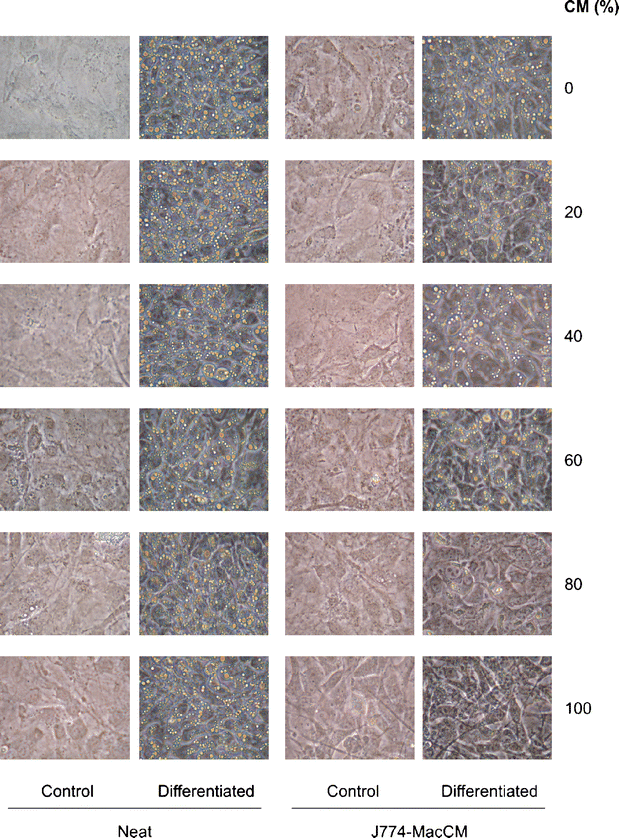
To initially evaluate the effect of macrophage-derived factors on adipogenesis, we used the established murine 3T3-L1 preadipose cell line model. These cells uniformly mature into round lipid-laden adipocytes upon exposure to a standardised differentiation protocol [24]. 3T3-L1 preadipocytes were induced to differentiate in the presence of various percentages of J774-MacCM or neat medium. J774-MacCM, but not neat medium, inhibited the differentiation of 3T3-L1 preadipocytes, gauged by cell rounding and lipid droplet formation (Fig. 1). The effect was dose-dependent, with a complete inhibition of adipose conversion observed with 80 and 100% J774-MacCM. This suggests that factors released by J774 macrophages interfere with adipocyte differentiation.
Fig. 1
Medium conditioned by J774 macrophages inhibits differentiation of 3T3-L1 preadipocytes, as assessed by morphology. 3T3-L1 preadipocytes were induced to differentiate in the presence of 0 to 100% of neat medium or J774-MacCM (CM). After 8 days, cultures were photographed at 400× magnification. Images shown are representative of five experiments
The inhibitory effect on adipogenesis was not due to cell toxicity. Viability of the 3T3-L1 cultures was not affected by J774-MacCM as measured by trypan blue dye exclusion (no cell death detected) and Hoechst staining. The percentage of apoptotic cells by Hoechst staining for preadipocytes and differentiated adipocytes in neat medium was 3.4 and 1.2%, respectively; for preadipocytes and differentiated adipocytes in MacCM, it was 1.9 and 2.0%, respectively.
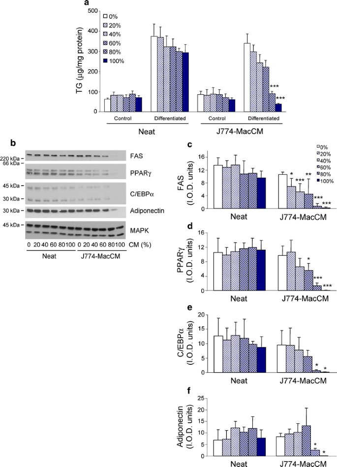
The usual increase in triacylglycerol accumulation was curtailed in a dose-dependent fashion when adipogenesis was induced in J774-MacCM, with a complete inhibition in the presence of 80 and 100% of J774-MacCM (Fig. 2a). Consistent with the morphological assessment and triacylglycerol response, the increase in protein expression of a variety of adipogenic markers was also inhibited by J774-MacCM, but not by neat medium (Fig. 2b–f). The differentiation-dependent expression of FAS appeared to be affected most by the presence of macrophage-derived factors, since a significant inhibition (47%; p<0.05) was observed in the presence of only 20% J774-MacCM. The induction of FAS and adiponectin, as well as of the key adipogenic transcription factors, PPARγ and C/EBPα, was completely abrogated in 100% J774-MacCM. MAPK protein expression served here as a loading control, since it does not change upon adipogenesis [25].
Fig. 2
Medium conditioned by J774 macrophages inhibits differentiation of 3T3-L1 preadipocytes, as assessed by adipogenic markers. 3T3-L1 preadipocytes were induced to differentiate in the presence of 0 to 100% of neat medium or J774-MacCM. a Triacylglycerol (TG) was extracted, quantified, and normalised to protein content. Results are expressed as the mean±SD of four independent experiments. b Solubilised protein from differentiated cultures was immunoblotted with antibodies against FAS, PPARγ, C/EBPα, adiponectin, or MAPK (loading control). Immunoblots shown are representative of five independent experiments. c–f Densitometric data for the differentiated samples from the five experiments, expressed as mean±SD. *p<0.05; **p<0.01; ***p<0.001 compared with respective neat condition
THP-1-MacCM inhibits 3T3-L1 adipocyte differentiation
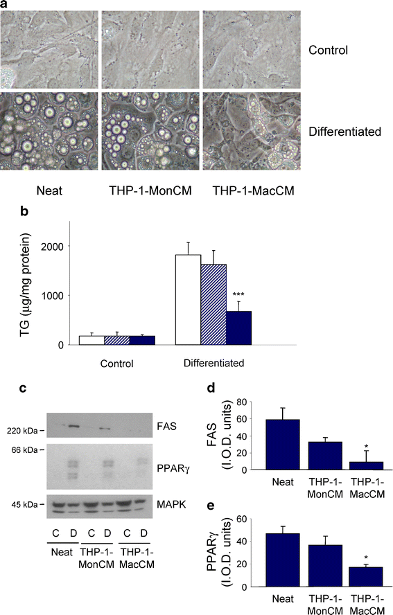
To verify that the negative regulation of adipogenesis by J774-MacCM is not particular to this murine macrophage cell line, we generated conditioned medium using the human THP-1 monocyte cell line. THP-1 monocytes were induced to differentiate into macrophages by a 24-h treatment with 100 nmol/l TPA, which was then removed prior to the preparation of conditioned medium. The effect on differentiation of these conditioned media compared with neat medium was then examined. The differentiation of preadipocytes in the neat RPMI medium was robust, and the resulting adipocytes exhibited a greater accumulation of triacylglycerol than that seen with standard differentiation performed in DMEM (Fig. 1). Nevertheless, THP-1-MacCM still inhibited 3T3-L1 adipogenesis, visualised as a reduction in rounded cells containing lipid droplets (Fig. 3a). This was confirmed biochemically, with a 65% (p<0.001) decrease in triacylglycerol accumulation (Fig. 3b), and an 85 and 62% inhibition of FAS and PPARγ induction, respectively (p<0.05; Fig. 3c,d). THP-1-MonCM had no significant effect on adipogenesis, although a non-significant downward trend was observed in the FAS and PPARγ induction, indicating that the factors that impaired adipogenesis in the conditioned medium were more specific to the macrophage than the monocyte phenotype.
Fig. 3
Medium conditioned by THP-1 macrophages inhibits differentiation of 3T3-L1 preadipocytes. 3T3-L1 preadipocytes were induced to differentiate in neat medium, THP-1-MonCM, or THP-1-MacCM for 8 days. a Cultures were photographed at 400× magnification. Images are representative of three independent experiments. b Triacylglycerol (TG) was extracted, quantified, and normalised to protein content. Results are expressed as the mean±SD of three independent experiments. Open bars, neat; hatched bars, THP-1-MonCM; solid bars, THP-1-MacCM. ***p<0.001 compared with differentiation in neat and THP-1-MonCM media. c Solubilised protein from control and differentiated cultures were immunoblotted with antibodies against FAS, PPARγ, or MAPK (loading control). Immunoblots representative of three independent experiments are shown. d, e Densitometric data for the differentiated samples from the three experiments, expressed as mean±SD. *p<0.05 compared with differentiation in neat medium
J774-MacCM inhibits human abdominal subcutaneous, but not omental adipocyte differentiation
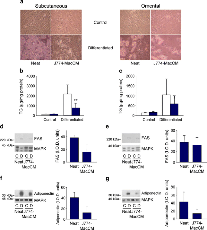
Subcutaneous and omental stromal preadipocytes were compared with respect to adipogenesis and their response to J774-MacCM. Similar to other adipogenic protocols containing PPARγ agonists (e.g. indomethacin—see Methods), we observed that subcutaneous preadipocytes differentiated somewhat better than their omental counterparts [20, 26]. Differentiation of the subcutaneous preadipocytes was moderately inhibited by J774-MacCM, as assessed morphologically (Fig. 4a). For these cells, there was a significant 70% inhibition (p<0.01) in triacylglycerol accumulation (Fig. 4b) and a 58 and 70% decrease in FAS (_p_=0.07) and adiponectin (_p_=0.13), respectively, that did not reach significance for either parameter (Fig. 4d,f). For J774-MacCM and omental preadipocytes, the changes in triacylglycerol accumulation (Fig. 4c) and FAS expression (Fig. 4e) were smaller, and neither was significant, whereas the adiponectin response resembled that of the subcutaneous preadipocytes (Fig. 4g).
Fig. 4
Medium conditioned by J774 macrophages inhibits differentiation of human abdominal subcutaneous preadipocytes. Human abdominal subcutaneous or omental preadipocytes were induced to differentiate in neat medium or J774-MacCM. a After 14 days, cultures were photographed at 400× magnification. Images are representative of four patient samples. b, c Triacylglycerol (TG) was extracted from subcutaneous (b) and omental (c) samples, quantified, and normalised to protein content. Results are expressed as the mean±SD of four patient samples. Open bars, neat; shaded bars, J774-MacCM. **p<0.01 compared with differentiation in neat medium. d–g Solubilised protein from control and differentiated subcutaneous (d, f) and omental (e, g) cultures was immunoblotted with antibodies against FAS (d, e) and adiponectin (f, g), or MAPK (loading control). Representative immunoblots from a single patient sample are shown. Densitometric data for the differentiated samples from four (FAS) or three (adiponectin) subcutaneous and omental patient samples are expressed as mean±SD
THP-1-MacCM inhibits human abdominal subcutaneous and omental adipocyte differentiation
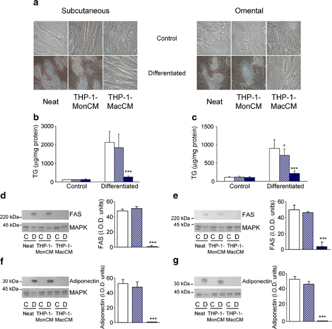
As described for the 3T3-L1 adipogenesis studies, we collected THP-1-MonCM and THP-1-MacCM to verify that the negative regulation of human adipogenesis was not particular to J774-MacCM. THP-1-MacCM strongly reduced the differentiation of subcutaneous preadipocytes compared with neat medium, as observed morphologically (Fig. 5a); this finding was confirmed by the 94% (p<0.001) inhibition of triacylglycerol accumulation (Fig. 5b), as well as by the 98% (p<0.001) and 99% (p<0.001) reduction in FAS and adiponectin protein expression, respectively (Fig. 5d,f). Unlike the studies with J774-MacCM, THP-1-MacCM inhibited omental adipogenesis to the same degree as it did subcutaneous adipogenesis (Fig. 5a,c,e,g). Adipogenesis was unaffected by THP-1-MonCM, except for a modest reduction in omental triacylglycerol accumulation, indicating that factors impairing human subcutaneous and omental adipogenesis were more specific to the macrophage phenotype (Fig. 5).
Fig. 5
Medium conditioned by THP-1 macrophages inhibits differentiation of human abdominal subcutaneous preadipocytes. Human subcutaneous or omental preadipocytes were induced to differentiate in neat medium, THP-1-MonCM, or THP-1-MacCM for 14 days. a Cultures were photographed at 400× magnification. Images are representative of four patient samples. b, c Triacylglycerol (TG) was extracted from subcutaneous (b) and omental (c) samples, quantified, and normalised to protein content. Results represent the mean±SD of four patient samples. d–g Solubilised protein from control and differentiated subcutaneous (d, f) and omental (e, g) cultures were immunoblotted with antibodies against FAS (d, e) and adiponectin (f, g), or MAPK (loading control). Representative immunoblots from a single patient sample are shown. Densitometric data for the differentiated samples from the four patients are expressed as mean±SD. *p<0.05 compared with differentiation in neat medium; ***p<0.001 compared with differentiation in neat medium and THP-1-MonCM. Open bars, neat; hatched bars, THP-1-MonCM; solid bars, THP-1-MacCM
Discussion
To determine whether macrophage-derived factors inhibit adipocyte differentiation, we used two models of adipogenesis, 3T3-L1 murine preadipocytes and human stromal preadipocytes isolated from the abdominal subcutaneous and omental fat depots. Our results demonstrate that MacCM, from either murine J774 or human THP-1 macrophages, impairs 3T3-L1 and human adipocyte differentiation.
Obesity-related adipose tissue dysfunctions include insulin resistance and aberrant regulation of inflammatory adipokines. Adipocytes and stromal preadipocytes produce these cytokines, and mechanisms to explain their inflamed state are being investigated [27]. Macrophages reside within adipose tissue, as a function of obesity and/or insulin resistance, and generate many of the same cytokines [9, 10, 28]. The attraction of monocytes to adipose tissue may be due to local production of monocyte chemoattractant protein-1 [13, 29]. Adipocytes enhance chemotaxis and diapedesis of monocytes, via the regulation of endothelial cell adhesion molecules, promoting their infiltration into adipose tissue as macrophages [12].
An adipogenic deficit in obesity may favour the development of adipocyte hypertrophy, which upreglates production of inflammatory adipokines associated with insulin resistance [5, 6]. Furthermore, the resulting limitation of the adipose tissue reservoir would promote lipid deposition in liver and skeletal muscle, leading to decreased insulin action in those tissues [3, 5]. The factors that curtail adipogenesis are not well defined, and macrophage-derived cytokines are potential paracrine candidates. Weight loss decreases the number of adipose tissue macrophages and reduces their inflammatory profile [30], and weight loss also enhances adipogenic C/EBPα protein expression in human stromal abdominal subcutaneous preadipocytes [31].
We observed a marked inhibition of 3T3-L1 adipocyte differentiation induced by either murine (J774) or human (THP-1) MacCM, without any evidence of cytotoxicity. THP-1-MacCM appeared slightly less potent with respect to 3T3-L1 adipogenesis. This may be because 3T3-L1 adipogenesis itself was more robust in RPMI medium than in the DMEM used for the J774-MacCM studies; the choice of these two media was dictated by the growth requirements of the two types of macrophages. In addition, a species-specific effect might result in murine J774-MacCM acting more efficiently on murine 3T3-L1 cells.
Immortalised 3T3-L1 preadipocytes are a popular and tractable experimental model, given their robust and uniform differentiation response [24, 32]. Nevertheless, their embryonic origin and their aneuploidy distinguish them from true preadipocytes, and so it is important to emphasise that we also studied stromal preadipocytes isolated from human abdominal subcutaneous or omental fat depots.
In response to J774-MacCM, differentiation of abdominal subcutaneous preadipocytes displayed a moderate inhibition, whereas the differentiation of omental stromal preadipocytes was less affected. The reason for the apparent depot-related susceptibility is not known. To minimise inter-subject variability, the subcutaneous and omental adipose tissue for these studies were paired samples. The maximal differentiation response of the omental preadipocytes was somewhat lower than that of subcutaneous preadipocytes as observed by others [20, 26]. Interestingly, omental/visceral adipose tissue contains more macrophages than does subcutaneous fat in obese rodents and humans [11]. Also adipose tissue possesses anatomical site-specific properties [33], and these could potentially influence interactions with macrophages.
In contrast, THP-1-MacCM completely suppressed human subcutaneous as well as omental adipogenesis, i.e. no depot-related responses were observed. The profile of secreted products may vary between these two macrophage models; alternatively, a species-specific effect of human THP-1-MacCM acting on human adipocyte differentiation may also account for its more potent and uniform inhibition.
There have been previous studies on individual cytokines known to be secreted by macrophages and on their effect on adipogenesis. TNFα was recognised early as an inhibitor of 3T3-L1 and human adipocyte differentiation [34, 35]. Interleukin 6 may have a similar role in subcutaneous adipose tissue differentiation [36]. In contrast, macrophage colony-stimulating factor promotes rabbit subcutaneous adipocyte hyperplasia, raising the possibility of enhanced adipogenesis [37]. Similarly, the human ortholog of mouse resistin, hFIZZ3, is expressed by macrophages, and stimulates human subcutaneous preadipocyte proliferation, yielding more adipocytes [22].
Therefore, cytokines studied in an isolated fashion yield pro- and anti-adipogenic effects. Our study, using MacCM, provides a more integrated system to ascertain the net effect of macrophage-derived products on preadipocytes. Of course, the nature of paracrine interactions between macrophages and adipose cells in vivo are likely to be more complex. Infiltrating macrophages could represent a special subpopulation of circulating monocytes, and it has even been suggested that adipose tissue macrophages may arise from adipose stromal cells [38]. Furthermore, the profile of cytokines secreted by resident macrophages may be influenced by adipose cells.
Our study demonstrates that J774- or THP-1-MacCM potently impairs 3T3-L1 and human adipogenesis in vitro. These results improve our understanding of macrophage and adipose cell interactions, and may provide a framework for further research in this rapidly developing field.
References
- Kershaw EE, Flier JS (2004) Adipose tissue as an endocrine organ. J Clin Endocrinol Metab 89:2548–2556
Article PubMed CAS Google Scholar - Hirsch J, Fried SK, Edens NK, Leibel RL (1989) The fat cell. Med Clin North Am 73:83–96
PubMed CAS Google Scholar - Danforth E Jr (2000) Failure of adipocyte differentiation causes type 2 diabetes mellitus? Nat Genet 26:13
Article PubMed CAS Google Scholar - Weyer C, Foley JE, Bogardus C, Tataranni PA, Pratley RE (2000) Enlarged subcutaneous abdominal adipocyte size, but not obesity itself, predicts type 2 diabetes independent of insulin resistance. Diabetologia 43:1498–1506
Article PubMed CAS Google Scholar - Heilbronn L, Smith SR, Ravussin E (2004) Failure of fat cell proliferation, mitochondrial function and fat oxidation results in ectopic fat storage, insulin resistance and type 2 diabetes mellitus. Int J Obesity 28:S12–S21
Article CAS Google Scholar - Le Lay S, Krief S, Farnier C et al (2001) Cholesterol, a cell size-dependent signal that regulates glucose metabolism and gene expression in adipocytes. J Biol Chem 276:16904–16910
Article PubMed Google Scholar - Lazar MA (2005) How obesity causes diabetes: not a tall tale. Science 307:373–375
Article PubMed CAS Google Scholar - Miles JM, Jensen MD (2005) Visceral adiposity is not casually related to insulin resistance. Diabetes Care 28:2326–2328
Article PubMed Google Scholar - Weisberg SP, McCann D, Desai M, Rosenbaum M, Leibel RL, Ferrante AWJ (2003) Obesity is associated with macrophage accumulation in adipose tissue. J Clin Invest 112:1796–1808
PubMed CAS Google Scholar - Xu H, Barnes GT, Yang Q et al (2003) Chronic inflammation in fat plays a crucial role in the development of obesity-related insulin resistance. J Clin Invest 112:1821–1830
PubMed CAS Google Scholar - Bouloumie A, Curat CA, Sengenes C, Lolmede K, Miranville A, Busse R (2005) Role of macrophage tissue infiltration in metabolic disease. Curr Opin Nutr Metab Care 8:347–354
Article CAS Google Scholar - Curat CA, Miranville A, Sengenes C et al (2004) From blood monocytes to adipose tissue-resident macrophages: induction of diapedesis by human mature adipocytes. Diabetes 53:1285–1292
Article PubMed CAS Google Scholar - Bruun JM, Lihn AS, Pedersen SB, Richelsen B (2005) Monocyte chemoattractant protein-1 release is higher in visceral than subcutaneous human adipose tissue (AT): implication of macrophages resident in the AT. J Clin Endocrinol Metab 90:2282–2289
Article PubMed CAS Google Scholar - Cinti S, Mitchell G, Barbatelli G et al (2005) Adipocyte death defines macrophage localization and function in adipose tissue of obese mice and humans. J Lipid Res 46:2347-2355
Article PubMed CAS Google Scholar - Gagnon A, Chen C-S, Sorisky A (1999) Activation of protein kinase B and induction of adipogenesis by insulin in 3T3-L1 preadipocytes. Contribution of phosphoinositide-3,4,5-trisphosphate versus phosphoinositide-3,4-bisphosphate. Diabetes 48:691–698
Article PubMed CAS Google Scholar - Laemmli UK (1970) Cleavage of structural proteins during the assembly of the head of bacteriophage T4. Nature 227:680–685
Article PubMed CAS Google Scholar - Magun R, Boone DL, Tsang BK, Sorisky A (1998) The effect of adipocyte differentiation on the capacity of 3T3-L1 cells to undergo apoptosis in response to growth factor deprivation. Int J Obes 22:567–571
Article CAS Google Scholar - Hauner H, Skurk T, Wabitsch M (2001) Cultures of human adipose precursor cells. Methods Mol Biol 155:239–247
PubMed CAS Google Scholar - Artemenko Y, Gagnon A, Aubin D, Sorisky A (2005) Anti-adipogenic effect of PDGF is reversed by PKC inhibition. J Cell Physiol 204:646–653
Article PubMed CAS Google Scholar - Adams M, Montague CT, Prins JB et al (1997) Activators of peroxisome proliferator-activated receptor have depot-specific effects on human preadipocyte differentiation. J Clin Invest 100:3149–3153
Article PubMed CAS Google Scholar - Hutley LJ, Newell FM, Joyner JM et al (2003) Effects of rosiglitazone and linoleic acid on human preadipocyte differentiation. Eur J Clin Invest 33:574–581
Article PubMed CAS Google Scholar - Ort T, Arjona AA, MacDougall JR et al (2005) Recombinant human FIZZ3/resistin stimulates lipolysis in cultured human adipocytes, mouse adipose explants, and normal mice. Endocrinology 146:2200–2209
Article PubMed CAS Google Scholar - Lehmann JM, Lenhard JM, Oliver BB, Ringold GM, Kliewer SA (1997) Peroxisome proliferator-activated receptors alpha and gamma are activated by indomethacin and other non-steroidal anti-inflammatory drugs. J Biol Chem 272:3406–3410
Article PubMed CAS Google Scholar - Ross SE, Hemati N, Longo KA et al (2000) Inhibition of adipogenesis by Wnt signaling. Science 289:950–953
Article PubMed CAS Google Scholar - Prusty D, Park BH, Davis KE, Farmer SR (2002) Activation of MEK/ERK signaling promotes adipogenesis by enhancing peroxisome proliferator-activated receptor gamma (PPAR) and C/EBPalpha gene expression during the differentiation of 3T3-L1 preadipocytes. J Biol Chem 277:46226–46232
Article PubMed CAS Google Scholar - Digby JE, Montague CT, Sewter CP et al (1998) Thiazolidinedione exposure increases the expression of uncoupling protein in cultured human preadipocytes. Diabetes 47:138–141
Article PubMed CAS Google Scholar - Rajala MW, Scherer PE (2003) The adipocyte—at the crossroads of energy homeostasis, inflammation, and atherosclerosis. Endocrinology 144:3765–3773
Article PubMed CAS Google Scholar - Di Gregorio GB, Yao-Borengasser A, Rasouli N et al (2005) Expression of CD68 and macrophage chemoattractant protein-1 genes in human adipose and muscle tissues. Diabetes 54:2305–2313
Article PubMed Google Scholar - Dahlman I, Kaaman M, Olsson T et al (2005) A unique role of monocyte chemoattractant protein-1 among chemokines in adipose tissue of obese subjects. J Clin Endocrinol Metab 90:5834–5840
Article PubMed CAS Google Scholar - Cancello R, Henegar C, Viguerie N et al (2005) Reduction of macrophage infiltration and chemoattractant gene expression changes in white adipose tissue of morbidly obese subjects after surgery-induced weight loss. Diabetes 54:2277–2286
Article PubMed CAS Google Scholar - Aubin D, Gagnon A, Grunder L, Dent R, Allen M, Sorisky A (2004) Adipogenic and antiapoptotic protein levels in human adipose stromal cells after weight loss. Obes Res 12:1231–1234
Article PubMed CAS Google Scholar - Spiegelman BM, Choy L, Hotamisligil GS, Graves RA, Tontonoz P (1993) Regulation of adipocyte gene expression in differentiation and syndromes of obesity/diabetes. J Biol Chem 268:6823–6826
PubMed CAS Google Scholar - Montague CT, O’Rahilly S (2000) The perils of portliness: causes and consequences of visceral obesity. Diabetes 49:883–888
Article PubMed CAS Google Scholar - Torti FM, Torti SV, Larrick JW, Ringold GM (1989) Modulation of adipocyte differentiation by tumor necrosis factor and transforming growth factor beta. J Cell Biol 108:1105–1113
Article PubMed CAS Google Scholar - Petruschke T, Hauner H (1993) Tumor necrosis factor-alpha prevents the differentiation of human adipocyte precursor cells and causes delipidation of newly developed fat cells. J Clin Endocrinol Metab 76:742–747
Article PubMed CAS Google Scholar - Sopasakis VR, Sandqvist M, Gustafson B et al (2004) High local concentrations and effects on differentiation implicate interleukin-6 as a paracrine regulator. Obes Res 12:454–460
PubMed CAS Google Scholar - Levine JA, Jensen MD, Eberhardt NL, O’Brien T (1998) Adipocyte macrophage colony-stimulating factor is a mediator of adipose tissue growth. J Clin Invest 101:1557–1564
Article PubMed CAS Google Scholar - Charriere G, Cousin B, Arnaud E et al (2003) Preadipocyte conversion to macrophage. Evidence of plasticity. J Biol Chem 278:9850–9855
Article PubMed CAS Google Scholar
