Changes in gene expression in beta cells after islet isolation and transplantation using laser-capture microdissection (original) (raw)
Introduction
Advances in human islet transplantation have increased the number of patients who achieve insulin-independence, yet there are still concerns about the survival and function of these islets in the liver [1, 2]. Even with islets being obtained from two to four donor pancreases, most patients continue to have impaired glucose tolerance [2]. A substantial amount of beta cell death must occur in the immediate transplantation period due to anoxia [3] and inflammation [4]. In addition, beta cell function may be impaired by even mild hyperglycaemia [5, 6] and hypoxia [7], and poorly understood factors related to the unnatural graft site. Over a period of 7–10 days islets are revascularised but have decreased oxygen tension compared with endogenous islets, which may in part be due to an abnormal distribution of the new vessels in the graft [8–10].
Laser-capture microdissection (LCM) makes it possible to obtain very small amounts of tissue from frozen sections, thus allowing collection of the beta cell-rich portions of islets from tissue sections. The RNA from these specimens can then be extracted and analysed for gene expression [11]. The beauty of LCM is that tissue can be selectively obtained from the beta cell-rich core of islets, leaving behind the confounding non-beta cells. We expect that islets damaged by the isolation procedure and transplantation process will have gene profile patterns different from those of endogenous islets. The aim of the present study was to investigate the changes in gene expression in beta cells using LCM before and after islet isolation and transplantation.
Materials and methods
Animals and induction of diabetes
For islet donors and recipients, inbred male B6AF1 mice (Taconic Farms, Germantown, NY, USA), aged 6–8 weeks, were used. Islets were classified into four groups: (1) islets in the pancreas (n = 7); (2) freshly isolated islets (n = 6); (3) islets cultured for 24 h in RPMI 1640 medium (Sigma-Aldrich, St Louis, MO, USA) with a glucose concentration of 200 mg/dl (n = 6); and (4) islet grafts (normal; n = 6, diabetic; n = 8). Transplantation recipients were either normal or diabetic, having been treated with an i.p. injection of streptozocin (STZ) (180 mg/kg body weight, dissolved in citrate buffer, pH 4.5; Sigma, St Louis, MO, USA). Before transplantation, diabetes was confirmed by the presence of hyperglycaemia; only those mice with glucose levels ≥19.4 mmol/l for at least 1 week were used as recipients. At no point were diabetic mice treated with exogenous insulin. All animals were maintained in accordance with US Government and local institutional guidelines.
Islet isolation and culture
The islet isolation method has been described previously [12]. Briefly, pancreases were distended with 3 ml cold M199 medium (Gibco BRL, Grand Island, NY, USA) containing 1.5 mg/dl of collagenase (type P; Boehringer Mannheim, Mannheim, Germany), excised, and incubated in a stationary bath at 37°C. Islets were separated by density gradients (Histopaque-1077; Sigma) and handpicked under a stereomicroscope. Islets were divided into samples of 250 and 500 islets for transplantation, whereas others, in groups of 500 islets, were cultured free-floating for 24 h in 5 ml culture medium consisting of RPMI 1640 supplemented with l-glutamine, benzyl penicillin (100 U/ml; Roche Diagnostics Scandinavia, Bormma, Sweden), and 10% FCS (Sigma-Aldrich).
Transplantation
Aliquots of islets were placed into 200 μl pipette tips (USA Scientific Plastic, Ocala, FL, USA) connected to a 1 ml Hamilton syringe (Hamilton Company, Reno, NV, USA) and were allowed to settle by gravity. After sedimentation, the tip was connected to polyethylene tubing (PE-50) and samples of 250 and 500 islets were transplanted under the kidney capsule of the normal and diabetic mice, respectively [13]. After transplantation, blood glucose levels and body weights were monitored daily during the first week after transplantation and then every other day. Blood was obtained from snipped tails, and glucose was measured with a glucose meter (One Touch II; Johnson & Johnson, Milpitas, CA, USA). Grafted islets were retrieved 3 weeks after transplantation.
Laser-capture microdissection
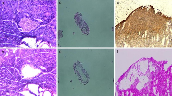
The central core of islets was microdissected using LCM as previously described [14]. Pancreatic tissues and islet grafts including kidney were embedded in Tissue Tek OCT medium (VWR Scientific Products, San Diego, CA, USA) and frozen at −80°C. To prepare frozen sections of fresh and cultured islets, tissue pellets were made by suspending islets in Eppendorf tubes, which were centrifuged at 300×g for 1 min. The supernatant fraction was then removed leaving as little excess medium as possible, and the pellets were embedded in Tissue Tek OCT medium. The Eppendorf tube was tapped lightly to dislodge the pellet, was placed on dry ice, and then stored at −80°C. Pellets were sectioned at 8 μm with a cryostat and mounted on untreated glass slides. For LCM, slides containing frozen sections were immediately fixed in 70% ethanol for 30 s and stained with haematoxylin and eosin (H&E), followed by dehydration steps in 70, 95 and 100% ethanol and a final 5-min dehydration step in xylene. Once air-dried, a PixCell II system from Arcturus Engineering (Mountain View, CA, USA) was used to microdissect islets from H&E-stained frozen tissue sections. For each specimen, at least 1,500 hits were used to obtain enough RNA for amplification. The amplitude and pulse duration were adjusted for each fixative as necessary to allow complete tissue capture using the 10 μm laser beam (Fig. 1).
Fig. 1
Laser-capture microdissection (LCM). Endogenous (a) and freshly isolated islet (c) H&E-stained frozen sections (8 μm). e Frozen section of a graft stained for insulin. b, d, f Adjacent sections that have undergone LCM. The holes created by the LCM procedure are clearly visible
RNA extraction and amplification
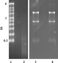
Total RNA from laser-captured cells was extracted using a modified protocol [11]. Briefly, after microdissection of each specimen, the transparent film and adherent cells were incubated with Buffer RLT (Qiagen, Valencia, CA, USA), extracted with phenol-chloroform-isoamyl alcohol, and precipitated with sodium acetate and glycogen carrier in isopropanol. After initial recovery and resuspension of the RNA pellet, DNA was removed by a 2-h incubation at 37°C with 10 units DNase (GenHunter, Nashville, TN, USA) in the presence of an RNase inhibitor (Life Technologies, Inc., Gaithersburg, MD, USA), followed by re-extraction and precipitation. Two rounds of amplification of RNA were performed using an RNA Amplification Kit (Arcturus). Because of the challenging characteristics of the pancreas, which has very high levels of RNase and protease activity, a stringent approach was used for the LCM technique. Thus, the amount of working time was reduced to the minimum (15 min per sample) in a dehumidified room at a constant temperature of 15°C. These precautions allowed us to obtain RNA of good quality. For the total RNA extracted by LCM, an A 260:A 280 ratio ranging between 1.7 and 2.0 and a concentration of 2.8–8.0 ng/μl were obtained using NanoDrop ND-1000 (Nano Drop Technologies, Inc., Wilmington, DE, USA). The quantity used for the amplification process varied between 28 and 80 ng and the antisense RNA (aRNA) obtained after two rounds of amplification was 7.4–62.9 μg (24.5 ± 14.5) with an A 260:A 280 ratio between 2.4 and 2.6. As expected from the amplification, the aRNA always appeared as a smear on the gel ranging from under 200 to over 600 bases in length (Fig. 2). This was very consistent for all amplifications with no differences being seen among the four tissue sources.
Fig. 2
Agarose electrophoresis of aRNA. Total RNA extracted from frozen sections of fresh islets was amplified. Gel electrophoresis was performed to confirm the quality of amplified aRNA product. Mouse total RNA from fresh (lane 3) and cultured islets (lane 4) show two clear bands (18 S; 1.9 kb, and 28 S; 4.7 kb). aRNA after amplification appears as a smear on the gel, ranging from under 200 to over 600 bases in length (lane 2). Amplified RNA from cultured islets showed the same smear on the gel (data not shown). Lane 1, RNA marker
cDNA synthesis and semiquantitative RT-PCR
To synthesise cDNA, 500 ng aRNA were reverse-transcribed in a 25-μl solution containing 5 μl first-strand buffer (Life Technologies), 10 mmol/l dithiothreitol, 1 mmol/l dNTP, 50 ng random hexamers, and 200 units of Superscript II RNase H− reverse transcriptase (Life Technologies). Reverse transcription reactions were incubated for 10 min at 25°C, 60 min at 42°C and 10 min at 95°C. cDNA products were diluted to a concentration corresponding to 20 ng starting RNA per 3 μl and stored at −80°C. PCRs were performed in 50-μl volumes consisting of 3 μl cDNA, 1.5–2.0 mmol/l MgCl2, 80–160 mmol/l dNTP, 40–500 nmol oligonucleotide primers, 92.5 kBq α-32P-labelled dCTP (111 TBq/mmol), 5 μl GeneAmp PCR buffer and 5 units AmpliTaq Gold DNA polymerase (Perkin-Elmer, Foster City, CA, USA). The Electronic supplementary material (ESM) Table 1 shows primers designed with Eugene version 2.2 software (Daniben Systems, Cincinnati, OH, USA). After two rounds of amplification, the produced aRNA is shorter and closer to the 3′ end than mRNA and thus requires primers designed within 300 bases from the poly-A tail with a product size less than 200 bases (Electronic Supplementary Material [ESM] Table 1). All reactions were performed in a 9700 Thermocycler (Perkin-Elmer), in which samples underwent a 10-min initial denaturing step to activate the DNA polymerase, followed by cycling of 1 min at 94°C, 1 min at the annealing temperature of each primer, and 1 min at 72°C. The final extension step was 10 min at 72°C. An internal control (housekeeping) gene (Ppia [probable iron-regulated peptidyl-prolyl _cis_-trans isomerise A, previously known as cyclophilin], Tbpl1 [TATA box binding protein-related factor] or Tuba2 [α-tubulin]) was used to normalise the input template cDNA to analyse relative expression of the gene of interest. Different internal control genes were used as required to match the amount of expression of the various tested genes. To be specific, Ppia was used for insulin (Ins) and glutathione peroxidase 1 (Gpx1); Tbpl1 was used for lactate dehydrogenase A (Ldha), haem oxygenase 1 (Hmox1, previously known as HO-1) and TNF receptor superfamily member 6 (Fas); and tuba2 was used for insulin promoter factor 1 (Ipf1, previously known as Pdx1), solute carrier family 2 (facilitated glucose transporter), member 2 (Slc2a2, previously known as GLUT2) and TNF receptor superfamily member 6 (Tnfaip3, previously known as A20). PCR products were separated on a 6% polyacrylamide gel in Tris-borate-EDTA buffer. Band intensity was measured with a PhosphoImager and quantified with ImageQuant software (Molecular Dynamics, Sunnyvale, CA, USA). Gene expression for control samples (see Fig. 4) was the ratio of the test gene expression to internal control expression and designated as 100%. Then the various experimental sample results were expressed as a percentage. Sequences of oligonucleotide primers and PCR conditions are available online (ESM Table 1).
Immunohistochemistry
Immunostaining of insulin, Tnfaip3 and Hmox1 protein was performed using the avidin–biotin complex (ABC) method (Vectastain ABC Kit; Vector Laboratories, Burlingame, CA, USA). Frozen tissue sections were fixed by immersion in acetone, incubated for 20 min and then incubated overnight at 4°C with rabbit anti-Tnfaip3 antibody (rabbit anti-human, a gift from S. Grey, Boston, MA, USA; 1:1,000 in PBS containing 1% BSA) and then for 30 min with biotinylated anti-rabbit IgG (Vector Laboratories; diluted 1:200). The same sections were stained for insulin (guinea-pig anti-human insulin, 1:200; Linco Research, St Charles, MO, USA). Staining for macrophages on frozen sections was done with anti-CD163 (mouse anti-rat 1:100 dilution; AbD Serotec Ltd, Oxford, UK). The secondary antibodies used for immunofluorescence were Texas Red-conjugated affinipure donkey anti-guinea-pig IgG (1:400; Jackson Immuno-Research, West Grove, PA, USA) for insulin and FITC-conjugated donkey anti-rabbit IgG and streptavidin-conjugated FITC (1:400; Jackson ImmunoResearch) for Tnfaip3. For Hmox1 immunostaining, tissues were fixed by immersion in 4% buffered formaldehyde, and then embedded in paraffin. Paraffin sections were incubated for 10 min with 3% H2O2 solution to block endogenous peroxidase and then overnight at 4°C with goat anti-Hmox1 antibody (Santa Cruz Biotechnology, Santa Cruz, CA, USA) diluted 1:1,000 in PBS containing 1% BSA, and for 30 min with biotinylated anti-goat IgG (Vector Laboratories) diluted 1:200. These sections were then incubated with ABC reagent for 30 min, and positive reactions were visualised by incubation with the peroxidase substrate solution containing 3,3′-diaminobenzidine tetrahydrochloride.
Statistical analysis
Results are presented as the mean±SE. Analyses were performed using an unpaired Student’s t test, or a one-way ANOVA with the post-test of Scheffé. Differences were considered statistically significant at p < 0.05.
Results
Blood glucose and body weight after transplantation
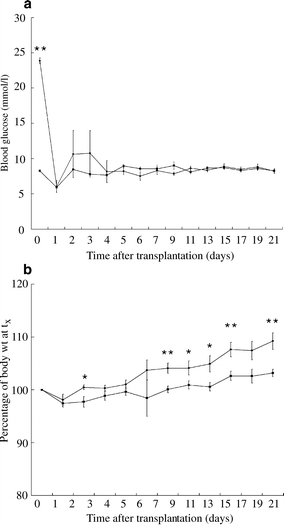
Islet transplantation in the diabetic group resulted in a return to near normoglycaemia. Figure 3 shows the changes in glucose levels and body weight after islet transplantation. One day after transplantation, glucose levels of both groups were significantly decreased (diabetic, n = 8: 23.8 ± 0.4 and 6.1 ± 0.9, vs non-diabetic, n = 6: 8.3 ± 0.3 and 5.9 ± 0.7 mmol/l, respectively, p < 0.01). During the first 3 days after transplantation, glucose levels in the diabetic group were decreased but unstable, after which there were no significant differences in glucose levels (Fig. 3a). Of note, rodent blood glucose levels obtained with glucose meters are 30–40% lower than plasma levels [15]. Body weight gain was expressed as the ratio of weight at each time-point to that at the time of transplantation. Weight gain in both groups was slightly decreased for the first few days after transplantation. In the diabetic mice, islet transplantation was followed by significant weight gain, while in normal mice gain tended to be less than that in diabetic mice (Fig. 3b).
Fig. 3
Blood glucose levels (a) and body weight (Body Wt) (b) of non-diabetic (squares), n = 6, and cured diabetic mice (circles), n = 8, were measured during the 3 weeks after islet transplantation (Tx). Values are means±SE. *p < 0.05, **p < 0.01 vs controls
Changes in expression of genes concerned with beta cell function after islet isolation and transplantation
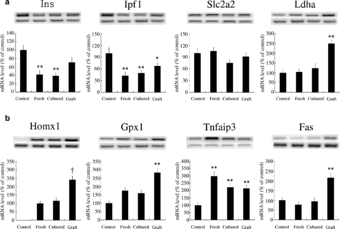
Representative gels from RT-PCR analysis comparing expression of genes related to beta cell function in fresh, cultured and transplanted islets are shown in Fig. 4a. Expression of specific genes was normalised to their internal control gene (Ppia, Tbpl1 or Tuba2) and quantified as a percentage of those from endogenous beta cell-rich tissue obtained from pancreases using LCM. Expression of Ins and Ipf1 was markedly decreased in fresh and isolated islets; mRNA level for Ins in fresh and cultured islets was decreased to 41.6 ± 9.4 and 38.2 ± 7.2% of normal, respectively (p < 0.01 compared with endogenous islets). In the graft of cured animals, insulin mRNA expression was decreased, but the difference compared with that of endogenous islets did not reach statistical significance (p = 0.12) by ANOVA, but did with the unpaired t test (p = 0.03). The mRNA level of Ipf1 decreased to the same extent as insulin in fresh and cultured islets. The normalisation of blood glucose in transplanted cured animals did not completely reverse the changes in levels of Ipf1 mRNA, which were decreased to 42.5 ± 9.2, 49.2 ± 6.4 and 67.6 ± 8.8% of normal in fresh islets, cultured islets and islet grafts of cured animals, respectively (p < 0.05 vs endogenous islets). Levels of Slc2a2 mRNA, the beta cell glucose transporter, were unchanged in isolated and transplanted islets of cured animals. Interestingly, expression of Ldha, normally present at low levels in beta cells, did not change in fresh and cultured islets; however, it was markedly increased (two- to threefold) in transplanted islets.
Fig. 4
Comparison of beta-cell function (a) and stress gene (b) mRNA levels in islets (endogenous islets [_control_], n = 7; freshly isolated islets [_fresh_], n = 6; cultured islets [_cultured_], n = 6; and islet grafts of cured animals [_graft_], n = 8). Values are means±SE. Hmox1 mRNA was not expressed in endogenous islets and is expressed as a per cent of fresh islets. *p < 0.05 and **p < 0.01 vs control for each gene; † p < 0.01 vs fresh islets for each gene
Changes in expression of stress genes after islet isolation and transplantation
Changes in stress gene mRNA expression in fresh, cultured and transplanted islets are shown in Fig. 4b. As previously noted [16], only minimal expression of Hmox1 mRNA was detected in endogenous islets, yet abundant expression was found in fresh and cultured islets, and even more in islet grafts. The mRNA levels for Gpx1 were higher in both fresh and cultured islets compared with endogenous islets (p < 0.01) and was further increased in transplanted islets (p < 0.05). Tnfaip3 was originally described as a TNF-α-induced anti-apoptotic gene in endothelial cells [17]. The expression level of Tnfaip3 was markedly increased immediately after islet isolation (twofold, p < 0.01) and remained higher in both cultured islets and transplanted islets in cured diabetic animals. In contrast to genes that are potentially anti-apoptotic, expression of the pro-apoptotic gene Fas was not changed after islet isolation but increased twofold in transplanted islets.
Comparison of beta cell gene expression in transplanted islets between cured and normal mice
To evaluate gene expression in transplanted islets, STZ-diabetic mice were transplanted with 500 islets, and non-diabetic mice with 250 islets. Glucose levels of these groups were not different from 1 week after transplantation until the end of the experiment. For both groups, gene expression of grafts was evaluated 3 weeks after the transplantations. Changes of beta cell function and stress genes in islet grafts are shown in Table 1. The expression of insulin mRNA in grafts of cured mice was not significantly lower than that of mice not diabetic prior to transplantation. We conclude from these two transplantation situations that although insulin expression of the grafts was marginally lower in the cured diabetes group than in endogenous islets, insulin expression in islet grafts is reasonably well maintained. Ipf1 mRNA expression was lower in the grafts of cured mice, but Slc2a2 and Ldha mRNA expression showed no significant difference between grafts from cured and normal mice.
Table 1 Comparison of islet gene expression in grafts of non-diabetic and diabetic cured mice
Comparison of stress gene expression in transplanted islets between cured and normal animals
Changes in stress gene mRNA levels in transplanted islets from normal and diabetic mice are shown in Table 1. The mRNA levels for Hmox1 and Gpx1 in transplanted islets of diabetic mice had the tendency to be increased compared with those of normal mice but were not statistically significant. Tnfaip3 mRNA expression in transplanted islets showed no difference between normal and diabetic mice. Similarly, Fas mRNA level was similar in both groups 3 weeks after transplantation.
Immunohistochemistry
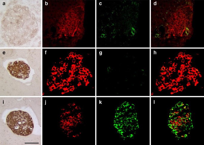
To verify that changes in mRNA levels were accompanied by parallel changes in protein level in beta cells, frozen sections of pancreas and fresh and cultured islets were stained for insulin and photographed at the same settings. Immunostaining for insulin in endogenous and fresh islets was clearly more intense than in islets cultured for 24 h in the medium with the standard glucose concentration of 11.1 mmol/l (Fig. 5). This fits with observations that insulin content of islets typically falls in the first days of culture [18]. Staining for Tnfaip3 was minimal in endogenous and freshly isolated islets but intense in cultured islets that also weakly stained for insulin. Increased staining for Hmox1 was observed in freshly isolated and cultured islets, revealing a clear increase of intensity compared with endogenous islets. Of note, message levels of Tnfaip3 and Hmox1 were increased in fresh islets, but protein increases were only seen for Hmox1. This could occur if protein synthesis of Tnfaip3 was slower than Hmox1, and/or that immunostaining for Tnfaip3 was less sensitive than for Hmox1. Because of concerns that inflammation in the grafts could have contributed to some of the gene expression signals, three grafts were harvested 2 weeks after transplantation of 500 islets into STZ-diabetic recipients. Frozen sections of these grafts were stained with the antibodies against macrophage marker CD163 and insulin. Spleen was the positive control. There were only very rare macrophages found in three grafts (data not shown), indicating that contamination of the LCM samples with macrophages is negligible.
Fig. 5
Staining for Hmox1 (a, e, i), ins (red), Tnfaip3 (green) and overlay (d, h, l) from frozen tissue sections of endogenous islets (a–d), fresh islets (e–h) and 24-h cultured islets (i–l). Magnification ×200; bar = 50 μm
Discussion
In both freshly isolated and cultured islets there are marked changes in the expression of beta cell genes concerned with insulin secretion and the stress response that could influence beta cell survival. During the process of islet isolation, chemical and mechanical injury occurs and this interruption of the vascular supply compounds the problem by producing hypoxia. Thanks to LCM, this is the first time that gene expression in beta cell tissue from isolated islets could be rigorously compared with that of islets in situ in the pancreas.
Expression of genes important for insulin synthesis in beta cells, insulin and Ipf1, were decreased in both freshly isolated and cultured islets. These reductions in gene expression seen in freshly isolated islets cannot be explained by a fall in transcription because insulin, and to a lesser extent Ipf1, message levels are stable, with their half-lives being many hours to days [19]. During the period of 3–4 h required for preparing fresh rodent islets, the fall of message levels must have been partially due to accelerated degradation of mRNA. Nevertheless, overnight-cultured islets are usually more compact because the extracellular spaces collapse in culture; the reduced intensity of staining for insulin from cultured islets can be explained by the degranulation that is seen after short-term culture.
It is of interest that the expressions of the glucose transporter Slc2a2 and of Ldha were not reduced. The practical importance of the reduction of insulin and Ipf1 message in islets prior to transplantation is that it may be one of various factors leading to reduced insulin secretion from grafts. It is of interest that in islet grafts retrieved 21 days after transplantation from mice with normalised glucose levels, there is still a reduction of Ipf1 message (p = 0.048) and a marginal change in insulin message (p = 0.115). These changes could be due to overwork of beta cells in this transplantation situation; as with humans [2], these mice might have had glucose intolerance. In our studies with minimal chronic hyperglycaemia, similar changes in beta cell gene expression were observed, including an increase in Ldha [5], as was found in the grafts of the present study. The Ldha increase may be part of a greater change in beta cell phenotype and may in itself contribute to altered function, since Ldha activity is usually very low in beta cells [20] and overexpression may have a deleterious influence [21]. Many questions remain about how well islets function when transplanted, with a number of studies in rodents suggesting that insulin secretion is impaired [22, 23].
Data on abnormalities of islet grafts continue to accumulate and could help explain the poor outcomes now found clinically. The oxygen content of islets in a graft site is considerably lower than that found in islets in their normal pancreatic location [9]; also, there is lower pH and a shift to non-oxidative glucose metabolism [24–26]. In addition, islet grafts have been found to have lower insulin content than normal islets [27].
Isolated islets also manifest a stress response as shown by markers including TNF-α, IL-1, IL-6, monocyte chemoattractant protein and tissue factor [28, 29]. Activation of the islet stress response may cause problems during the peritransplantation period by promoting the instant blood-mediated inflammatory reaction, which may enhance immune destruction of transplanted islets [29]. Some of this stress response may be due to the endotoxin content of collagenase preparations [28, 30], but mechanical insults and hypoxia could also be playing a role. Accumulation of intracellular reactive oxygen species after hypoxia can increase antioxidant enzymes in vitro, which can protect islets from oxidative injury [31, 32].
In the present study, evidence for a stress response was found with elevations in the expressions of Hmox1, Gpx1 and Tnfaip3. Interestingly, Fas expression was not elevated in isolated or cultured islets, but was in grafts 3 weeks after transplantation. The stress response is partially mediated by activation of nuclear factor κB, which leads to expression of both pro- and anti-apoptotic factors in islets [16, 33]. Therefore, even though the present experiments only show increased expression of anti-apoptotic genes in isolated and cultured islets, had a full array of stress genes been measured, activated pro-apoptotic genes might have been found. As for concerns that inflammatory cells in the graft might account for changes, we think this is unlikely because we find no evidence for such a reaction in these syngeneic grafts. On balance, it may be that Hmox1, Gpx1 and Tnfaip3 are playing important roles in allowing isolated islets to survive. Hmox1 overexpression has been found to protect islets from apoptosis [34, 35]. Expression of Tnfaip3 in islets, as in other cell types confers resistance to cytokine-induced apoptosis [36, 37]. The finding of increased Fas expression in the beta cell-rich tissue of grafts raises important questions about whether transplanted islets might be more susceptible to killing by the immune system.
In summary, using the novel approach of LCM we have been able to measure gene expression in beta cell-rich islet tissue obtained from pancreas, isolated islets and grafts and found important changes in gene expression that could have both adverse and protective effects upon islet function. Genetically modifying islets to overcome these adverse changes in gene expression could result in better transplantation outcomes.
References
- Shapiro AMJ, Lakey JR, Ryan EA et al (2000) Islet transplantation in seven patients with type 1 diabetes mellitus using a glucocorticoid-free immunosuppressive regimen. N Engl J Med 343:230–238
Article PubMed CAS Google Scholar - Ryan EA, Lakey JR, Paty BW et al (2002) Successful islet transplantation: continued insulin reserve provides long-term glycemic control. Diabetes 51:2148–2157
Article PubMed CAS Google Scholar - Davalli AM, Scaglia L, Zangen DH, Hollister J, Bonner-Weir S, Weir GC (1996) Vulnerability of islets in the immediate posttransplantation period. Diabetes 45:1161–1167
Article PubMed CAS Google Scholar - Bennet W, Sundberg B, Groth CG et al (1999) Incompatibility between human blood and isolated islets of Langerhans: a finding with implications for clinical intraportal islet transplantation? Diabetes 48:1907–1914
Article PubMed CAS Google Scholar - Laybutt DR, Glandt M, Xu G et al (2003) Critical reduction in beta-cell mass results in two distinct outcomes over time. Adaptation with impaired glucose tolerance or decompensated diabetes. J Biol Chem 278:2997–3005
Article PubMed CAS Google Scholar - Weir GC, Bonner-Weir S (2004) Five stages of evolving beta-cell dysfunction during progression to diabetes. Diabetes 53(Suppl 3):S16–S21
Article PubMed CAS Google Scholar - Dionne KE, Colton CK, Yarmuch ML (1993) Effect of hypoxia on insulin secretion by isolated rat and canine islets of Langerhans. Diabetes 42:12–21
Article PubMed CAS Google Scholar - Menger MD, Yamauchi J, Vollmar B (2001) Revascularization and microcirculation of freely grafted islets of Langerhans. World J Surg 25:509–515
Article PubMed CAS Google Scholar - Carlsson PO, Palm F (2002) Oxygen tension in isolated transplanted rat islets and in islets of rat whole-pancreas transplants. Transpl Int 15:581–585
Article PubMed CAS Google Scholar - Carlsson PO, Palm F, Mattsson G (2002) Low revascularization of experimentally transplanted human pancreatic islets. J Clin Endocrinol Metab 87:5418–5423
Article PubMed CAS Google Scholar - Emmert-Buck MR, Bonner RF, Smith PD et al (1996) Laser capture microdissection. Science 274:998–1001
Article PubMed CAS Google Scholar - Montana E, Bonner-Weir S, Weir GC (1993) Beta cell mass and growth after syngeneic islet cell transplantation in normal and streptozocin-diabetic C57BL/6 mice. J Clin Invest 91:780–787
Article PubMed CAS Google Scholar - Davalli AM, Ogawa Y, Scaglia L et al (1995) Function, mass, and replication of porcine and rat islets transplanted into diabetic nude mice. Diabetes 44:104–111
Article PubMed CAS Google Scholar - Sgroi DC, Teng S, Robinson G, LeVangie R, Hudson JR Jr, Elkahloun AG (1999) In vivo gene expression profile analysis of human breast cancer progression. Cancer Res 59:5656–5661
PubMed CAS Google Scholar - Weitgasser R, Davalli AM, Weir GC (1999) Measurement of glucose concentrations in rats: differences between glucose meter and plasma laboratory results. Diabetologia 42:256 (Letter)
Article PubMed CAS Google Scholar - Laybutt DR, Kaneto H, Hasenkamp W et al (2002) Increased expression of antioxidant and antiapoptotic genes in islets that may contribute to beta-cell survival during chronic hyperglycemia. Diabetes 51:413–423
Article PubMed CAS Google Scholar - Opipari AW Jr, Hu HM, Yabkowitz R, Dixit VM (1992) The A20 zinc finger protein protects cells from tumor necrosis factor cytotoxicity. J Biol Chem 267:12424–12427
PubMed CAS Google Scholar - Rabinovitch A, Cuendet GS, Sharp GWG, Renold AE, Mintz DM (1978) Relation of insulin release to cyclic AMP content in rat pancreatic islets maintained in tissue culture. Diabetes 27:766–773
PubMed CAS Google Scholar - Tillmar L, Carlsson C, Welsh N (2002) Control of insulin mRNA stability in rat pancreatic islets. Regulatory role of a 3′-untranslated region pyrimidine-rich sequence. J Biol Chem 277:1099–1106
Article PubMed CAS Google Scholar - Sekine N, Cirulli V, Regazzi R et al (1994) Low lactate dehydrogenase and high mitochondrial glycerol phosphate dehydrogenase in pancreatic B-cells. J Biol Chem 269:4895–4902
PubMed CAS Google Scholar - Zhao C, Rutter GA (1998) Overexpression of lactate dehydrogenase A attenuates glucose-induced insulin secretion in stable MIN-6 beta-cell lines. FEBS Lett 430:213–216
Article PubMed CAS Google Scholar - Shi CL, Taljedal IB (1996) Dynamics of glucose-induced insulin release from mouse islets transplanted under the kidney capsule. Transplantation 62:1312–1318
Article PubMed CAS Google Scholar - Ogawa Y, Noma Y, Davalli AM et al (1995) Loss of glucose-induced insulin secretion and GLUT2 expression in transplanted B-cells. Diabetes 44:75–79
Article PubMed CAS Google Scholar - Mattsson G, Jansson L, Nordin A, Carlsson PO (2003) Impaired revascularization of transplanted mouse pancreatic islets is chronic and glucose-independent. Transplantation 75:736–739
Article PubMed Google Scholar - Carlsson PO, Nordin A, Palm F (2003) pH is decreased in transplanted rat pancreatic islets. Am J Physiol Endocrinol Metab 284:E499–E504
PubMed CAS Google Scholar - Carlsson PO, Kiuru A, Nordin A et al (2002) Microdialysis measurements demonstrate a shift to nonoxidative glucose metabolism in rat pancreatic islets transplanted beneath the renal capsule. Surgery 132:487–494
Article PubMed Google Scholar - Taljedal IB, Shi CL, Rooth P (1994) Functional aspects of mouse islets transplanted to the kidney. Diabetologia 37(Suppl 2):S112–S116
Article PubMed Google Scholar - Vargas F, Vives-Pi M, Somoza N et al (1998) Endotoxin contamination may be responsible for the unexplained failure of human pancreatic islet transplantation. Transplantation 65:722–727
Article PubMed CAS Google Scholar - Moberg L, Olsson A, Berne C et al (2003) Nicotinamide inhibits tissue factor expression in isolated human pancreatic islets: implications for clinical islet transplantation. Transplantation 76:1285–1288
Article PubMed CAS Google Scholar - Jahr H, Pfeiffer G, Hering BJ, Federlin K, Bretzel RG (1999) Endotoxin-mediated activation of cytokine production in human PBMCs by collagenase and Ficoll. J Mol Med 77:118–120
Article PubMed CAS Google Scholar - Lepore DA, Shinkel TA, Fisicaro N et al (2004) Enhanced expression of glutathione peroxidase protects islet beta cells from hypoxia-reoxygenation. Xenotransplantation 11:53–59
Article PubMed Google Scholar - Tiedge M, Lortz S, Munday R, Lenzen S (1998) Complementary action of antioxidant enzymes in the protection of bioengineered insulin-producing RINm5F cells against the toxicity of reactive oxygen species. Diabetes 47:1578–1585
Article PubMed CAS Google Scholar - Eizirik DL, Mandrup-Poulsen T (2001) A choice of death—the signal-transduction of immune-mediated beta-cell apoptosis. Diabetologia 44:2115–2133
Article PubMed CAS Google Scholar - Pileggi A, Molano RD, Berney T et al (2001) Heme oxygenase-1 induction in islet cells results in protection from apoptosis and improved in vivo function after transplantation. Diabetes 50:1983–1991
Article PubMed CAS Google Scholar - Tobiasch E, Gunther L, Bach FH (2001) Heme oxygenase-1 protects pancreatic beta cells from apoptosis caused by various stimuli. J Investig Med 49:566–571
Article PubMed CAS Google Scholar - Grey ST, Arvelo MB, Hasenkamp W, Bach FH, Ferran C (1999) A20 inhibits cytokine-induced apoptosis and nuclear factor kappaB-dependent gene activation in islets. J Exp Med 190:1135–1146
Article PubMed CAS Google Scholar - Ferran C, Stroka DM, Badrichani AZ et al (1998) A20 inhibits NF-kappaB activation in endothelial cells without sensitizing to tumor necrosis factor-mediated apoptosis. Blood 91:2249–2258
PubMed CAS Google Scholar
