Exercise Improves the Function of Endothelial Cells by MicroRNA (original) (raw)
Abstract
Vascular diseases induced by diabetes and obesity (e.g., atherosclerosis) are associated with insulin resistance (IR), which leads to endothelial cell dysfunction due to metabolic disorder and oxidative stress. Research conducted by Cai showed that exercise prevented the formation of aortic plaque by regulating miR-429 and its target resistin, thus might be a novel potential therapeutic strategy for cardiovascular diseases.
Introduction
Diabetes is a common disease. It was estimated that there were 425 million diabetics in 2017 worldwide. The rising prevalence of obesity further leads to a rapid increase in the number of diabetes cases. It is well known that unhealthy lifestyles, including lack of exercise, can lead to cardiovascular diseases. In particular, macrovascular diseases, such as atherosclerosis (AS), are usually associated with hyperglycemia and hyperinsulinemia.
A previous study has shown that physical activity is able to reverse and prevent diabetes-related AS by stabilizing atherosclerotic plaque [[1](/article/10.1007/s12265-018-9855-4#ref-CR1 "Cai, Y., Xie, K. L., Zheng, F., & Liu, S. X. (2018). Aerobic exercise prevents insulin resistance through the regulation of miR-492/resistin axis in aortic endothelium. Journal of Cardiovascular Translational Research.
                https://doi.org/10.1007/s12265-018-9828-7

              .")\]. Similarly, in a recent study, Cai et al. proved that swimming exercise was helpful to inhibit the formation of aortic plaques \[[1](/article/10.1007/s12265-018-9855-4#ref-CR1 "Cai, Y., Xie, K. L., Zheng, F., & Liu, S. X. (2018). Aerobic exercise prevents insulin resistance through the regulation of miR-492/resistin axis in aortic endothelium. Journal of Cardiovascular Translational Research.

                https://doi.org/10.1007/s12265-018-9828-7

              .")\]. At this stage, it has been confirmed that physical activity is an important way to prevent and treat cardiovascular diseases \[[2](/article/10.1007/s12265-018-9855-4#ref-CR2 "Wang, L., Lv, Y., Li, G., & Xiao, J. (2018). MicroRNAs in heart and circulation during physical exercise. Journal of Sport and Health Science., 7(4), 433–441.")\]. Although the underlying mechanisms for the beneficial effects of exercise in cardiovascular diseases remain elusive, strong evidence of the benefits of exercise in AS lead to its widespread use in AS treatment.
Risk Factors for Cardiovascular Disease
Diabetes is caused by insufficient insulin resistance (IR) in the maintenance of blood glucose homeostasis. IR is frequently associated with endothelial dysfunction, which may cause further cardiovascular diseases. Decades of studies have shown that IR-induced accumulation of reactive oxygen species (ROS) inhibits endothelial NO synthase (eNOS), and sufficient bioavailability of eNOS is critical in cardiovascular health status [3].
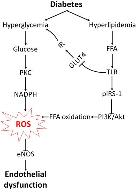
Overproduction of ROS is associated with diabetes-induced vascular complications. In the presence of diabetes, IR-induced hyperglycemia contributes to the activation of NADPH oxidase and protein kinase C (PKC), and subsequently, increases the generation of downstream ROS [3]. (Fig. 1 left half).
Fig. 1
The pathway of diabetes-induced endothelial dysfunction. PKC, protein kinase C; NADPH, nicotinamide adenine dinucleotide phosphate; ROS, reactive oxygen species; eNOS, endothelial NO synthase; FFA, free fatty acid; TLR, Toll-like receptor; pIRS-1, phosphorylation of insulin receptor substrate-1; PI3K, phosphatidylinositol-4,5-bisphosphate 3-kinase; Akt, protein kinase B; GLUT4, glucose transporter 4; IR, insulin resistance
Moreover, hyperglycemia is not the only symptom of diabetes, for instance, type 2 diabetes is usually accompanied by hyperlipidemia. In Cai’s study, ApoE-/- mice fed with a high-fat diet had significantly thickened vascular and increased plaque volume, in addition to lipid metabolism disorders with elevated levels of free fatty acid (FFA), total cholesterol (TC), triglyceride (TG) and low-density lipoprotein (LDL), and decreased high-density lipoprotein (HDL) [[1](/article/10.1007/s12265-018-9855-4#ref-CR1 "Cai, Y., Xie, K. L., Zheng, F., & Liu, S. X. (2018). Aerobic exercise prevents insulin resistance through the regulation of miR-492/resistin axis in aortic endothelium. Journal of Cardiovascular Translational Research.
                https://doi.org/10.1007/s12265-018-9828-7

              .")\]. The increased plasma FFA concentration binds Toll-like receptor (TLR) and induces phosphorylation of insulin receptor substrate-1 (IRS-1) and then downregulates glucose transporter 4 (GLUT4), which ultimately causes IR and hyperglycemia \[[3](/article/10.1007/s12265-018-9855-4#ref-CR3 "Paneni, F., Beckman, J. A., Creager, M. A., & Cosentino, F. (2013). Diabetes and vascular disease: pathophysiology, clinical consequences, and medical therapy: Part I. European Heart Journal., 34(31), 2436–2443.")\] (Fig. [1](/article/10.1007/s12265-018-9855-4#Fig1) right half). Hyperlipidemia-induced IR also plays a role in inhibiting PI3-kinase/Akt pathway, leading to increased generation of ROS by oxidation of surplus FFA \[[3](/article/10.1007/s12265-018-9855-4#ref-CR3 "Paneni, F., Beckman, J. A., Creager, M. A., & Cosentino, F. (2013). Diabetes and vascular disease: pathophysiology, clinical consequences, and medical therapy: Part I. European Heart Journal., 34(31), 2436–2443.")\] (Fig. [1](/article/10.1007/s12265-018-9855-4#Fig1) bottom).
MicroRNA Mediates the Beneficial Effects of Exercise
The progression of diabetes is generally considered to be irreversible; nevertheless, diabetes-induced cardiovascular diseases can be controlled. It has been shown that exercise helps to inhibit IR and to enhance endothelial function by increasing the activity of eNOS [[1](/article/10.1007/s12265-018-9855-4#ref-CR1 "Cai, Y., Xie, K. L., Zheng, F., & Liu, S. X. (2018). Aerobic exercise prevents insulin resistance through the regulation of miR-492/resistin axis in aortic endothelium. Journal of Cardiovascular Translational Research.
                https://doi.org/10.1007/s12265-018-9828-7

              .")\]. Therefore, in the context of AS, Cai et al. suggested that swimming could reduce hyperlipidemia and decrease the volume of plaques. In addition, miR-429 has also been found to be upregulated by swimming \[[1](/article/10.1007/s12265-018-9855-4#ref-CR1 "Cai, Y., Xie, K. L., Zheng, F., & Liu, S. X. (2018). Aerobic exercise prevents insulin resistance through the regulation of miR-492/resistin axis in aortic endothelium. Journal of Cardiovascular Translational Research.

                https://doi.org/10.1007/s12265-018-9828-7

              .")\]. miRNAs are small noncoding RNAs which has been widely recognized to be crucially involved in the development and progression of AS. Some of them, such as miR-126-5p, miR-26a, and miR-19a, attenuate endothelial cell proliferation and/or inhibit endothelial apoptosis by regulating the target of endothelial cells, thus suppressing the genesis of atherosclerosis \[[4](/article/10.1007/s12265-018-9855-4#ref-CR4 "Feinberg, M. W., & Moore, K. J. (2016). MicroRNA regulation of atherosclerosis. Circulation Research., 118(4), 703–720.")\]. In contrast, other miRNAs, including miR-155, miR-92a, and miR-33, prompts the progression of AS, which turn out to enhance the inflammatory response or disturb lipid metabolism in macrophages in an AS animal model \[[4](/article/10.1007/s12265-018-9855-4#ref-CR4 "Feinberg, M. W., & Moore, K. J. (2016). MicroRNA regulation of atherosclerosis. Circulation Research., 118(4), 703–720.")\].
MiR-429, as a major microRNA in miR-200 family, has been mainly investigated in cancer therapies, for its regulatory roles on downstream factors including VEGFA, moesin, and PKCα [5]. Recent evidences demonstrated that miR-429 would be a potential mediator of glucose responses involved in endothelial cell function. However, the regulation mechanism of its downstream effector is still relatively vague. In the study conducted by Cai et al., high-fat diet caused a downregulation of miR-492 and an increase in the expression level of resistin in the aortic endothelial of AS model mice. They proved that miR-492 directly affect against resistin. Resistin, as a determinant of obesity, can indirectly lead to insulin resistance and contribute to the early development of AS. Additionally, they also found for the first time that exercise can increase the expression level of miR-492, which proved once again miRNAs’ important role in the protective influence of exercise training on the cardiovascular system. However, the other target genes and its upstream triggers remain to be explored. In addition, the current study has not provided evidences about the role of miR-429 in vivo. However, administration of miR-492 and its targets in a signaling pathway that regulates insulin resistance might be new intervention agents in improving cardiovascular diseases, which may contribute to potential therapeutic strategy [3].
Conclusion
In the diabetic population, IR, hyperglycemia, and hyperlipidemia have been suggested to aggravate oxidative stress and dysfunction caused by energy substrate metabolic disorder. This study shows that exercise-mediated miRNAs inhibited the development of diabetes-induced AS by protecting the endothelial cell function.
References
- Cai, Y., Xie, K. L., Zheng, F., & Liu, S. X. (2018). Aerobic exercise prevents insulin resistance through the regulation of miR-492/resistin axis in aortic endothelium. Journal of Cardiovascular Translational Research. https://doi.org/10.1007/s12265-018-9828-7.
Article Google Scholar - Wang, L., Lv, Y., Li, G., & Xiao, J. (2018). MicroRNAs in heart and circulation during physical exercise. Journal of Sport and Health Science., 7(4), 433–441.
Article Google Scholar - Paneni, F., Beckman, J. A., Creager, M. A., & Cosentino, F. (2013). Diabetes and vascular disease: pathophysiology, clinical consequences, and medical therapy: Part I. European Heart Journal., 34(31), 2436–2443.
Article CAS Google Scholar - Feinberg, M. W., & Moore, K. J. (2016). MicroRNA regulation of atherosclerosis. Circulation Research., 118(4), 703–720.
Article CAS Google Scholar - Humphries, B., & Yang, C. (2015). The microRNA-200 family: small molecules with novel roles in cancer development, progression and therapy. Oncotarget, 6(9), 6472–6498.
Article Google Scholar
Funding
Our work was supported by the grants from the National Natural Science Foundation of China (81722008, 91639101, and 81570362 to JJ Xiao), Innovation Program of Shanghai Municipal Education Commission (2017-01-07-00-09-E00042 to JJ Xiao), the grant from Science and Technology Commission of Shanghai Municipality (17010500100 to JJ Xiao), and the development fund for Shanghai talents (to JJ Xiao).
Author information
Authors and Affiliations
- Cardiac Regeneration and Ageing Lab, Institute of Cardiovascular Sciences, School of Life Science, Shanghai University, Shanghai, 200444, China
Minjun Xu, Yi Duan & Junjie Xiao
Authors
- Minjun Xu
You can also search for this author inPubMed Google Scholar - Yi Duan
You can also search for this author inPubMed Google Scholar - Junjie Xiao
You can also search for this author inPubMed Google Scholar
Corresponding author
Correspondence toJunjie Xiao.
Ethics declarations
Conflict of Interest
The authors declare that they have no conflict of interest.
Research Involving Human Participants and/or Animals
This article does not contain any studies with human participants or animals performed by any of the authors.
Informed Consent
This article does not contain any studies with human participants.
Additional information
Associate Editor Joost Sluijter oversaw the review of this article
Publisher’s Note
Springer Nature remains neutral with regard to jurisdictional claims in published maps and institutional affiliations.
Provenance:
Comment on: Ying Cai, Kanng-Ling Xie, Fan Zheng, Sui-Xin Liu. Aerobic exercise prevents insulin resistance through the regulation of miR-492/resistin axis in aortic endothelium. J Cardiovasc Transl Res. 2018 Sep 19. doi: 10.1007/s12265-018-9828-7
Rights and permissions
About this article
Cite this article
Xu, M., Duan, Y. & Xiao, J. Exercise Improves the Function of Endothelial Cells by MicroRNA.J. of Cardiovasc. Trans. Res. 12, 391–393 (2019). https://doi.org/10.1007/s12265-018-9855-4
- Received: 27 November 2018
- Accepted: 07 December 2018
- Published: 02 January 2019
- Issue Date: October 2019
- DOI: https://doi.org/10.1007/s12265-018-9855-4
