Sidestream dark field imaging: an improved technique to observe sublingual microcirculation (original) (raw)
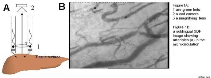
Sublingual orthogonal polarization spectral (OPS) imaging has revealed the central role of the microcirculation in the pathophysiology, outcome and treatment of sepsis by its ability to visualize the microcirculation in great detail under clinical conditions. Of particular importance in this context has been the response of the smallest microvessels, the capillaries. OPS imaging illuminates the tissues with polarized green light and measures the reflected light from the tissue surface after filtering out the polarized portion of the reflected light. This filters out the surface reflection of the tissues and allows the visualization of the underlying microcirculation. Due to the reflected and emitted light passing down the same light guide (mainstream), however, OPS imaging is highly sensitive to internal scatter of light. This results in limited visualization of the capillaries due to blurring. The technique also requires high-powered bulky light sources, limiting its utility in difficult conditions such as emergency medicine. In this communication we introduce sidestream dark field (SDF) imaging as a new way of clinical observation of the microcirculation. In this modality a light guide imaging the microcirculation is surrounded by light-emitting diodes (Fig. 1A, 1) of a wavelength (530 nm) absorbed by the hemoglobin of erythrocytes so that they can be clearly observed as flowing cells. Covered by a disposable cap (not shown) the probe is placed on tissue surfaces. The concentrically placed light-emitting diodes at the tip of the probe directly penetrate deep into the tissue illuminating the microcirculation. By not being in direct optical contact with the sensing central core of the probe, no direct surface reflections interfere with the image of the microcirculation. A five or 10 times magnifying lens (Fig. 1A, 3) projects the image onto a video camera (Fig. 1A, 2). This way of observing the microcirculation provides clear images of the capillaries without blurring. The deeper sublingual arterioles can also be clearly observed (Fig. 1B, a). Improved image quality allows better computer automatic analysis of the images and the low energy requirement of SDF imaging further enhances its utility by allowing battery and/or portable computer operation. It is expected that SDF imaging will provide an improved imaging modality of the microcirculation in various clinical scenarios.
Figure 1
(A) 1, green light-emitting diodes; 2, ccd camera; 3, magnifying lens. (B) Sublingual sidestream dark field image showing arterioles (a) in the microcirculation.
Author information
Authors and Affiliations
- Academic Medical Center, Amsterdam, The Netherlands
C Ince
Additional information
Competing interest
CI is CSO of Micro Vision Medical.
