Facet Joints (original) (raw)
Description[edit | edit source]
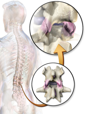
Illustration highlighting facet joint articulation between two vertebrae
Also known as the zygapophyseal or apophyseal joint, is a synovial joint between the superior articular process of one vertebra and the inferior articular process of the vertebra directly above it. There are two facet joints in each spinal motion segment.
The facet joints are situated between the pedicle and lamina of the same vertebra and form the articular pillars that act to provide structural stability to the vertebral column as a whole.
Together with the disc, the bilateral facet joints transfer loads and guide and constrain motions in the spine due to their geometry and mechanical function[1].
Articulating Surfaces[edit | edit source]
Articular facet on the superior process of the vertebra below with the articular facet on the inferior articular process of the vertebra above. The superior facet of the inferior vertebra is rather flat in the cervical and thoracic regions and more convex in the lumbar region. The opposing inferior facet of the superior vertebra is concave and forms an arch with its apex pointing towards the vertebral body
Facets joints have different orientations depending on the area:
Cervical Region = 45 degrees; frontal plane; all movements are possible such as flexion, extension, lateral flexion, and rotation.
The articulating facets in the cervical vertebrae face 45 degrees to the transverse plane and lie parallel to the frontal plane, with the superior articulating process facing posterior and up and the inferior articulating processes facing anteriorly and down.
Thoracic Region = 60 degrees; frontal plane; lateral flexion and rotation; no flexion/extension
The facet joints between adjacent thoracic vertebrae are angled at 60° to the transverse plane and 20° to the frontal plane, with the superior facets facing posterior and a little up and laterally and the inferior facets facing anteriorly, down, and medially
Lumbar Region = 90 degrees; sagittal plane; only flexion and extension.
The facet joints in the lumbar region lie in the sagittal plane; the articulating facets are at right angles to the transverse plane and 45° to the frontal plane. The superior facets face medially, and the inferior facets face laterally. This changes at the lumbosacral junction, where the apophyseal joint moves into the frontal plane and the inferior facet on L5 faces front. This change in orientation keeps the vertebral column from sliding forward on the sacrum[2].
Ligaments and Joint Capsule[edit | edit source]
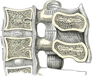
The posterior ligamentous complex acts to stabilise the vertebral column and hold the facet joints of the neighbouring vertebrae in fixed relation with each other. It is made up of the following structures:
- posterior longitudinal ligament
- facet joint capsule - Like other synovial joints in the body, they are surrounded by a capsule of connective tissue that produces synovial fluid to nourish and lubricate the joint. Each facet joint is innervated by two small nerves (dually innervated) – paired medial branches of the posterior ramus (dorsal primary rami) of the spinal nerves. The facet receives a medial branch from the spinal nerve above the facet and the nerve below. When there is degeneration or inflammation within a facet joint, pain activates the medial branch nerve. These nerves do not control sensations or muscles in the limbs.[4]
- ligamentum flavum
- interspinous ligament
- supraspinous ligament
Motions Available[edit | edit source]
The movements of each spinal segment are limited by anatomical structures such as ligaments, intervertebral discs, and facets. Specifically, anatomical structures cause the coupling of motions of the spine, that is, movements occur simultaneously. Flexion, extension, translation, axial rotation, and lateral bending are physiologically coupled. The exact pattern of coupling depends on the regional variations of anatomical structures. In the cervical and upper thoracic spine, side bending is coupled with axial rotation in the same direction. In the lumbar spine, lateral bending is coupled with axial rotation in the opposite direction. In the middle and lower thoracic spine, the coupling pattern is inconsistent. However, the pattern of coupling will change depending on which movement is initiated first. In the lumbar spine, lateral bending will be coupled with axial rotation in the same direction if lateral bending is the first movement. Conversely, if axial rotation is the first movement, it will be coupled with lateral bending in the opposite direction.[5]
Function[edit | edit source]
Guide and limit movement of the spinal motion segment. Contribute to stability of each motion segment.
Clinical significance[edit | edit source]
Osteoarthritis (OA) of the spine involves the facet joints (located in the posterior aspect of the vertebral column) and are the only true synovial joints between adjacent spinal levels. Facet joint osteoarthritis (FJ OA) is widely prevalent in older adults, and is a common cause of back and neck pain. The prevalence of facet-mediated pain in clinical populations increases with increasing age, suggesting that FJ OA might have a particularly important role in older adults with spinal pain[6]. See also Facet Joint Syndrome
Diagnostic positive facet joint block can indicate facet joints as the source of chronic spinal pain. These patients may benefit from specific interventions to eliminate facet joint pain such as neurolysis, by radiofrequency or cryoablation[7]. A 2013 study looking at available evidence of lumbar facet joint injections and the physiotherapy treatments, concluding that lumbar facet joint injections create a short period when pain is reduced. Physiotherapy treatments including land-based lower back mobility exercise and soft tissue massage may be of benefit during this time to improve the longer-term outcomes of patients with chronic low back pain.[8] See also Lumbar Facet Joint Injections , Lumbar Facet Syndrome, Cervical Osteoarthritis
References[edit | edit source]
- ↑ Nicolas V. Jaumard, William C. Welch, and Beth A. Winkelstein1. Spinal Facet Joint Biomechanics and Mechanotransduction in Normal, Injury and Degenerative Conditions. J Biomech Eng. 2011 July; 133(7): 71010–NaN.
- ↑ Hamill, Joseph, and Kathleen M. Knutzen. Biomechanical Basis of Human Movement, 3rd Ed., USA: Lippincott Williams & Wilkins, 2009.
- ↑ medeasy 3D Facet Orientation || Cervical, Thoracic, Lumbar Spine #OMM #COMLEX #WeDaBest Available from: https://www.youtube.com/watch?v=8zQv2wWcrF0&feature=youtu.be (last accessed 14.11.2019)
- ↑ Ainsworth institute of pain management Understanding Anatomy: FACET JOINTS Available from: https://ainsworthinstitute.com/patient-information/anatomy/facet-joints/ (last accessed 13.11.2019)
- ↑ Fitness Medico Italiano Biomechanics of The Spine Available from: http://fitnessmedicoitaliano.it/wp-content/uploads/2018/03/Banton-Spine-Biomechanics.pdf (last accessed 13.11.2019)
- ↑ Gellhorn AC, Katz JN, Suri P. Osteoarthritis of the spine: the facet joints. Nature Reviews Rheumatology. 2013 Apr;9(4):216. Available from: https://www.ncbi.nlm.nih.gov/pmc/articles/PMC4012322/ (last accessed 13.11.2019)
- ↑ Perolat R, Kastler A, Nicot B, Pellat JM, Tahon F, Attye A, Heck O, Boubagra K, Grand S, Krainik A. Facet joint syndrome: from diagnosis to interventional management. Insights into imaging. 2018 Oct 1;9(5):773-89. Available from: https://link.springer.com/article/10.1007/s13244-018-0638-x (last accessed 13.11.2019)
- ↑ Chambers H. Physiotherapy and lumbar facet joint injections as a combination treatment for chronic low back pain. A narrative review of lumbar facet joint injections, lumbar spinal mobilizations, soft tissue massage and lower back mobility exercises. Musculoskeletal Care. 2013 Jun;11(2):106-20. Available from: https://www.ncbi.nlm.nih.gov/pubmed/23468052 (last accessed 13.11.2019)

