The Role of Influenza A Virus Hemagglutinin Residues 226 and 228 in Receptor Specificity and Host Range Restriction (original) (raw)
Abstract
Influenza A viruses can be isolated from a variety of animals, but their range of hosts is restricted. For example, human influenza viruses do not replicate in duck intestine, the major replication site of avian viruses in ducks. Although amino acids at positions 226 and 228 of hemagglutinin (HA) of the H3 subtype are known to be important for this host range restriction, the contributions of specific amino acids at these positions to restriction were not known. Here, we address this issue by generating HAs with site-specific mutations of a human virus that contain different amino acid residues at these positions. We also let ducks select replication-competent viruses from a replication-incompetent virus containing a human virus HA by inoculating animals with 1010.5 50% egg infectious dose of the latter virus and identified a mutation in the HA. Our results showed that the Ser-to-Gly mutation at position 228, in addition to the Leu-to-Gln mutation at position 226 of the HA of the H3 subtype, is critical for human virus HA to support virus replication in duck intestine.
Influenza A viruses can be isolated from various animals, including humans, pigs, horses, wild waterfowl, chickens, passerine birds, sea mammals, minks, and camels (27). Of these host animals, wild waterfowl are the principal reservoir of influenza A viruses. In fact, all of the viruses currently circulating in other animal species are thought to have originated from wild waterfowl (27). However, under experimental conditions, avian influenza viruses do not replicate efficiently in humans (3); similarly, human viruses do not replicate efficiently in ducks (7, 10, 29). Such studies indicate host range restriction of influenza viruses. The viruses that caused the 1957 and 1968 influenza pandemics were reassortant viruses of human and avian influenza viruses (22, 26). Avian influenza virus genes were somehow introduced into the human populations, breaking through the host range restriction. To elucidate the mechanism by which pandemic influenza virus strains are generated, we must first understand the molecular basis of host range restriction of influenza virus and how such restriction is breached.
The hemagglutinin (HA) of influenza A viruses is a major surface glycoprotein that is responsible for attachment of the virus to the cell surface of oligosaccharide receptors. All influenza A viruses recognize oligosaccharides containing terminal sialic acid (SA) as receptors; however, human viruses preferentially recognize SA linked to galactose by α2,6 linkages (SAα2,6Gal), whereas avian viruses preferentially recognize SAα2,3Gal (12, 17, 24). The amino acids that make up the receptor-binding site (RBS) are highly conserved, even among the HAs of different subtypes of avian influenza virus; however, those of human viruses display distinct variability (12). In particular, the residues at positions 138, 190, 194, 225, 226, and 228 are highly conserved in the avian RBS, whereas human HAs harbor substitutions at these positions. In H2 and H3 influenza virus strains, residues at positions 226 and 228 in the HA correlate with the preferential recognition of the SA-Gal linkage by HA and the host species from which the virus was isolated. HAs with Leu at position 226 (Leu-226) and Ser-228 (human viruses) preferentially recognize SAα2,6Gal, whereas those with Gln-226 and Gly-228 (avian and equine viruses) recognize SAα2,3Gal (4). Moreover, the HA plays an important role in host range restriction of influenza virus. For example, a reassortant virus possessing only the HA gene from human A/Udorn/307/72 (Udorn) (H3N2) virus and the rest of its genes from A/mallard/New York/6750/78 (Mal/NY) (H2N2) virus does not grow in the duck intestine, the major site of avian influenza virus replication in this animal (7). However, two mutations, Leu-to-Gln at position 226 and Ser-to-Gly at position 228 (but not the 226 mutation alone) allow the human virus HA to support virus replication in duck intestine (13). These findings demonstrate the importance of these residues for receptor specificity and for host range restriction of the virus. However, the contribution of the 228 mutation alone to changes in viral properties remains unknown.
Recently, we showed that agglutination of erythrocytes from different animal species can be used to assess the receptor specificity of influenza A viruses (9). Human viruses, including those known to preferentially recognize SAα2,6Gal, agglutinated erythrocytes from chicken, ducks, guinea pigs, and sheep but not those from horses or cows; however, avian and equine viruses, including those known to preferentially recognize SAα2,3Gal, agglutinated all of these erythrocytes. Fluorescence-activated cell sorting (FACS) analysis of the erythrocytes with SA-Gal linkage-specific lectins demonstrated that horse erythrocytes contained mostly SAα2,3Gal and hardly any SAα2,6Gal, whereas human and chicken erythrocytes contained both SAα2,3Gal and SAα2,6Gal. Because more than 97% of SA in horse erythrocytes is the _N_-glycolyl form and no _N_-glycolyl SA exists in human and chicken erythrocytes (25), the above findings suggest that avian but not human viruses recognize _N_-glycolyl SA linked to galactose by α2,3 linkages, which are abundant on horse, but not human and chicken, erythrocytes.
In this study, we focused on the restriction of human influenza viruses in ducks, because previous studies have shown that duck influenza viruses replicate in duck intestine efficiently (replicating up to 106 50% egg infectious dose [EID50]/g), whereas replication of human viruses in this site is undetectable (10), providing a clear-cut system. Although host range restriction of influenza viruses is polygenic (1, 7, 21, 23), we focused on the contribution of HA in this study. Our aim was to determine which amino acids at positions 226 and 228 of the H3 HA are specifically required for replication of the virus in duck intestine. To this end, we made mutant human virus HAs with substitutions at positions 226 and/or 228 and tested their receptor specificity with horse, human, and chicken erythrocytes. We also generated transfectant influenza viruses containing some of these HA mutants and tested their ability to replicate in ducks. In addition, we sought mutations that convert human virus HA, which does not support virus replication in duck intestine, to a form that would support virus replication in this site, by inoculating a large amount of a replication-incompetent virus in duck intestine and allowing the ducks to select replication-competent viruses.
Influenza A virus Udorn (H3N2), Mal/NY (H2N2), A/Puerto Rico/8/34 (H1N1), and a reassortant virus, R4 (7), that possesses a mutant Udorn HA (Leu-to-Gln mutation at position 226 [L226Q]) and the rest of its genes from the Mal/NY virus, were obtained from the repository at St. Jude Children’s Research Hospital. Udorn and Mal/NY viruses have been isolated and passaged in eggs. Seal/E1 virus, a variant of A/Seal/Massachusetts/1/80 (H7N7), was obtained from Rudolf Rott (15). This virus grows in Madin-Darby canine kidney (MDCK) cells in the presence of elastase (2 μg/ml; Calbiochem, La Jolla, Calif.) but not trypsin due to a mutation at its HA cleavage site (15). A reassortant virus, Seal/E1-Mal/NY, containing the Seal/E1 HA gene and the rest of its genes from Mal/NY, was made as previously described (9).
A plasmid, pUd72HA-39, containing the Udorn HA gene was constructed as described by Huddleston and Brownlee (8). To generate transfectant viruses, a plasmid, pGt3UH23, was constructed by PCR (18) amplification of the Udorn HA gene flanked by the _Ksp_632I site and the T3 RNA polymerase promoter sequence by using pUd72HA-39 as a template. The PCR product was cloned into a pGEM7 vector as described previously (5). The HA genes mutated at codons 226 and/or 228 were generated by PCR (18). The 332-bp PCR products (spanning the _Xba_I site at nucleotide 732 and the _Stu_I site at nucleotide 1064) that contained the desired mutations were used to replace the corresponding region of pGt3UH23. For HA expression in cells, the HA genes were cloned into the _Asp_718 and the _Sph_I sites of pCAGGS/MCS (11), which contains the chicken β-actin promoter (14).
Plasmids, used to generate transfectant viruses (e.g., pGt3UH23), were digested with _Ksp_632I, and their ends were filled in with Klenow fragment as described previously (5). To generate an HA-ribonucleoprotein (RNP) complex, we transcribed the plasmids in vitro with T3 RNA polymerase in the presence of nucleoprotein and polymerase proteins. The HA-RNP complex was then transfected into 80% confluent Madin-Darby bovine kidney (MDBK) cells that had been infected 1 h earlier with Seal/E1-Mal/NY reassortant virus at a multiplicity of infection of one. Eighteen hours after transfection, the culture supernatants were collected and clarified by centrifugation (10,000 × g, 30 s). Transfectant viruses were selected by infection of MDCK cells in the presence of trypsin. Because the HA of the helper Seal/E1-Mal/NY reassortant is cleaved by elastase but not by trypsin, transfectant viruses have a growth advantage over Seal/E1-Mal/NY virus grown in the presence of trypsin. The transfectant viruses obtained were plaque purified three times on MDCK cells, and a final tissue culture stock was used to inoculate 11-day-old embryonated chicken eggs. Allantoic fluid stocks of viruses were stored at −70°C. The entire HA gene of each transfectant virus was sequenced by using an automated sequencer (Applied Biosystem Inc., Foster City, Calif.).
Three 3-month-old Peking ducks (Ridgeway Hatcheries) were orally inoculated with 106 EID50 of each transfectant virus (1 ml/duck, three ducks/virus) (12). Three days postinoculation, the ducks were sacrificed and their colons were removed. The tissue was homogenized, resuspended in phosphate-buffered saline (PBS), and inoculated into 11-day-old embryonated chicken eggs for virus isolation. The sequence of the HA1 portion of the gene of virus isolated from at least one duck inoculated with each virus was determined.
Effects of HA amino acid residues 226 and 228 on cell surface expression and receptor specificity.
Residues 226 and 228 in the human influenza virus HA of H2 and H3 subtypes are both thought to be important for host range restriction and receptor specificity of the virus (4). However, the exact contributions that these residues make to these viral properties were unknown. The Leu-to-Gln mutation at position 226 of a human virus HA (X31 strain) alters its receptor specificity from SAα2,6Gal to SAα2,3Gal (17). While this mutation alone is not sufficient to convert another human virus HA (Udorn strain) to a form of HA that supports virus replication in duck intestine, an additional mutation at residue 228 from Ser to Gly does permit this conversion (13). Although the majority of duck virus HAs possess Gly at position 228, some have Arg or Ser (2). To understand the effects of different amino acid residues at position 228 on the receptor specificity of HA, we made a number of human Udorn virus HA mutants that had different amino acids at this position with and without the Leu-to-Gln mutation at position 226 (Table 1). Mutants were designated by a single amino acid code according to their amino acid residues at positions 226 and 228. For example, a mutant possessing Gln at position 226 and Cys at position 228 was designated QC.
TABLE 1.
Properties of HA mutants
| HA | Amino acid at position: | Cell surface expressiona | Adsorptionb of erythrocytes from: | Rescued by reverse geneticsc | Ability to support viral replication in duck intestined | |||
|---|---|---|---|---|---|---|---|---|
| 226 | 228 | Chickens | Humans | Horses | ||||
| Wild type | Leu | Ser | ++ | ++ | ++ | + | + | − |
| LN | Leu | Asn | − | NT | NT | NT | NT | NA |
| LC | Leu | Cys | ++ | − | − | − | NT | NA |
| LV | Leu | Val | ++ | ++ | ++ | − | − | NA |
| LK | Leu | Lys | ++ | + | ++ | − | − | NA |
| LT | Leu | Thr | ++ | ++ | ++ | − | − | NA |
| LA | Leu | Ala | + | +++ | +++ | + | − | NA |
| LR | Leu | Arg | ++ | + | ++ | − | − | NA |
| LG | Leu | Gly | ++ | ++ | ++ | + | NT | NA |
| LD | Leu | Asp | +++ | − | − | − | NT | NA |
| QS (R4) | Gln | Ser | ++ | ++ | ++ | +++ | + | − |
| QN | Gln | Asn | +++ | + | + | − | NT | NA |
| QC | Gln | Cys | + | + | + | − | − | NA |
| QV | Gln | Val | ++ | +++ | +++ | +++ | − | NA |
| QK | Gln | Lys | +++ | − | + | + | NT | NA |
| QT | Gln | Thr | + | − | − | − | NT | NA |
| QA | Gln | Ala | ++ | + | + | + | + | − |
| QR | Gln | Arg | ++ | + | ++ | ++ | + | − |
| QG (R2) | Gln | Gly | ++ | +++ | +++ | +++ | + | + |
| QD | Gln | Asp | ++ | − | − | + | NT | NA |
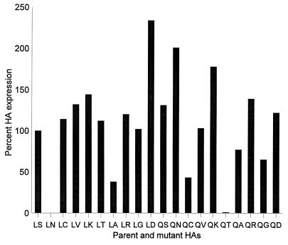
For HA to function, it has to be transported to the cell surface. We therefore examined the relative cell surface expression levels of our HA mutants and found some variations (Fig. 1). FACS analysis showed that many of the mutant HAs were expressed on the cell surface at levels comparable to that of the wild type but that others, including LA, QC, QA, and QG, were expressed at significantly lower levels on the cell surface. The QT mutant was consistently detected on the cell surface at just above the background level, whereas the LN mutant was negative for cell surface expression.
FIG. 1.
Cell surface expression of mutant HAs analyzed by FACS. Cos-1 cells (60% confluency) were transfected with 2 μg of purified plasmid DNA per well of a 6-well tissue culture plate with Lipofectamine (Gibco). The cells and transfection mixture were incubated for 5 h at 37°C, after which the transfection medium was replaced with 2 ml of Opti-Mem medium (Life Technologies, Inc.) containing 5% fetal calf serum (FCS). The cells were then incubated for 40 h at 37°C. Cells expressing HA were washed with PBS, treated with Vibrio cholerae sialidase (5.5 milliunits/ml; Life Technologies, Inc.) for 1 h at 37°C (which abolishes the susceptibility of cells to influenza virus infection) to remove sialic acid from the HA, which interferes with receptor recognition (17), and then maintained in suspension following trypsinization. Cells were spun down, washed twice with PBS containing 10% FCS, and then resuspended in PBS containing 10% FCS and an anti-H3 HA monoclonal antibody (S11/4, S28/1, S37/2, and 121/1) pool (diluted 1:400). After a 1-h incubation at 4°C, the cells were washed twice with PBS containing 10% FCS, and then incubated with fluorescein isothiocynate-labeled goat anti-mouse immunoglobulin (diluted 1:20 in PBS containing 10% FCS) (Boehringer Mannheim Biochemicals) for 30 min at 4°C. The cells were washed twice as before and then fixed in PBS containing 3.7% paraformaldehyde for 20 min at 4°C. Cells were centrifuged, resuspended in PBS containing 10% FCS, and stored at 4°C until analyzed by FACS with a FACScan (Becton Dickinson). The relative expression levels presented are based on mean values of fluorescence intensity. Experiments were repeated three times, and representative data are shown.
To confirm that our failure to detect some of the mutants was not due to a lack of expression per se, we analyzed HA expression by Western blot analysis of cell lysates. The results showed that all of the HAs, with the exception of the LN mutant, were expressed at least 50% of the wild-type level (data not shown). Because the LN mutant was expressed at only 15% of the wild-type level and was not transported to the cell surface (Fig. 1), this mutant was excluded from subsequent assays.
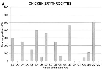
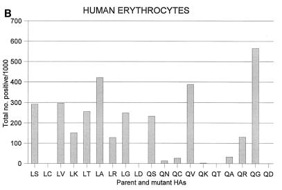
We next tested these HA mutants for their ability to bind to chicken (Fig. 2A) and human (Fig. 2B) erythrocytes. Mutations to Lys, Thr, or Arg at position 228 (LK, LT, and LR) appreciably reduced hemadsorption activity, whereas those to Cys or Asp (LC and LD mutants) completely abolished activity. Other mutations at position 228 reduced or enhanced the activity slightly, depending on the erythrocytes used. Although chicken and human erythrocytes exhibited similar profiles in SA-Gal linkage-specific lectin assays, which suggests comparable α2,3 and α2,6 linkage proportions, some mutants differentially bound these erythrocytes; for example, the LK and the LR mutants both bound to chicken erythrocytes less efficiently than to human erythrocytes.
FIG. 2.
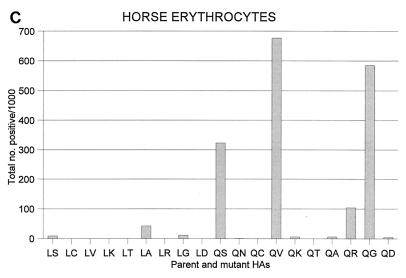
Effects of mutations at positions 226 and/or 228 on the hemadsorbing activity of human virus HA. Cos-1 cells were transfected with plasmid expressing HA. At 40 h posttransfection, they were washed twice with PBS containing 10% FCS and incubated with chilled 1% erythrocyte suspensions (chicken [in house], human [type O, St. Jude Children’s Research Hospital blood bank], or horse [Rockland]) in PBS. After a 1-h incubation at 4°C, the cells were washed at least five times with PBS and rinsed with methanol to remove erythrocytes that were nonspecifically bound. The cells were then air dried and stained with a 1:20 dilution of Giemsa stain (Sigma) for 15 min at 4°C. The numbers of hemadsorption-positive cells were recorded by examining five randomly selected microscopic fields that consisted of approximately 500 cells. Experiments were performed three times with similar results.
We then analyzed the effect of the 228 mutations in conjunction with the L226Q mutation. The L226Q mutation alone (i.e., the QS mutant) did not affect chicken or human erythrocyte binding appreciably. However, when coupled with the L226Q mutation, the HA activity detected with the S228K or S228T mutation alone (i.e., the LK or LT mutant, respectively) was completely abolished (QK and QT mutants [Fig. 2]). Interestingly, two mutants, QV and QG, bound both erythrocytes more efficiently than did wild-type (LS) HA. One mutant, QG, is the same as the HA of R2 virus, which supports viral replication in ducks (13). The QV mutation, however, has not been found in nature. HA possessing the L226Q and S228R mutations (i.e., the QR mutant) exists in some avian H3 viruses (2); however, this mutant did not bind either type of erythrocyte efficiently. These results indicate that limited alterations at positions 226 and 228 of HA are accommodated to permit HA function.
Because avian, but not human, influenza viruses hemagglutinate horse erythrocytes (9), we also tested the ability of our mutants to hemadsorb these erythrocytes (Fig. 2C). None of the mutants that had a mutation only at position 228 adsorbed horse erythrocytes efficiently. However, the mutants that had an alteration at position 228 as well as the L226Q mutation and adsorbed either chicken or human erythrocytes (QV, QR, and QG mutants) also adsorbed horse erythrocytes. Interestingly, the L226Q mutation alone (i.e., QS mutant) made the human Udorn virus HA adsorb these erythrocytes. However, an additional mutation at position 228, from Ser to Val or Gly, made this HA adsorb horse erythrocytes extremely well. The finding that the QG mutant (i.e., the R2 mutant) hemadsorbs horse erythrocytes efficiently is consistent with the fact that most avian H3 HAs contain these residues (2). However, although the HA that possesses Val at position 228 has not been found in nature, it adsorbed these erythrocytes extremely well. Interestingly, even though the avian viruses A/duck/Hokkaido/8/80 (H3N8) and A/mallard/New York/6874/78 (H3N2) possess Gln at position 226 and Arg at position 228 and agglutinate horse erythrocytes, the ability of the QR mutant to bind to these erythrocytes was not as strong as those of the QV and QG mutants. These results show that the L226Q mutation causes the human Udorn virus HA to recognize sialyloligosaccharides on horse erythrocytes and that the additional mutation, S228G, significantly enhances this ability.
Replication of transfectant viruses possessing mutations at positions 226 and 228 in HA.
Because avian, but not human, influenza viruses agglutinate horse erythrocytes efficiently (9), we asked whether the mutant human Udorn HAs, which adsorb these erythrocytes, support virus replication in duck intestine. To this end, we attempted to generate transfectant viruses comprised of the mutant Udorn HA gene and the rest of its genes from Mal/NY. To generate such viruses, we first made a reassortant virus, Seal/E1-Mal/NY, that possesses the HA gene from Seal/E1 and the rest of its genes from the Mal/NY virus. The HA of Seal/E1-Mal/NY reassortant virus is derived from the Seal/E1 virus and is cleaved by elastase but not by trypsin. Therefore, transfectant viruses that possess HA that is cleavable by trypsin have a growth advantage in the presence of this protease. Using this selection method, we attempted to generate viruses possessing the LA, LT, LV, LK, LR, QN, QA, QR, QV, QC, and QG HAs as well as the wild-type HA. We were, however, only able to obtain viruses possessing the wild-type, QA, QR, or QG mutant HA, which were designated Udorn-Mal/NY, QA-Mal/NY, QR-Mal/NY, and QG-Mal/NY, respectively. These results suggest that the HA mutants that were not rescued do not support virus replication to the level at which the current reverse genetics system operates.
We then examined the replication of transfectant viruses in MDCK cells, embryonated chicken eggs, and ducks. All of the rescued viruses efficiently replicated in MDCK cells (ranging from 107.1 to 107.8 PFU/ml) and eggs (ranging from 107.3 to 108.1 EID50). Upon inoculation into ducks, the wild-type (Udorn-Mal/NY; thus, the same as R4 [13]) virus did not replicate in intestine, whereas QG-Mal/NY (thus, the same as R2) virus was able to replicate in ducks, consistent with previous reports (13). The QA-Mal/NY virus did not replicate in duck intestine and neither did QR-Mal/NY, even though viruses possessing Gln-226 and Arg-228 have been isolated from ducks (A/duck/Hokkaido/8/80 [H3N8] and A/mallard/New York/6874/78 [H3N2] [2]). It may be that the Gly-to-Arg mutation at position 228 occurred in the HAs of A/duck/Hokkaido/8/80 and A/mallard/New York/6874/78 during their propagation in eggs and that these viruses no longer replicated in duck intestine. To exclude this possibility, we sequenced the HA gene of A/duck/Hokkaido/8/80 (H3N8) and confirmed the Gln at position 226 and Arg at position 228. We then inoculated A/duck/Hokkaido/8/80 into ducks and found that the virus indeed replicated in the intestines of all three ducks tested. Together, these results suggest that residues other than 226 and 228 contribute to the replication of A/duck/Hokkaido/8/80 virus in duck intestine.
Importance of Gly-228 for virus replication in duck intestine.
Previous studies have shown that although a virus (R4) that possesses a single mutation at position 226 (from Leu to Gln) in the human Udorn virus HA does not replicate in duck intestine, another mutation at position 228 from Ser to Gly allows the virus to grow (13). Is this Ser-to-Gly mutation at position 228 the only change that converts the 226 mutant to one that supports virus replication in ducks? To answer this question, we inoculated a large amount (1010.5 EID50/duck) of R4 virus into the rectum of ducks through the cloaca. The cloacal inoculation was done to efficiently introduce a large amount of virus into the major site of virus replication in ducks without substantially reducing virus titers in the stomach, as would occur with oral inoculation. Assuming the mutation rate of influenza virus to be approximately 10−4 (16), we anticipated that a virus with an additional mutation that supports virus replication in ducks would be selected by this approach. Upon inoculation of the virus into 10 ducks, we recovered virus from the intestines of 8 ducks 3 days after inoculation. Each of the recovered viruses (a total of eight) was orally inoculated into two ducks to test its replicative ability in intestine. Of the eight viruses tested, seven replicated in duck intestine. Upon sequencing the entire HA gene of all eight viruses, we found that the seven that replicated in duck intestine upon oral inoculation had the Ser-to-Gly mutation at position 228 (thus, they were the same as the QG mutant). The one that did not replicate in ducks had no additional mutation (i.e., it was identical to R4). These results indicate that the Ser-to-Gly mutation at position 228 of a human virus HA possessing the L226Q mutation is indeed the most critical mutation to support virus replication in duck intestine.
In the current study, we have identified specific combinations of amino acid changes that affect the ability of human virus HA to support virus replication in ducks. Our results show that the residues at positions 226 and 228 are both essential for HA to support this function. The importance of the L226Q change was shown by the lack of horse erythrocyte binding ability (a characteristic of human, but not avian, influenza viruses [9]), with the S228G mutation alone. The critical role of Gly-228 in viral replication in duck intestine was demonstrated by the consistent selection of mutant viruses possessing this change during replication in ducks. Moreover, the presence of Gln-226 and Gly-228 correlated with the high affinity of HA for horse erythrocytes. Thus, these findings establish critical roles for these amino acids in receptor recognition and in host range restriction of influenza virus.
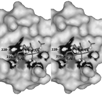
It is unclear at the molecular level how the presence of Gln-226 and Gly-228 results in the high affinity of HA for SAα2,3Gal-terminated receptors of equine erythrocytes and why this affinity decreases with an amino acid substitution in either of these positions. Significant favorable interactions are known to occur between the avian virus HA and the penultimate galactose moiety linked to sialic acid by α2,3, but not α2,6 linkage, and Leu-226 and Ser-228, specific for the human virus HA, are known to abrogate these interactions (12). According to the structural models of the complexes of the X31 human influenza virus HA with sialyloligosaccharides (19, 30) (Fig. 3), amino acid substitutions at positions 226 and 228 can affect the hydrogen bond formation and van der Waals interactions between the sialic acid moiety and the protein. We can therefore speculate that the presence of both Gln-226 and Gly-228 is required for the proper orientation of the sialic acid moiety in the avian virus RBS for the optimal (energetically favorable) fit of the α2,3-linked Gal and that a mutation in either position could slightly reorient the whole sialyloligosaccharide in the RBS and destroy this fit.
FIG. 3.
Positions of residues 226 and 228 in the RBS of influenza virus HA in relation to the Neu5Ac2,3Gal moiety. Only the relevant portion of the human virus X31 HA complexed with 3′ sialylactose (1HGG structure, Brookhaven Protein Databank [19]) and the Neu5Acα2-3Gal moiety of 3′ sialyl lactose (heavy atoms, stick presentation) are shown for clarity. Atoms of sialic acid (NAN) and galactose (GAL) that are thought to participate in hydrogen bonding with HA (19) are shown as small white balls; atoms of the protein that participate in these hydrogen bonds are shown as white on black background. Substitution 226S→G results in a loss of a hydrogen bond between the hydroxyl group of serine and the O-9 hydroxyl of sialic acid, whereas mutation 226L→Q results in new hydrogen bonds between the amide side chain of glutamine and the C-8 hydroxyl and carboxylic groups of Neu5Ac (30). This figure was generated with a WebLab viewer (Molecular Simulations, Inc., San Diego, Calif.).
Our data explain why avian virus HAs possess Gln at position 226 (2). Irrespective of the amino acid at position 228, HAs with Leu-226 do not exhibit characteristics of avian virus HAs (e.g., binding to horse erythrocytes [11]). This finding indicates that Gln-226 is indispensable for the avian virus receptor-binding phenotype. In fact, all avian virus HAs examined thus far possess Gln-226 (2). Regarding the residue at position 228, A/duck/Hokkaido/8/80 possesses Arg-228 but replicates in ducks. By contrast, the QR-Mal/NY virus failed to replicate in ducks. The HA of A/duck/Hokkaido/8/80 virus has Gly at position 229, whereas all of the other avian H3 HAs have Arg at this position (2). It may be that this amino acid substitution compensates for the effect of the G228R mutation in this duck virus.
What do our findings mean with respect to pandemic awareness? Our results suggest that if we find changes at residues 226 and 228 in the HA of avian viruses that have transmitted to mammals such as pigs (6, 20) and horses (28), then these viruses may have acquired the ability to recognize receptors in human trachea. Therefore, during the surveillance of viruses in nonhuman animals, it is particularly important to pay attention to the amino acid residues at these positions. Genes other than HA are also known to contribute to the host range restriction of influenza A viruses. Such genes include PB2 (1, 23), NA (7), and nucleoprotein (21). However, the molecular mechanisms by which these other genes affect the host range of influenza viruses have yet to be elucidated. A better understanding of such mechanisms would provide knowledge important for preventing future influenza pandemics.
Acknowledgments
We thank Rudolf Rott for Seal/E1 virus, Robert G. Webster for monoclonal antibodies, and Susan Watson for editing the manuscript.
This study was supported in part by a Public Health Service research grant (AI33898) from the National Institute of Allergy and Infectious Disease, by a CAST program from the National Research Council, by a Cancer Center Support (CORE) grant, and by the American Lebanese Syrian Associated Charities (ALSAC).
REFERENCES
- 1.Almond J W. A single gene determines the host range of influenza virus. Nature. 1977;270:617–618. doi: 10.1038/270617a0. [DOI] [PubMed] [Google Scholar]
- 2.Bean W J, Schell M, Katz J, Kawaoka Y, Naeve C, Gorman O, Webster R G. Evolution of the H3 influenza virus hemagglutinin from human and nonhuman hosts. J Virol. 1992;66:1129–1138. doi: 10.1128/jvi.66.2.1129-1138.1992. [DOI] [PMC free article] [PubMed] [Google Scholar]
- 3.Beare A S, Webster R G. Replication of avian influenza viruses in humans. Arch Virol. 1991;119:37–42. doi: 10.1007/BF01314321. [DOI] [PubMed] [Google Scholar]
- 4.Connor R J, Kawaoka Y, Webster R G, Paulson J C. Receptor specificity in human, avian, and equine H2 and H3 influenza virus isolates. Virology. 1994;205:17–23. doi: 10.1006/viro.1994.1615. [DOI] [PubMed] [Google Scholar]
- 5.Enami M, Luytjes W, Krystal M, Palese P. Introduction of site-specific mutations into the genome of influenza virus. Proc Natl Acad Sci USA. 1990;87:3802–3805. doi: 10.1073/pnas.87.10.3802. [DOI] [PMC free article] [PubMed] [Google Scholar]
- 6.Guan Y, Shortridge K F, Krauss S, Li P H, Kawaoka Y, Webster R G. Emergence of avian H1N1 influenza viruses in pigs in China. J Virol. 1996;70:8041–8046. doi: 10.1128/jvi.70.11.8041-8046.1996. [DOI] [PMC free article] [PubMed] [Google Scholar]
- 7.Hinshaw V S, Webster R G, Naeve C W, Murphy B R. Altered tissue tropism of human-avian reassortant influenza viruses. Virology. 1983;128:260–263. doi: 10.1016/0042-6822(83)90337-9. [DOI] [PubMed] [Google Scholar]
- 8.Huddleston J A, Brownlee G G. The sequence of the nucleoprotein gene of human influenza A virus strain, A/NT/60/68. Nucleic Acids Res. 1982;10:1029–1037. doi: 10.1093/nar/10.3.1029. [DOI] [PMC free article] [PubMed] [Google Scholar]
- 9.Ito T, Suzuki Y, Mitnaul L, Vines A, Kida H, Kawaoka Y. Receptor specificity of influenza A viruses correlates with the agglutination of erythrocytes from different animal species. Virology. 1997;227:493–499. doi: 10.1006/viro.1996.8323. [DOI] [PubMed] [Google Scholar]
- 10.Kida H, Yanagawa R, Matsuoka Y. Duck influenza lacking evidence of disease signs and immune response. Infect Immun. 1980;30:547–553. doi: 10.1128/iai.30.2.547-553.1980. [DOI] [PMC free article] [PubMed] [Google Scholar]
- 11.Kobasa D, Rodgers M E, Wells K, Kawaoka Y. Neuraminidase hemadsorption activity, conserved in avian influenza A viruses, does not influence viral replication in ducks. J Virol. 1997;71:6706–6713. doi: 10.1128/jvi.71.9.6706-6713.1997. [DOI] [PMC free article] [PubMed] [Google Scholar]
- 12.Matrosovich M N, Gambaryan A S, Teneberg S, Piskarev V E, Yamnikova S S, Lvov D K, Robertson J S, Karlsson K A. Avian influenza A viruses differ from human viruses by recognition of sialyloligosaccharides and gangliosides and by a higher conservation of the HA receptor-binding site. Virology. 1997;233:224–234. doi: 10.1006/viro.1997.8580. [DOI] [PubMed] [Google Scholar]
- 13.Naeve C W, Hinshaw V S, Webster R G. Mutations in the hemagglutinin receptor-binding site can change the biological properties of an influenza virus. J Virol. 1984;51:567–569. doi: 10.1128/jvi.51.2.567-569.1984. [DOI] [PMC free article] [PubMed] [Google Scholar]
- 14.Niwa H, Yamamura K, Miyazaki J. Efficient selection for high-expression transfectants by a novel eukaryotic vector. Gene. 1991;108:193–200. doi: 10.1016/0378-1119(91)90434-d. [DOI] [PubMed] [Google Scholar]
- 15.Orlich M, Linder D, Rott R. Trypsin-resistant protease activation mutants of an influenza virus. J Gen Virol. 1995;76:625–633. doi: 10.1099/0022-1317-76-3-625. [DOI] [PubMed] [Google Scholar]
- 16.Portner A, Webster R G, Bean W J. Similar frequencies of antigenic variation in Sendai, vesicular stomatis, and influenza A viruses. Virology. 1980;104:235–238. doi: 10.1016/0042-6822(80)90382-7. [DOI] [PubMed] [Google Scholar]
- 17.Rogers G N, Paulson J C. Receptor determinants of human and animal influenza virus isolates: differences in receptor specificity of the H3 hemagglutinin based on species of origin. Virology. 1983;127:361–373. doi: 10.1016/0042-6822(83)90150-2. [DOI] [PubMed] [Google Scholar]
- 18.Saiki R K, Gelfand D H, Stoffel S, Scharf S J, Higuchi R, Horn G T, Mullis K B, Erlich H A. Primer-directed enzymatic amplification of DNA with a thermostable DNA polymerase. Science. 1988;239:487–491. doi: 10.1126/science.2448875. [DOI] [PubMed] [Google Scholar]
- 19.Sauter N K, Glick G D, Crowther R L, Park S-J, Eisen M B, Skehel J J, Knowles J R, Wiley D C. Crystallographic detection of a second ligand binding site in influenza virus hemagglutinin. Proc Natl Acad Sci USA. 1992;89:324–328. doi: 10.1073/pnas.89.1.324. [DOI] [PMC free article] [PubMed] [Google Scholar]
- 20.Scholtissek C, Burger H, Bachmann P A, Hannoun C. Genetic relatedness of hemagglutinins of the H1 subtype of influenza A viruses isolated from swine and birds. Virology. 1983;129:521–523. doi: 10.1016/0042-6822(83)90194-0. [DOI] [PubMed] [Google Scholar]
- 21.Scholtissek C, Burger H, Kistner O, Shortridge K F. The nucleoprotein as a possible major factor in determining host specificity of influenza H3N2 viruses. Virology. 1985;147:287–294. doi: 10.1016/0042-6822(85)90131-x. [DOI] [PubMed] [Google Scholar]
- 22.Scholtissek C, Rohde W, Von Hoyningen V, Rott R. On the origin of the human influenza virus subtype H2N2 and H3N2. Virology. 1978;87:13–20. doi: 10.1016/0042-6822(78)90153-8. [DOI] [PubMed] [Google Scholar]
- 23.Subbarao E K, London W, Murphy B R. A single amino acid in the PB2 gene of influenza A virus is a determinant of host range. J Virol. 1993;67:1761–1764. doi: 10.1128/jvi.67.4.1761-1764.1993. [DOI] [PMC free article] [PubMed] [Google Scholar]
- 24.Suzuki Y. Gangliosides as influenza virus receptors. Variation of influenza viruses and their recognition of the receptor sialo-sugar chains. Prog Lipid Res. 1994;33:429–457. doi: 10.1016/0163-7827(94)90026-4. [DOI] [PubMed] [Google Scholar]
- 25.Suzuki Y, Matsunaga M, Matsumoto M. N-Acetylneuraminyllactosylceramide, GM3-NeuAc, a new influenza A virus receptor which mediates the adsorption-fusion process of viral infection. J Biol Chem. 1985;260:1362–1365. [PubMed] [Google Scholar]
- 26.Webster R G. On the origin of pandemic influenza viruses. Curr Top Microbiol Immunol. 1972;59:75–105. doi: 10.1007/978-3-642-65444-2_3. [DOI] [PubMed] [Google Scholar]
- 27.Webster R G, Bean W J, Gorman O T, Chambers T M, Kawaoka Y. Evolution and ecology of influenza A viruses. Microbiol Rev. 1992;56:152–179. doi: 10.1128/mr.56.1.152-179.1992. [DOI] [PMC free article] [PubMed] [Google Scholar]
- 28.Webster R G, Guo Y. New influenza virus in horses. Nature. 1991;351:527. doi: 10.1038/351527a0. [DOI] [PubMed] [Google Scholar]
- 29.Webster R G, Yakhno M A, Hinshaw V S, Bean W J, Murti K G. Intestinal influenza: replication and characterization of influenza viruses in ducks. Virology. 1978;84:268–278. doi: 10.1016/0042-6822(78)90247-7. [DOI] [PMC free article] [PubMed] [Google Scholar]
- 30.Weis W, Brown J H, Cusack S, Paulson J C, Skehel J J, Wiley D C. Structure of the influenza virus haemagglutinin complexed with its receptor, sialic acid. Nature. 1988;333:426–431. doi: 10.1038/333426a0. [DOI] [PubMed] [Google Scholar]
