Hypoxic pulmonary vasoconstriction: mechanisms and controversies (original) (raw)
Abstract
The pulmonary circulation differs from the systemic in several important aspects, the most important being that pulmonary arteries constrict to moderate physiological (∼20–60 mmHg _P_O2) hypoxia, whereas systemic arteries vasodilate. This phenomenon is called hypoxic pulmonary vasoconstriction (HPV), and is responsible for maintaining the ventilation–perfusion ratio during localized alveolar hypoxia. In disease, however, global hypoxia results in a detrimental increase in total pulmonary vascular resistance, and increased load on the right heart. Despite many years of study, the precise mechanisms underlying HPV remain unresolved. However, as we argue below, there is now overwhelming evidence that hypoxia can stimulate several pathways leading to a rise in the intracellular Ca2+ concentration ([Ca2+]i) in pulmonary artery smooth muscle cells (PASMC). This rise in [Ca2+]i is consistently found to be relatively small, and HPV seems also to require rho kinase-mediated Ca2+ sensitization. There is good evidence that HPV also has an as yet unexplained endothelium dependency. In this brief review, we highlight selected recent findings and ongoing controversies which continue to animate the study of this remarkable and unique response of the pulmonary vasculature to hypoxia.
HPV and [Ca2+]i
It is well accepted that the elevation of vascular smooth muscle cell (VSMC) [Ca2+]i is an early and critical element leading to HPV, and involves Ca2+ entry across the cell membrane. However, the Ca2+ channels and signal transduction pathways involved, as well as the oxygen sensor(s) responsible for their activation, are hotly debated. Currently, the best documented hypothesis for HPV proposes that Ca2+ entry is mediated primarily via voltage-dependent L-type channels (e.g. McMurtry et al. 1976), as a result of hypoxia-induced inhibition of voltage-gated K+ channels (KV channels) and consequent depolarization. According to this scheme, KV channel inhibition is caused by a decrease in the ambient intracellular concentration of H2O2 which results when mitochondrial electron transport, and consequently the production of superoxide ion, falls due to the lack of O2. There is an enormous body of evidence supporting this hypothesis, which has been presented in a number of authoritative reviews (Weir & Archer, 1995 Weir & Archer, 1995); Archer & Michelakis, 2002; Moudgil et al. 2005; Mauban et al. 2005.
However, others have presented evidence that hypoxia increases the levels of H2O2 and other reactive oxygen species in HPV (see the review by Waypa and Schumacker, 2005). Morever, it is noteworthy that both in vivo studies of HPV in humans (e.g. Naeije et al. 1982; Burghuber, 1987) and in vitro studies of HPV in isolated human pulmonary arteries (Hoshino et al. 1988; Ohe et al. 1992; Demiryurek et al. 1993; Savineau et al. 1995) show that blockers of L-type Ca2+ channels, which according to this model should abolish HPV, produce a variable and in some cases rather small inhibition of this response. Work from our laboratory (Robertson et al. 2000_a_) and many others has similarly demonstrated the relative insensitivity of HPV to L-type Ca2+ channel blockers in animal models ranging from the perfused lung to isolated pulmonary artery (PA) and PASMC (see reviews by Ward & Aaronson, 1999; Ward et al. 2005). Moreover, a substantial and growing literature indicates that drugs which block non-voltage-gated Ca2+-elevating pathways such as the release of Ca2+ from intracellular stores and the opening of Ca2+-permeable cation channels, also attenuate, and in some cases abolish, HPV.
Key evidence that the release of intracellular Ca2+ stores plays an important role in HPV was presented by Jabr et al. (1997) in isolated dog PA. They found that HPV in these arteries was strongly inhibited by depletion of ryanodine- and caffeine-sensitive Ca2+ stores, yet was enhanced when Ca2+ stores were depleted with thapsigargin or cyclopiazonic acid (CPA). From this, they concluded that hypoxia was releasing ryanodine-sensitive Ca2+ stores, and that some of this Ca2+ was being buffered by a separate, CPA-sensitive store. Similarly, Liu et al. (2001) found that ryanodine pretreatment abolished HPV in isolated rabbit and pig intrapulmonary arteries, and Morio & McMurtry (2002) showed that ryanodine suppressed HPV in perfused rat lung. Wilson et al. (2001) have presented extensive evidence that cyclic ADP ribose, which acts on ryanodine receptors to stimulate Ca2+ release, is elevated by hypoxia in small pulmonary arteries. Moreover, the sustained (phase 2) contraction to hypoxia in these arteries was completely suppressed by the cyclic ADP ribose antagonist 8-Br-cADPR, a fascinating and potentially pivotal observation which, however, awaits independent verification by other laboratories.
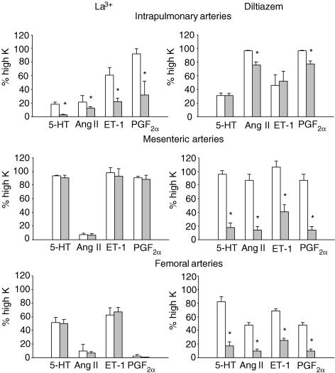
The release of intracellular Ca2+ stores triggers store-operated Ca2+ entry (SOCE) in PASMC as in many other types of cell and it would therefore be expected that hypoxia-induced Ca2+ release should activate SOCE. We have reported that contraction in rat intrapulmonary arteries (IPA) appears to be more sensitive to SOCE activated by store depletion than it is in systemic arteries of similar size (Snetkov et al. 2003). This unique property of IPA is also observed with regard to multiple agonists. Figure 1 demonstrates the effect of diltiazem (10 μm), which blocks L-type Ca2+ channels, and La3+ (10 μm), which is mainly selective for SOCE (Lin et al. 2004), on contractions to 5-HT, angiotensin II, endothelin-1, and PGF2α in IPA, mesenteric and femoral arteries of the rat. For each agonist, IPA are much more susceptible to block by La3+ and less sensitive to diltiazem than are both types of systemic arteries, indicating the general importance of voltage-independent mechanisms in the excitation–contraction coupling in IPA.
Figure 1. Contractions caused by a range of vasoconstrictors in rat intrapulmonary arteries (IPA) are more sensitive to block by La3+ (10 μM), but less sensitive to block by diltiazem (10 μM) compared with those evoked in third or fourth order arterial branches of two systemic arteries, the mesenteric (MA) and femoral (FA) arteries.
The amplitude of contraction is expressed as a percentage of that caused by depolarization with 80 mm K+ solution. Control responses are shown as open bars, and responses in the presence of La3+ and diltiazem are shown as grey bars. Asterisks indicate a significant (P < 0.05) effect of La3+ or diltiazem. Experiments with diltiazem were carried out using Krebs solution, whereas those with La3+ used a Hepes-buffered solution (Snetkov et al. 2003) to avoid La3+ precipitation. Note that Hepes itself often suppressed contraction to some extent. Concentrations for 5-HT (3–10 μm), endothelin 1 (0.1 μm) and PGF2α (30 μm) were chosen to give a response which was 80–90% of the maximum obtained for the agonist in each artery. A maximal angiotensin II concentration (0.3 μm) was used because the response to this agonist tended to be small and transient. Each bar represents 4–6 experiments using arteries from different rats. The resting tension for the each type of artery was set to correspond closely to its internal pressure in vivo, as described in Robertson et al. (2000_b_).
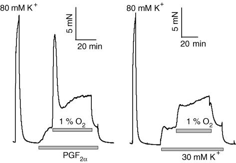
HPV in isolated IPA from rat and rabbit exhibits a biphasic profile, with an initial transient contraction (phase 1) followed by a slow progressive increase in force development (phase 2) (see Fig. 2). We reported that phase 1 of HPV was strongly suppressed by both the Ca2+ store-releasing agent CPA and the SOCE blocker La3+, and suggested that phase 1 HPV is mainly due to SOCE (Robertson et al. 2000_b_). Direct evidence for the involvement of SOCE in the immediate hypoxia-induced response to HPV has recently been presented by Ng et al. (2005) and Wang et al. (2005) in PASMC from the dog and rat, respectively. The latter study was particularly significant as it was soon followed by a companion report in which the effects of the agents (LaCl3, NiCl2 and SKF-96365) shown to inhibit hypoxia-induced SOCE in PASMC were tested on HPV in the isolated perfused rat lung (Weigand et al. 2005). It was found that concentrations of these agents sufficient to block SOCE in the cells also abolished HPV in the perfused lungs, yet had no effect on the increase in pulmonary artery pressure caused by depolarization. Together, these papers provide compelling evidence that SOCE, or a cation conductance with a similar pharmacological profile, plays an important role in HPV.
Figure 2. Hypoxic pulmonary vasoconstriction in rat IPA is influenced by the nature of the pretone agent used to enhance it.
When 30 mm K+ was used as a pretone-inducing agent, the response was essentially monophasic, whereas when PGF2α (10 μm) was used for this purpose, the response consisted of an initial transient (phase 1) contraction superimposed on a sustained contraction which continued to increase for many minutes (phase 2). The response to 80 mm K+ on the left of each trace represents a maximal contraction, and is included to show the relative magnitude of HPV.
Given the evidence that both voltage-dependent and -independent Ca2+-elevating pathways contribute to HPV, is it possible to ascribe a predominant role to either of these? Another important observation emerging from these reports was that HPV in perfused lung was abolished by antagonists of both pathways (Weigand et al. 2005), whereas the hypoxia-induced rise in [Ca2+]i in PASMC was only partially blocked by the L-type Ca2+ inhibitors nifedipine (Wang et al. 2005) and nisoldipine (Ng et al. 2005). Assuming that the SOCE blockers were acting selectively, the most straightforward interpretation of these findings would seem to be that SOCE causes both a direct influx of Ca2+ and a depolarization which promotes Ca2+ entry via L-type channels, and that both mechanisms are required to raise [Ca2+]i sufficiently to elicit HPV. However, this possibility requires further study, ideally utilizing measurements of [Ca2+]i in perfused lungs.
HPV, the endothelium and Ca2+ sensitization
The hypoxia-induced rise in [Ca2+]i in PASMC, particularly when recorded for more than a few minutes, has consistently been observed to be small, typically in the order of 100 nm or less (Salvaterra & Goldman, 1993; Cornfield et al. 1994; Archer et al. 1998; Liu et al. 2001; Ng et al. 2005; Wang et al. 2005). Measurements in permeabilized arteries (Kitazawa et al. 1989; Crichton et al. 1997) suggest that simply raising [Ca2+]i by this amount is unlikely to cause much contraction. In addition, during sustained HPV in IPA, force development continues to increase for tens of minutes, whereas [Ca2+]i quickly attains a constant level (Robertson et al. 1995, 2003). We therefore proposed that Ca2+ sensitization of the myofilaments contributes to HPV, especially when hypoxia is sustained (Robertson et al. 1995). In a collaborative study with Mark Evans and Michelle Dipp, we later demonstrated that Y-27632, an inhibitor of rho kinase, suppressed HPV in IPA and perfused lungs from the rat with IC50 values of ∼300 and ∼60 nm, respectively (Robertson et al. 2000_a_).
As demonstrated initially in pig pulmonary arteries by Holden & McCall (1984), HPV, especially if maintained, is generally (but not universally –Marshall & Marshall, 1992; Archer et al. 2004) found to be potentiated by, or to require the presence of, an intact endothelium (see review by Aaronson et al. 2002). Removal of the endothelium does not suppress the rise in [Ca2+]i during sustained HPV in rat IPA (Robertson et al. 2003). However, this procedure causes a suppression of contraction which is indistinguishable from that caused by Y-27632, suggesting that hypoxia may cause the release from the endothelium of a constricting factor which causes Ca2+ sensitization in the PASMC by stimulating rho kinase. Direct evidence for the existence of such a factor was provided by the study of Gaine et al. (1998), who demonstrated that loss of sensitivity to hypoxia in endothelium-denuded pig proximal PA rings was reversed by insertion into the preparation of a pulmonary valve leaflet, a source of endothelial cells. Robertson et al. (2001) subsequently showed that salt solution which had been used to perfuse isolated rat lungs under hypoxic but not normoxic conditions, contained one or more substances which could cause contraction of IPA with no concomitant rise in [Ca2+]i. Although an obvious candidate for such a substance is endothelin, combined blockade of ETA and ETB receptors did not suppress phase 2 HPV in rat IPA (Robertson et al. 2003), and HPV in salt solution-perfused lungs is also generally seen to be unaffected by endothelin receptor antagonists (Aaronson et al. 2002). Thus, it would seem that the identity of this putative endothelium-derived substance remains to be established.
However, endothelin does seem to be important for HPV, since a number of in vivo studies of HPV have found that it is suppressed by blockade of ETA receptors. The role of endothelin in HPV was clarified by the elegant study of Sato et al. (2000), who demonstrated that responsiveness to hypoxia, which was lost in perfused lung during combined ETA/ETB receptor blockade, was restored when angiotensin II was added to the perfusion solution. The authors suggested that endothelin was acting as a non-specific priming stimulus which was strongly magnifying the response to hypoxia, in this case by inhibiting KATP channels to cause depolarization. This concept was corroborated by Liu et al. (2001), who found that the loss of HPV in pig distal PA caused by endothelial denudation was prevented by treatment with a very low concentration of endothelin 1.
The effect of pretone on HPV
These studies serve to highlight the common finding that the response to hypoxia in isolated arteries and salt solution-perfused lungs is strongly enhanced by, or in some cases requires, a low-level preconstriction of the preparation by an agonist or some other stimulus (e.g. McMurtry, 1984; Rodman et al. 1989). This dependence of HPV on ‘pretone’ is perhaps not surprising if hypoxia is indeed associated with a rather small rise in [Ca2+]i in PASMC, but it does introduce a further complication into the debate about the mechanisms causing HPV, especially since the requirement for pretone is not always observed (Dipp et al. 2001; Evans & Dipp, 2002; Archer et al. 2004). This variability between results is in part probably species related, for example HPV in feline PA consistently does not require pretone (Harder et al. 1985; Marshall & Marshall, 1992), although in some cases divergent results have been obtained by different laboratories working in similar preparations from the same species (Hoshino et al. 1988; Ohe et al. 1992; Robertson et al. 2000_b_; Dipp et al. 2001). It is also possible that relatively small differences in the extent to which different workers stretch small pulmonary arteries in order to record isometric tension development might influence HPV as suggested by the work of Ozaki et al. (1998). It is regrettable that few laboratories utilize the method of Mulvany & Halpern (1977) to adjust the resting wall tension to a value close to that which would exist in vivo in the low-pressure pulmonary circulation. Instead arteries are often stretched to produce the maximum response to agonists, which is likely to result in artefactually high wall tensions and the possible activation of stretch-sensitive channels.
The nature of the pretone stimulus is capable of shaping HPV to some extent, as shown in Fig. 2. When PGF2α is used to provide pretone, HPV assumes the biphasic profile which we and others have previously described (similar results are obtained with U-46619 or sphingosylphosphorylcholine as pretone-inducing agents, but are not shown). Conversely, when a moderately elevated K+ concentration (∼30 mm) is used as the pretone-inducing agent, the initial transient phase HPV is smaller or non-existent, closely resembling that often observed in perfused lungs. Perhaps the initial rapid phase of HPV (phase 1) is due to agonist-induced loading of a Ca2+ store which is then released by hypoxia. However, it is noteworthy that the slow rise in force seems to occur independently of the type of pretone inducer used, and has also been observed in the absence of pretone both in precapillary resistance PA and IPA (Evans & Dipp, 2002; Archer et al. 2004).
The effect of pretone on HPV, and its use by many laboratories, raises important questions which go to the heart of ongoing controversies about the mechanisms underlying this unique response. Could the use of pretone cause fundamental changes in the nature of the response of pulmonary arteries to hypoxia, thereby biasing results? It is possible, for example, that pretone-inducing stimuli or cell isolation cause membrane depolarization which in effect substitutes for an inhibitory effect of hypoxia on KV channels, thus artefactually enhancing the apparent importance of non-voltage-dependent mechanisms. This does not seem likely in rat IPA, since HPV evoked in the absence of pretone was abolished by a combination of ryanodine and caffeine, which had no effect on depolarization-induced contraction (Evans & Dipp, 2002), but it could occur in other preparations. On the other hand, there is good reason to believe that pulmonary artery smooth muscle in vivo is somewhat depolarized and constricted by endothelin release (Sato et al. 2000) or some factor present in plasma (McMurtry, 1984), so that the properties of HPV in the presence of pretone may be more physiologically relevant than those in its absence. In either case, elucidation of the precise mechanisms by which pretone enables HPV is likely to provide new insights into this complex and fascinating response.
References
- Aaronson PI, Robertson TP, Ward JP. Endothelium-derived mediators and hypoxic pulmonary vasoconstriction. Respir Physiol Neurobiol. 2002;132:107–120. doi: 10.1016/s1569-9048(02)00053-8. [DOI] [PubMed] [Google Scholar]
- Archer S, Michelakis E. The mechanism(s) of hypoxic pulmonary vasoconstriction: potassium channels, redox O2 sensors, and controversies. News Physiol Sci. 2002;17:131–137. doi: 10.1152/nips.01388.2002. [DOI] [PubMed] [Google Scholar]
- Archer SL, Souil E, nh-Xuan AT, Schremmer B, Mercier JC, El YA, Nguyen-Huu L, Reeve HL, Hampl V. Molecular identification of the role of voltage-gated K+ channels, Kv1.5 and Kv2.1, in hypoxic pulmonary vasoconstriction and control of resting membrane potential in rat pulmonary artery myocytes. J Clin Invest. 1998;101:2319–2330. doi: 10.1172/JCI333. [DOI] [PMC free article] [PubMed] [Google Scholar]
- Archer SL, Wu XC, Thebaud B, Nsair A, Bonnet S, Tyrrell B, McMurtry MS, Hashimoto K, Harry G, Michelakis ED. Preferential expression and function of voltage-gated, O2-sensitive K+ channels in resistance pulmonary arteries explains regional heterogeneity in hypoxic pulmonary vasoconstriction: ionic diversity in smooth muscle cells. Circ Res. 2004;95:308–318. doi: 10.1161/01.RES.0000137173.42723.fb. [DOI] [PubMed] [Google Scholar]
- Burghuber OC. Nifedipine attenuates acute hypoxic pulmonary vasoconstriction in patients with chronic obstructive pulmonary disease. Respiration. 1987;52:86–93. doi: 10.1159/000195309. [DOI] [PubMed] [Google Scholar]
- Cornfield DN, Stevens T, McMurtry IF, Abman SH, Rodman DM. Acute hypoxia causes membrane depolarization and calcium influx in fetal pulmonary artery smooth muscle cells. Am J Physiol. 1994;266:L469–L475. doi: 10.1152/ajplung.1994.266.4.L469. [DOI] [PubMed] [Google Scholar]
- Crichton CA, Smith GC, Smith GL. α-Toxin-permeabilised rabbit fetal ductus arteriosus is more sensitive to Ca2+ than aorta or main pulmonary artery. Cardiovasc Res. 1997;33:223–229. doi: 10.1016/s0008-6363(96)00171-x. [DOI] [PubMed] [Google Scholar]
- Demiryurek AT, Wadsworth RM, Kane KA, Peacock AJ. The role of endothelium in hypoxic constriction of human pulmonary artery rings. Am Rev Respir Dis. 1993;147:283–290. doi: 10.1164/ajrccm/147.2.283. [DOI] [PubMed] [Google Scholar]
- Dipp M, Nye PC, Evans AM. Hypoxic release of calcium from the sarcoplasmic reticulum of pulmonary artery smooth muscle. Am J Physiol Lung Cell Mol Physiol. 2001;281:L318–L325. doi: 10.1152/ajplung.2001.281.2.L318. [DOI] [PubMed] [Google Scholar]
- Evans AM, Dipp M. Hypoxic pulmonary vasoconstriction: cyclic adenosine diphosphate-ribose, smooth muscle Ca2+ stores and the endothelium. Respir Physiol Neurobiol. 2002;132:3–15. doi: 10.1016/s1569-9048(02)00046-0. [DOI] [PubMed] [Google Scholar]
- Gaine SP, Hales MA, Flavahan NA. Hypoxic pulmonary endothelial cells release a diffusible contractile factor distinct from endothelin. Am J Physiol. 1998;274:L657–L664. doi: 10.1152/ajplung.1998.274.4.L657. [DOI] [PubMed] [Google Scholar]
- Harder DR, Madden JA, Dawson CA. A membrane electrical mechanism for hypoxic vasoconstriction of small pulmonary arteries from the cat. Chest. 1985;88:233–235. doi: 10.1378/chest.88.4_supplement.233s. [DOI] [PubMed] [Google Scholar]
- Holden WE, McCall E. Hypoxia-induced contractions of porcine pulmonary artery strips depend on intact endothelium. Exp Lung Res. 1984;7:101–112. doi: 10.3109/01902148409069671. [DOI] [PubMed] [Google Scholar]
- Hoshino Y, Obara H, Kusunoki M, Fujii Y, Iwai S. Hypoxic contractile response in isolated human pulmonary artery: role of calcium ion. J Appl Physiol. 1988;65:2468–2474. doi: 10.1152/jappl.1988.65.6.2468. [DOI] [PubMed] [Google Scholar]
- Jabr RI, Toland H, Gelband CH, Wang XX, Hume JR. Prominent role of intracellular Ca2+ release in hypoxic vasoconstriction of canine pulmonary artery. Br J Pharmacol. 1997;122:21–30. doi: 10.1038/sj.bjp.0701326. [DOI] [PMC free article] [PubMed] [Google Scholar]
- Kitazawa T, Kobayashi S, Horiuti K, Somlyo AV, Somlyo AP. Receptor-coupled, permeabilized smooth muscle. Role of the phosphatidylinositol cascade, G-proteins, and modulation of the contractile response to Ca2+ J Biol Chem. 1989;264:5339–5342. [PubMed] [Google Scholar]
- Lin MJ, Leung GP, Zhang WM, Yang XR, Yip KP, Tse CM, Sham JS. Chronic hypoxia-induced upregulation of store-operated and receptor-operated Ca2+ channels in pulmonary arterial smooth muscle cells: a novel mechanism of hypoxic pulmonary hypertension. Circ Res. 2004;95:496–505. doi: 10.1161/01.RES.0000138952.16382.ad. [DOI] [PubMed] [Google Scholar]
- Liu Q, Sham JS, Shimoda LA, Sylvester JT. Hypoxic constriction of porcine distal pulmonary arteries: endothelium and endothelin dependence. Am J Physiol Lung Cell Mol Physiol. 2001;280:L856–L865. doi: 10.1152/ajplung.2001.280.5.L856. [DOI] [PubMed] [Google Scholar]
- Marshall C, Marshall BE. Hypoxic pulmonary vasoconstriction is not endothelium dependent. Proc Soc Exp Biol Medical. 1992;201:267–270. doi: 10.3181/00379727-201-43506. [DOI] [PubMed] [Google Scholar]
- McMurtry IF. Angiotensin is not required for hypoxic constriction in salt solution-perfused rat lungs. J Appl Physiol. 1984;56:375–380. doi: 10.1152/jappl.1984.56.2.375. [DOI] [PubMed] [Google Scholar]
- McMurtry IF, Davidson AB, Reeves JT, Grover RF. Inhibition of hypoxic pulmonary vasoconstriction by calcium antagonists in isolated rat lungs. Circ Res. 1976;38:99–104. doi: 10.1161/01.res.38.2.99. [DOI] [PubMed] [Google Scholar]
- Mauban JR, Remillard CV, Yuan JX. Hypoxic pulmonary vasoconstriction: role of ion channels. J Appl Physiol. 2005;98:415–420. doi: 10.1152/japplphysiol.00732.2004. [DOI] [PubMed] [Google Scholar]
- Morio Y, McMurtry IF. Ca2+ release from ryanodine-sensitive store contributes to mechanism of hypoxic vasoconstriction in rat lungs. J Appl Physiol. 2002;92:527–534. doi: 10.1152/jappl.2002.92.2.527. [DOI] [PubMed] [Google Scholar]
- Moudgil R, Michelakis ED, Archer SL. Hypoxic pulmonary vasoconstriction. J Appl Physiol. 2005;98:390–403. doi: 10.1152/japplphysiol.00733.2004. [DOI] [PubMed] [Google Scholar]
- Mulvany MJ, Halpern W. Contractile properties of small arterial resistance vessels in spontaneously hypertensive and normotensive rats. Circ Res. 1977;41:19–26. doi: 10.1161/01.res.41.1.19. [DOI] [PubMed] [Google Scholar]
- Naeije R, Melot C, Mols P, Hallemans R. Effects of vasodilators on hypoxic pulmonary vasoconstriction in normal man. Chest. 1982;82:404–410. doi: 10.1378/chest.82.4.404. [DOI] [PubMed] [Google Scholar]
- Ng LC, Wilson SM, Hume JR. Mobilization of sarcoplasmic reticulum stores by hypoxia leads to consequent activation of capacitative Ca2+ entry in isolated canine pulmonary arterial smooth muscle cells. J Physiol. 2005;563:409–419. doi: 10.1113/jphysiol.2004.078311. [DOI] [PMC free article] [PubMed] [Google Scholar]
- Ohe M, Ogata M, Katayose D, Takishima T. Hypoxic contraction of pre-stretched human pulmonary artery. Respir Physiol. 1992;87:105–114. doi: 10.1016/0034-5687(92)90103-4. [DOI] [PubMed] [Google Scholar]
- Ozaki M, Marshall C, Amaki Y, Marshall BE. Role of wall tension in hypoxic responses of isolated rat pulmonary arteries. Am J Physiol. 1998;275:L1069–L1077. doi: 10.1152/ajplung.1998.275.6.L1069. [DOI] [PubMed] [Google Scholar]
- Robertson TP, Aaronson PI, Ward JP. Hypoxic vasoconstriction and intracellular Ca2+ in pulmonary arteries: evidence for PKC-independent Ca2+ sensitization. Am J Physiol. 1995;268:H301–H307. doi: 10.1152/ajpheart.1995.268.1.H301. [DOI] [PubMed] [Google Scholar]
- Robertson TP, Aaronson PI, Ward JP. Ca2+ sensitization during sustained hypoxic pulmonary vasoconstriction is endothelium dependent. Am J Physiol Lung Cell Mol Physiol. 2003;284:L1121–L1126. doi: 10.1152/ajplung.00422.2002. [DOI] [PubMed] [Google Scholar]
- Robertson TP, Dipp M, Ward JPT, Aaronson PI, Evans MA. Inhibition of sustained vasoconstriction by Y-27632 in isolated intrapulmonary arteries and perfused lung of the rat. Br J Pharmacol. 2000a;131:5–9. doi: 10.1038/sj.bjp.0703537. [DOI] [PMC free article] [PubMed] [Google Scholar]
- Robertson TP, Hague D, Aaronson PI, Ward JP. Voltage-independent calcium entry in hypoxic pulmonary vasoconstriction of intrapulmonary arteries of the rat. J Physiol. 2000b;525:669–680. doi: 10.1111/j.1469-7793.2000.t01-1-00669.x. [DOI] [PMC free article] [PubMed] [Google Scholar]
- Robertson TP, Ward JP, Aaronson PI. Hypoxia induces the release of a pulmonary-selective, Ca2+-sensitising, vasoconstrictor from the perfused rat lung. Cardiovasc Res. 2001;50:145–150. doi: 10.1016/s0008-6363(01)00192-4. [DOI] [PubMed] [Google Scholar]
- Rodman DM, Yamaguchi T, O'Brien RF, McMurtry IF. Hypoxic contraction of isolated rat pulmonary artery. J Pharmacol Exp Ther. 1989;248:952–959. [PubMed] [Google Scholar]
- Salvaterra CG, Goldman WF. Acute hypoxia increases cytosolic calcium in cultured pulmonary arterial myocytes. Am J Physiol. 1993;264:L323–L328. doi: 10.1152/ajplung.1993.264.3.L323. [DOI] [PubMed] [Google Scholar]
- Sato K, Morio Y, Morris KG, Rodman DM, McMurtry IF. Mechanism of hypoxic pulmonary vasoconstriction involves ETA receptor-mediated inhibition of KATP channel. Am J Physiol Lung Cell Mol Physiol. 2000;278:L434–L442. doi: 10.1152/ajplung.2000.278.3.L434. [DOI] [PubMed] [Google Scholar]
- Savineau JP, De Gonzalez La FP, Marthan R. Cellular mechanisms of hypoxia-induced contraction in human and rat pulmonary arteries. Respir Physiol. 1995;99:191–198. doi: 10.1016/0034-5687(94)00091-d. [DOI] [PubMed] [Google Scholar]
- Snetkov VA, Aaronson PI, Ward JP, Knock GA, Robertson TP. Capacitative calcium entry as a pulmonary specific vasoconstrictor mechanism in small muscular arteries of the rat. Br J Pharmacol. 2003;140:97–106. doi: 10.1038/sj.bjp.0705408. [DOI] [PMC free article] [PubMed] [Google Scholar]
- Von Euler US, Liljestrand G. Observations on the pulmonary arterial blood pressure in the cat. Acta Physiol Scand. 1946;12:301–320. [Google Scholar]
- Wang J, Shimoda LA, Weigand L, Wang W, Sun D, Sylvester JT. Acute hypoxia increases intracellular [Ca2+] in pulmonary arterial smooth muscle by enhancing capacitative Ca2+ entry. Am J Physiol Lung Cell Mol Physiol. 2005;288:L1059–L1069. doi: 10.1152/ajplung.00448.2004. [DOI] [PubMed] [Google Scholar]
- Ward JP, Aaronson PI. Mechanisms of hypoxic pulmonary vasoconstriction: Can anyone be right? Respir Physiol. 1999;115:261–271. doi: 10.1016/s0034-5687(99)00025-0. [DOI] [PubMed] [Google Scholar]
- Ward JP, Robertson TP, Aaronson PI. Capacitative calcium entry: a central role in hypoxic pulmonary vasoconstriction? Am J Physiol Lung Cell Mol Physiol. 2005;289:L2–L4. doi: 10.1152/ajplung.00101.2005. [DOI] [PubMed] [Google Scholar]
- Waypa GB, Schumacker PT. Hypoxic pulmonary vasoconstriction: redox events in oxygen sensing. J Appl Physiol. 2005;98:404–414. doi: 10.1152/japplphysiol.00722.2004. [DOI] [PubMed] [Google Scholar]
- Weigand L, Foxson J, Wang J, Shimoda LA, Sylvester JT. Inhibition of hypoxic pulmonary vasoconstriction by antagonists of store-operated Ca2+ and nonselective cation channels. Am J Physiol Lung Cell Mol Physiol. 2005;289:L5–L13. doi: 10.1152/ajplung.00044.2005. [DOI] [PubMed] [Google Scholar]
- Weir EK, Archer SL. The mechanism of acute hypoxic pulmonary vasoconstriction: the tale of two channels. FASEB J. 1995;9:183–189. doi: 10.1096/fasebj.9.2.7781921. [DOI] [PubMed] [Google Scholar]
- Wilson HL, Dipp M, Thomas JM, Lad C, Galione A, Evans AM. Adp-ribosyl cyclase and cyclic ADP-ribose hydrolase act as a redox sensor. a primary role for cyclic ADP-ribose in hypoxic pulmonary vasoconstriction. J Biol Chem. 2001;276:11180–11188. doi: 10.1074/jbc.M004849200. [DOI] [PubMed] [Google Scholar]

