Effects of eight growth factors on the differentiation of cells derived from human embryonic stem cells (original) (raw)
Abstract
Human embryonic stem (ES) cells are pluripotent cells derived from the inner cell mass of in vitro fertilized human blastocysts. We examined the potential of eight growth factors [basic fibroblast growth factor (bFGF), transforming growth factor β1 (TGF-β1), activin-A, bone morphogenic protein 4 (BMP-4), hepatocyte growth factor (HGF), epidermal growth factor (EGF), β nerve growth factor (βNGF), and retinoic acid] to direct the differentiation of human ES-derived cells in vitro. We show that human ES cells that have initiated development as aggregates (embryoid bodies) express a receptor for each of these factors, and that their effects are evident by differentiation into cells with different epithelial or mesenchymal morphologies. Differentiation of the cells was assayed by expression of 24 cell-specific molecular markers that cover all embryonic germ layers and 11 different tissues. Each growth factor has a unique effect that may result from directed differentiation and/or cell selection, and we can divide the overall effects of the factors into three categories: growth factors (Activin-A and TGFβ1) that mainly induce mesodermal cells; factors (retinoic acid, EGF, BMP-4, and bFGF) that activate ectodermal and mesodermal markers; and factors (NGF and HGF) that allow differentiation into the three embryonic germ layers, including endoderm. None of the growth factors directs differentiation exclusively to one cell type. This analysis sets the stage for directing differentiation of human ES cells in culture and indicates that multiple human cell types may be enriched in vitro by specific factors.
Human embryonic stem (ES) cells were recently derived from human blastocysts and shown to form differentiated embryonic tumors (teratomas) when injected into muscles or testis of severe combined immunodeficiency (SCID) mice (1, 2). In addition, human ES cells spontaneously form embryoid bodies (EBs) composed of three embryonic germ layers, on their in vitro aggregation (3). Similarly, human embryonic germ cell lines were established from fetal primordial germ cells, and they were also shown to spontaneously differentiate in vitro (4).
ES cells are unique in their ability to grow indefinitely in culture while retaining a normal karyotype. Consequently, human ES cells may be useful as a source of cells for transplantation in numerous pathologies, and as a component in biomedical engineering. In addition, the study of human ES cells may enlighten us on early stages of human development. To investigate these potentials, we have begun to explore the regulation and manipulation of differentiation of human ES cells in culture.
In the past years, several soluble factors were shown to direct differentiation of mouse ES cells; e.g., IL-3 directs cells to become macrophages, mast cells or neutrophils (5); IL-6 directs cells to the erythroid lineage (6); retinoic acid induces neuron formation (7, 8); and transforming growth factor (TGF)-β1 induces myogenesis (7, 9). In several studies, differentiation was induced in the ES cells by culturing them on different feeder layers (10, 11). However, in most experiments, the growth factors and inducing agents are not directly applied to ES cells, but rather to aggregates of ES cells or simple embryoid bodies. These simple EBs form after leukemia inhibitory factor (LIF) is removed and the cells are cultured for 2–5 days, during which time they form intercellular contacts and may initiate signaling and spontaneous differentiation. For example, the differentiation of neuronal and muscle cells was achieved by exposing cells from simple EBs to retinoic acid (7, 8) and TGF-β (7, 9), respectively.
None of the examined factors exclusively directs differentiation to only one cell type, but rather alters the relative proportions of a specific cell type. An alternative strategy to obtain a uniform population of differentiated cells is the selection or sorting of specific cells from within a heterogeneous population (12–14).
The differentiation of human ES cells into embryoid bodies or teratomas is spontaneous and uncontrolled; i.e., the experimenter cannot determine which cell types will form in vivo or_in vitro_ (1–3). As a first step toward achieving the directed differentiation of human ES cells, we analyzed the effects of eight different growth factors on embryonic cells in culture. By using assays for 24 cell-specific molecular markers, we were able to point out the effects of the examined growth factors on the differentiation status of 11 tissues derived from human ES cells.
Materials and Methods
Cell Culture.
Human ES cells [H9 clone (1)] were grown on mouse embryo fibroblasts in 80% KnockOut DMEM, an optimized Dulbecco's modified Eagle's medium for ES cells (GIBCO/BRL), 20% KnockOut SR, a serum-free formulation (GIBCO/BRL), 1 mM glutamine (GIBCO/BRL), 0.1 mM β-mercaptoethanol (Sigma), 1% nonessential amino acids stock (GIBCO/BRL), 4 ng/ml basic fibroblast growth factor (bFGF) (GIBCO/BRL), and 103 units/ml LIF (GIBCO/BRL). LIF helps retain the hES cells in an undifferentiated state. We used 0.1% gelatin (Merck) to cover the tissue culture plates. To induce formation of EBs, ES cells were transferred by using 0.1%/1 mM trypsin/EDTA (GIBCO/BRL) to plastic Petri dishes to allow their aggregation and prevent adherence to the plate. Human EBs were grown in the same culture medium, except that it lacked LIF and bFGF. The EBs were cultured for 5 days and then dissociated with trypsin and plated on tissue culture plates coated with 50 μg/ml Fibronectin (Boehringer Mannheim). In each experiment, about 107 ES cells were transferred to a 100-mm Petri dish to allow creation of EBs and then plated again after 5 days on a 100-mm tissue cultute plate as monolayer. The cells were grown in the presence of the following human recombinant growth factors: bFGF (9), 10 ng/ml (GIBCO/BRL); TGF-β1 (9), 2 ng/ml (R&D Systems); activin-A (15), 20 ng/ml (R&D Systems); bone morphogenic protein 4 (BMP-4) (15, 16), 10 ng/ml (R&D Systems); hepatocyte growth factor (HGF)/scatter factor (17), 20 ng/ml (R&D Systems); epidermal growth factor (EGF) (17), 100 ng/ml (R&D Systems); β nerve growth factor (NGF) (18), 100 ng/ml (R&D Systems); and retinoic acid (RA) (8, 19), 1 μM (Sigma). Under these conditions, the differentiated embryonic (DE) cells were grown for another 10 days. All of the examined growth factors are absent from the commercially available KnockOut serum replacement in which the embryonic cells were cultured.
Reverse Transcription (RT)-PCR Analysis.
Total RNA was extracted by using Atlas Pure Total RNA Labeling Kit (CLONTECH). cDNA was synthesized from 1 μg total RNA, by using Advantage RT-for-PCR Kit (CLONTECH). cDNA samples were subjected to PCR amplification with DNA primers selective for the human genes. For each gene, the DNA primers were derived from different exons to ensure that the PCR product represents the specific mRNA species and not genomic DNA. PCR was performed by using the CLONTECH AdvanTaq Plus RT-PCR kit and by using a two-step cycle at 68°C.
Primers were synthesized for the following human genes: neurofilament heavy chain (NFH), phosphoprotein enriched in astrocytes (PEA-15), ultra high sulfur keratin (keratin), dopamine β hydroxylase (DβH), follicular stimulating hormone (FSH), enolase_,_ myosin light polypeptide 2 (myosin), cartilage matrix protein (CMP),renin, kallikrein, wilms tumor 1 (WT1), β-globin,δ-globin, cardiac actin (cACT), albumin, _α_1 anti-trypsin (α1AT),lipase, amylase, PDX-1,insulin, glucagon, surfactant, parathyroid hormone (PTH),_α-_feto protein (αFP), retinoic acid receptor type α (RARα), hepatocyte growth factor receptor (c-MET), nerve growth factor receptor (NGFR), fibroblast growth factor receptor type I (FGFRI), transforming growth factor receptor type II (TGFRII), epidermal growth factor receptor (EGFR), activin receptor type IIB (ACTRIIB), bone morphogenic protein type II (BMPRII), octamer binding protein 4 (OCT4), glyceraldehyde 3-phosphate dehydrogenase (G3PD), and β-actin.
A full description of the primers and their expected products appears in our web site:http://www.ls.huji.ac.il/∼nissimb/factors_es/primers.html.
Results
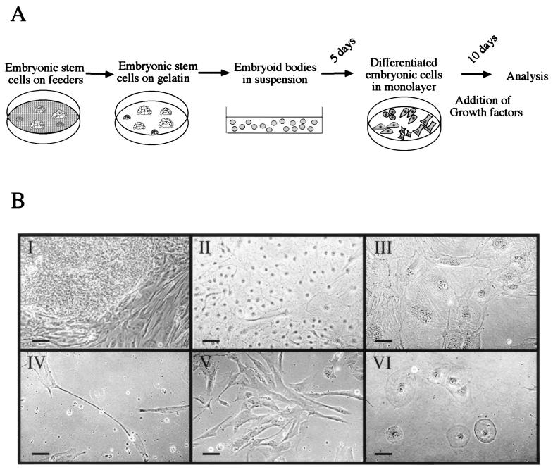
To direct the differentiation of human ES cells in vitro, we examined the effects of various growth factors on simple EBs derived from the human ES cells. For this purpose, we established a protocol in which human ES cells are initially grown on a feeder layer of mouse embryonic fibroblasts (MEF) and then transferred to gelatin-coated plates and cultured further to reduce the number of murine cells in the culture. Differentiation into EBs was initiated by transfer to Petri dishes. After 5 days, the EBs were dissociated, and cells were cultured as a monolayer forming DE cells. This protocol allows for initial differentiation of the human ES cells as aggregates and further differentiation as a monolayer wherein cells can be exposed to exogenous growth factors. A similar protocol is commonly used to direct differentiation of mouse ES cells into specific cell types, such as neuronal or muscle cells (7–9). In this case, the human DE cells were cultured for 10 days in the presence of 8 different growth factors: NGF, bFGF, activin-A, TGF-β1, HGF, EGF, BMP4, and RA (Fig.1A).
Figure 1.
Induced differentiation of human ES-derived cells in culture. (A) A schematic representation of the differentiation protocol (see Materials and Methods). (B) Various morphologies of ES cells before and after induced differentiation: I, a colony of ES cells on feeders; II–VI, differentiated cells cultured in the presence of HGF, activin-A, RA, bFGF, or BMP-4, respectively. Note the small size of ES cells in the colony in comparison with the size of the differentiated cells. Scale bar = 100 μm.
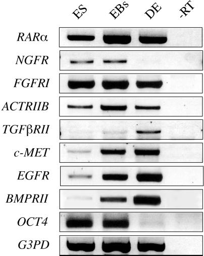
We first tested for the presence of growth factor receptors at the stage when growth factors were to be added to the culture (5-day-old EBs). RNA from human ES cells, 5-day-old EBs, and 10-day-old DE cells was isolated and analyzed by RT-PCR by using primers specific to_RARα_, NGFR, FGFRI,ACTRIIB, TGFRII, c-MET,EGFR, and BMPRII. As control, we have also examined the expression of OCT-4, a marker of the undifferentiated cells, and of G3PD, a housekeeping gene. All eight examined receptors are expressed in 5-day-old EBs, and most are expressed in ES and DE cells (Fig.2). Notably, four of the receptors (TGFβRII, c-MET, EGFR, and BMPRII) are expressed at very low levels in human ES cells. The low levels of expression may result from partial differentiation in the ES culture as the expression of these receptors increases after culture and further differentiation.
Figure 2.
Expression of receptors for various growth factors in human embryonic cells. RNA samples from ES cells, 5-day-old EBs, and 10-day-old DE cells were analyzed by RT-PCR for expression of specific receptors. As a control, RNA of 5-day-old EBs was analyzed without the prior generation of cDNA (−RT). For abbreviations of receptors used, see_Materials and Methods_.
Because growth factor receptors are expressed in 5-day-old EBs, we could examine the effects of the corresponding ligands on differentiation. After several days of culture on plastic and continuous exposure to growth factors, the cells acquired different morphologies (Fig. 1B). Without growth factors, cells spontaneously differentiated into many different types of colonies, whereas the addition of growth factors produced more mature cell morphologies, such as syncitial myocytes and neuronal cells. Compared with control DE cells, the growth factor-treated cultures were more homogenous, and up to half of the culture contained one or two cell types. For example, large populations of small cells with pronounced nuclei are found in cultures treated with HGF (Fig. 1B, I), muscle-like syncitiums in the activin-A-treated cells (Fig.1B, III), neuronal-like cells in the RA-treated culture (Fig. 1B, IV), fibroblast-like cells in the cultures treated with bFGF (Fig. 1B, V), and large round cells in cultures treated with BMP-4 (Fig. 1B, VI). These varied cell morphologies suggest that specific programs are initiated as a result of growth factor treatment.
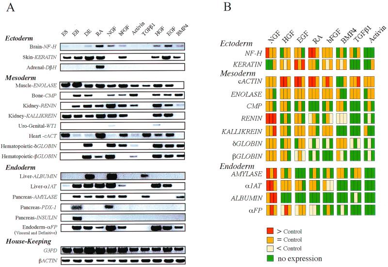
The differentiation induced by growth factors was further examined by determining the expression of a panel of 24 cell-specific genes by using RT-PCR. RNA was isolated from ES cells, 20-day-old EBs, 10-day-old DE cells, and DE cells that had been treated with either RA, NGF, bFGF, activin-A, TGF-β1, HGF, EGF, and BMP4. cDNA was generated from the various samples, and transcripts were amplified by specific primers to these genes. For each gene, the two DNA primers were derived from different exons to ensure that the PCR product represents the specific mRNA species and not genomic DNA. In addition, the correct product of amplification from the primers was ascertained by amplifying cDNAs from a variety of human adult or embryonic tissues (CLONTECH, MTC Panels) (results not shown). Of the 24 cell-specific markers, 16 were expressed in the treated DE cells. Most transcripts were also expressed in the control DE cells (see Fig.3A), indicating that many different cell types develop from ES cells without a requirement of exogenous growth factors. We also note that human ES cell cultures express some cell-specific genes, indicating that there is partial spontaneous differentiation in the cultures.
Figure 3.
(A) Analysis of expression of cell-specific genes in human ES cells treated with various growth factors. RNA from ES, 20-day-old EBs, and DE cells treated with the different growth factors was analyzed by RT-PCR for expression of seventeen cell-specific genes and two housekeeping genes. The genes were categorized by their embryonic germ layer (24). [Note that cartilage may derive from the ectodermal neural crest as well as from the mesoderm (24)]. For abbreviations of the various gene primers used, see Materials and Methods. (B) Schematic representation of the growth factor effects on gene expression. Results are shown from three experiments on the effects of NGF, HGF, EGF, RA, bFGF, BMP4, TGF-β1, or activin-A on the expression of 13 genes. The results are color-coded: orange represents similar expression to the control (no growth factor), red represents induction of at least 2-fold, yellow represents inhibition of 2-fold or more, and green represent no detectable expression.
Several conclusions can be drawn comparing the expression pattern of cell-specific markers in the different treatments to that of the control DE cells. Some markers are expressed only in the presence of a specific growth factor. For example, the adrenal marker_DβH_ is expressed only when the cells are treated with retinoic acid, and the uro-genital marker WT-1 is expressed when either NGF or HGF are added (Fig. 3A). In contrast, muscle-specific enolase is expressed under all conditions except after BMP-4 exposure (Fig. 3A). Some growth factors, e.g., NGF, induce expression of a variety of transcripts indicative of cells from all three germ layers whereas other factors, e.g., TGF-β1, lead to the production of a relatively reduced set of specific transcripts. Such an effect may result from direct repression of differentiation or from selection against specific cell types. Such potential selection is especially relevant to TGF-β, which was shown to inhibit epithelial proliferation (20).
Each of the growth factor induction experiments was repeated at least three times. RT-PCR analysis was performed, and a schematic representation of the results is shown in Fig. 3B. Induction or repression of gene expression is color-coded, and the growth factors were ordered based on their overall induction or repression of cell-specific expression. Thus, on the left is the factor (NGF) that induces the greatest variety of cell-specific gene expression of the factors tested, and on the right are the factors (BMP-4, activin-A, and TGF-β1) that mostly repress or fail to direct differentiation of as many cell types.
Discussion
ES cells are pluripotent cells capable of differentiating into many cell types. The recent isolation of human ES cells has attracted considerable attention because of the promise of using these cells or their derivatives for research in developmental biology and medical applications, including cellular transplantation. One obvious goal is to manipulate the differentiation of human ES cells so that a uniform population of precursors or fully differentiated cells can be obtained_in vivo_ or in vitro. Previous work with murine ES cells showed that several types of cells can be enriched in culture either by the addition of growth factors (5–9, 21, 22) or by introduction of transcription factors (23). Whereas conditions that direct differentiation of mouse ES cells into neuronal, hematopoietic, or cardiac cells have been published (5–9), a broad analysis of the effects of various growth factors on differentiation into multiple cell types has not been performed.
It has already been shown that the human ES cells have the capacity to differentiate into derivatives of all three germ layers either in vivo or in vitro (3), but such differentiation is spontaneous and unregulated. Our purpose here was to make an initial and broad scan for the effects of various growth factors and to assess their ability to regulate human ES cell differentiation. The protocol devised for inducing human ES differentiation consisted of an aggregation step that allows complex signaling to occur between the cells (somewhat resembling the gastrulation process) and a dissociation step, after 5 days as EBs, thereby allowing exposure to exogenous growth factors. At the stage of 5-day-old EBs, at least some of the cells are not terminally differentiated, as is evident from the expression of OCT-4, a marker for undifferentiated ES cells. To determine whether the growth factors we chose to employ could affect the human ES cells, we first tested for the expression of the their respective receptors. As one might expect for pluripotent stem cells, they evidently express a wide range of receptors for growth factors.
The ability to induce specific differentiation was initially evident as morphological changes in the DE cell cultures. After 10 days incubation with a growth factor, DE cells were more homogenous and displayed a larger proportion of specific cell types, as compared with controls not treated with growth factors. To assess their differentiation by gene expression, we analyzed the transcription of 24 tissue-specific markers from all three germ layers and sublayers. Our results show that 16 of the 24 examined genes are expressed in the DE cells, representing 11 different tissues (Fig. 3). These data allow us to specify factors that can direct differentiation into specific cell types. For example, both NGF and RA induce differentiation into neuronal cells whereas only RA allowed expression of adrenal markers, and only NGF induced expression of endodermal markers. The effects of the growth factors on the overall representation of the different cells may result from their direct differentiation or from cell selection either by promoting proliferation or by inducing apoptosis of specific cell types.
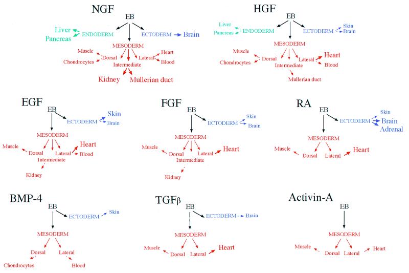
An interpretation of lineage-specific differentiation induced by the various growth factors is summarized in Fig.4. Clustering the growth factors based on their effects on differentiation reveals three groups. The first group (TGF-β1 and activin-A) appear to inhibit endodermal and ectodermal cells, but allow differentiation into mesodermal (muscle) cells. The second group includes factors that allow or induce differentiation into ectoderm as well as mesodermal cells (RA, bFGF, BMP-4, and EGF), whereas the third group (NGF and HGF) allows differentiation into all three embryonic lineages including endoderm.
Figure 4.
An interpretation of the effects of various growth factors on in vitro differentiation of human ES-derived cells. Thick arrows and large fonts indicate an induction compared with the control (no growth factor) for genes specific to the tissue indicated. Dashed arrows represent a decrease in expression. Disappearance of the cell-specific transcripts is symbolized by elimination of the corresponding tissue symbol.
Comparing spontaneous with growth factor-induced differentiation suggests that most of the growth factors inhibit differentiation of specific cell types and that this inhibitory effect is more pronounced than an induction effect. Such an inhibitory effect suggests that specific differentiation might also be achieved by using growth factor inhibitors, such as follistatin or noggin, to act on any endogenous growth factors that might be produced during differentiation. The work presented here shows that none of the eight growth factors tested directs a completely uniform and singular differentiation of cells. At the same time, it can be concluded that certain classes of growth factors are useful in achieving differentiation of specific germ layers. These results represent an initial step toward achieving fully directed cell differentiation and open the way to combining growth factor incubation with selection methods.
Acknowledgments
We thank Olga Martinez for excellent technical assistance. The study was partially supported by funds from the Herbert Cohn Chair (N.B), by the Howard Hughes Medical Institute (D.A.M), and by the Juvenile Diabetes Foundation (N.B. and D.A.M)
Abbreviations
ES
embryonic stem
EB
embryoid body
TGF
transforming growth factor
LIF
leukemia inhibitory factor
bFGF
basic fibroblast growth factor
BMP
bone morphogenic protein
HGF
hepatocyte growth factor
EGF
epidermal growth factor
βNGF
β nerve growth factor
RA
retinoic acid
DE
differentiated embryonic
RT-PCR
reverse transcription PCR
References
- 1.Thomson J A, Itskovitz-Eldor J, Shapiro S S, Waknitz M A, Swiergiel J J, Marshall V S, Jones J M. Science. 1998;282:1145–1147. doi: 10.1126/science.282.5391.1145. [DOI] [PubMed] [Google Scholar]
- 2.Reubinoff B E, Pera M F, Fong C Y, Trounson A, Bongso A. Nat Biotechnol. 2000;18:399–404. doi: 10.1038/74447. [DOI] [PubMed] [Google Scholar]
- 3.Itskovitz-Eldor J, Schuldiner M, Karsenti D, Eden A, Yanuka O, Amit M, Soreq H, Benvenisty N. Mol Med. 2000;6:88–95. [PMC free article] [PubMed] [Google Scholar]
- 4.Shamblott M J, Axelman J, Wang S, Bugg E M, Littlefield J W, Donovan P J, Blumenthal P D, Huggins G R, Gearhart J D. Proc Natl Acad Sci USA. 1998;95:13726–13731. doi: 10.1073/pnas.95.23.13726. [DOI] [PMC free article] [PubMed] [Google Scholar]
- 5.Wiles M V, Keller G. Development. 1991;111:259–267. doi: 10.1242/dev.111.2.259. [DOI] [PubMed] [Google Scholar]
- 6.Biesecker L G, Emerson S G. Exp Hematol. 1993;21:774–778. [PubMed] [Google Scholar]
- 7.Slager H G, Van Inzen W, Freund E, Van den Eijnden-Van Raaij A J, Mummery C L. Dev Genet. 1993;14:212–224. doi: 10.1002/dvg.1020140308. [DOI] [PubMed] [Google Scholar]
- 8.Bain G, Kitchens D, Yao M, Huettner J E, Gottlieb D I. Dev Biol. 1995;168:342–357. doi: 10.1006/dbio.1995.1085. [DOI] [PubMed] [Google Scholar]
- 9.Rohwedel J, Maltsev V, Bober E, Arnold H H, Hescheler J, Wobus A M. Dev Biol. 1994;164:87–101. doi: 10.1006/dbio.1994.1182. [DOI] [PubMed] [Google Scholar]
- 10.Nakano T, Kodama H, Honjo T. Science. 1994;265:1098–1101. doi: 10.1126/science.8066449. [DOI] [PubMed] [Google Scholar]
- 11.Palacios R, Golunski E, Samaridis J. Proc Natl Acad Sci USA. 1995;92:7530–7534. doi: 10.1073/pnas.92.16.7530. [DOI] [PMC free article] [PubMed] [Google Scholar]
- 12.Klug M G, Soonpaa M H, Koh G Y, Field L J. J Clin Invest. 1996;98:216–224. doi: 10.1172/JCI118769. [DOI] [PMC free article] [PubMed] [Google Scholar]
- 13.Kolossov E, Fleischmann B K, Liu Q, Bloch W, Viatchenko-Karpinski S, Manzke O, Ji G J, Bohlen H, Addicks K, Hescheler J. J Cell Biol. 1998;143:2045–2056. doi: 10.1083/jcb.143.7.2045. [DOI] [PMC free article] [PubMed] [Google Scholar]
- 14.Li M, Pevny L, Lovell-Badge R, Smith A. Curr Biol. 1998;8:971–974. doi: 10.1016/s0960-9822(98)70399-9. [DOI] [PubMed] [Google Scholar]
- 15.Hollnagel A, Oehlmann V, Heymer J, Ruther U, Nordheim A. J Biol Chem. 1999;274:19838–19345. doi: 10.1074/jbc.274.28.19838. [DOI] [PubMed] [Google Scholar]
- 16.Wiles M V, Johansson B M. Exp Cell Res. 1999;247:241–248. doi: 10.1006/excr.1998.4353. [DOI] [PubMed] [Google Scholar]
- 17.Rubin J S, Chan A M, Bottaro D P, Burgess W H, Taylor W G, Cech A C, Hirschfield D W, Wong J, Miki T, Finch P W, et al. Proc Natl Acad Sci USA. 1991;88:415–419. doi: 10.1073/pnas.88.2.415. [DOI] [PMC free article] [PubMed] [Google Scholar]
- 18.Wobus A M, Grosse R, Schoneich J. Biomed Biochim Acta. 1988;47:965–973. [PubMed] [Google Scholar]
- 19.Bain G, Ray W J, Yao M, Gottlieb D I. Biochem Biophys Res Commun. 1996;223:691–694. doi: 10.1006/bbrc.1996.0957. [DOI] [PubMed] [Google Scholar]
- 20.Moses H L, Yang E Y, Pietenpol J A. Cell. 1990;63:245–247. doi: 10.1016/0092-8674(90)90155-8. [DOI] [PubMed] [Google Scholar]
- 21.Brustle O, Jones K N, Learish R D, Karram K, Choudhary K, Wiestler O D, Duncan I D, McKay R D. Science. 1999;285:754–756. doi: 10.1126/science.285.5428.754. [DOI] [PubMed] [Google Scholar]
- 22.Gutierrez-Ramos J C, Palacios R. Proc Natl Acad Sci USA. 1992;89:9171–9175. doi: 10.1073/pnas.89.19.9171. [DOI] [PMC free article] [PubMed] [Google Scholar]
- 23.Levinson-Dushnik M, Benvenisty N. Mol Cell Biol. 1997;17:3817–3822. doi: 10.1128/mcb.17.7.3817. [DOI] [PMC free article] [PubMed] [Google Scholar]
- 24.Gilbert S F. Developmental Biology. Sunderland, MA: Sinauer; 1997. p. 342. [Google Scholar]


