The discovery of nitric oxide and its role in vascular biology (original) (raw)
Abstract
Nitric oxide (NO) is a relative newcomer to pharmacology, as the paper which initiated the field was published only 25 years ago. Nevertheless its impact is such that to date more than 31,000 papers have been published with NO in the title and more than 65,000 refer to it in some way. The identification of NO with endothelium-derived relaxing factor and the discovery of its synthesis from L-arginine led to the realisation that the L-arginine: NO pathway is widespread and plays a variety of physiological roles. These include the maintenance of vascular tone, neurotransmitter function in both the central and peripheral nervous systems, and mediation of cellular defence. In addition, NO interacts with mitochondrial systems to regulate cell respiration and to augment the generation of reactive oxygen species, thus triggering mechanisms of cell survival or death. This review will focus on the role of NO in the cardiovascular system where, in addition to maintaining a vasodilator tone, it inhibits platelet aggregation and adhesion and modulates smooth muscle cell proliferation. NO has been implicated in a number of cardiovascular diseases and virtually every risk factor for these appears to be associated with a reduction in endothelial generation of NO. Reduced basal NO synthesis or action leads to vasoconstriction, elevated blood pressure and thrombus formation. By contrast, overproduction of NO leads to vasodilatation, hypotension, vascular leakage, and disruption of cell metabolism. Appropriate pharmacological or molecular biological manipulation of the generation of NO will doubtless prove beneficial in such conditions.
Keywords: L-arginine, blood pressure, endothelial dysfunction, free radicals, mitochondria, nitric oxide
Early work
Furchgott & Zawadzki (1980) first described endothelium-dependent relaxation, a phenomenon whereby acetylcholine relaxes isolated preparations of blood vessels only if the vascular endothelium lining the vessels is present and intact. Subsequent studies revealed that acetylcholine and other agents (including bradykinin, histamine and 5-hydroxytryptamine) release a transferable factor (endothelium-derived relaxing factor, EDRF) which is unstable, acts via stimulation of the soluble guanylate cyclase and is inhibited by haemoglobin and methylene blue (Furchgott et al., 1984).
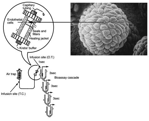
We at the Wellcome Research Laboratories in Beckenham became interested in this ephemeral substance in early 1985. Previous experience of our group in the identification of thromboxane synthase and the discovery of prostacyclin (see Moncada, 2005) convinced us that we should use bioassay experiments in cascades in order to carry out more detailed quantitative pharmacology and try to elucidate the structure of EDRF. We therefore decided not to compare the responses of vascular strips with and without endothelium; instead we proceeded to culture porcine aortic endothelial cells on microcarrier beads, perfused them inside a modified chromatography column and used the effluent to superfuse vascular tissues denuded of endothelium as a detection system for EDRF (Figure 1).
Figure 1.
Bioassay system used to detect the release of EDRF from endothelial cells. Porcine aortic endothelial cells were grown in culture on microcarrier beads (approximately 70 _μ_m), which were packed into a modified chromatographic column. Inset shows an electron micrograph of a bead covered in endothelial cells. The beads were perfused with Krebs buffer at 37°C and the perfusate was allowed to flow over the bioassay tissues (four rabbit aortae denuded of endothelium). The time taken for the superfusate to reach the first tissue was 1 s and the gap between each subsequent tissue was 3 s. From Gryglewski et al. (1986a), with permission.
This combination of cell culture with a bioassay cascade was a success in that we were able to demonstrate the release of EDRF from the endothelial cells after stimulation with bradykinin. This was detected by four rabbit aortic strips, denuded of endothelium and pre-contracted with U46619 or phenylephrine. The relaxation of the aortic strips caused by EDRF was progressively attenuated down the cascade (half-life <7 s) and was not affected by indomethacin, which prevents the synthesis of the vasodilator prostacyclin (Gryglewski et al., 1986a). Using this bioassay method, we went on to make two significant observations. Firstly, we demonstrated that superoxide (O2−) anions are involved in the inactivation of EDRF (Gryglewski et al., 1986b). Secondly, we showed that several of the described inhibitors of EDRF were compounds with redox properties, which generated O2− in solution and that this was the mechanism by which they inhibited EDRF (Moncada et al., 1986). This latter observation showed that all the assumptions about the nature of EDRF based on results with pharmacological inhibitors with a ‘known' mechanism of action were most probably wrong. All those compounds, independent of their pharmacological classification, shared a property, namely redox activity, which explained their action as EDRF inhibitors/blockers. Haemoglobin was the exception to this in that it proved to inactivate EDRF by binding to it. Our experiments also led us to suspect that EDRF might be a free radical and therefore focused the search for its chemical structure.
In 1987, at a symposium in Rochester, Minnesota on Mechanisms of Vasodilatation, Bob Furchgott proposed that EDRF might be nitric oxide (NO) (Furchgott, 1988). This proposal was based on the observations that superoxide dismutase (SOD, which removes O2−) protected EDRF from rapid inactivation and that haemoglobin selectively inhibited EDRF (Furchgott et al., 1984), as well as on a study of the transient relaxations of endothelium-denuded rings of rabbit aorta to ‘acidified' inorganic nitrite (NO2−) solutions. Ignarro et al. (1988) also made a similar suggestion. The proposal sounded extremely interesting, since NO was not known to be generated in mammals, let alone to be synthesised for a specific biological purpose. Back in Beckenham, Richard Palmer located a commercial source of NO, made an aqueous solution of it and compared its actions on a bioassay cascade with those produced by the material that we were releasing from the vascular endothelial cells in culture. Despite the technical difficulties related to the low solubility of NO in water, its reactivity with oxygen and its instability, it was soon apparent to us that EDRF and NO resembled one another in several pharmacological tests, including their half-life down the bioassay cascade, stabilisation by SOD and inhibition by haemoglobin (Palmer et al., 1987).
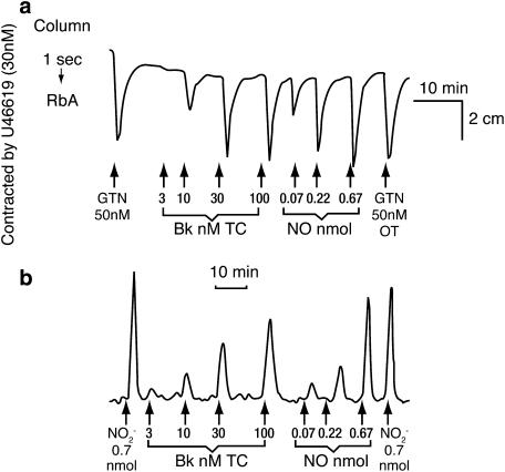
Although the comparative pharmacology of EDRF and NO on vascular strips convinced us of the identity of EDRF as NO, we were interested in measuring NO using methods other than bioassay. There are several chemical methods for measuring the breakdown products of NO (NO2− and nitrate, NO3−). However, we wanted to measure NO itself as it was released from the cells following stimulation. Richard Palmer identified a potential method, used in both the car and food industries, based on a specific chemiluminescent signal which is generated when NO interacts with ozone. Using this technique, modified to detect very low quantities of NO, we demonstrated that NO was indeed generated from vascular endothelial cells when stimulated with bradykinin. Furthermore, the quantities released were sufficient to account for the actions of EDRF (Figure 2; Palmer et al., 1987).
Figure 2.
Detection of exogenous and endogenous NO. (a) Bioassay. The rabbit aorta was relaxed in a concentration-dependent manner by EDRF released from the endothelial cells by bradykinin (BK, TC) and by NO (OT), as in Figure 1. (b) Chemiluminescence. EDRF was released by bradykinin from a replicate column of the cells used in the bioassay. The amounts of both EDRF (endogenously-produced NO) and of exogenously-applied authentic NO which relaxed the bioassay tissue were also detectable by chemiluminescence. From Palmer et al. (1987), with permission.
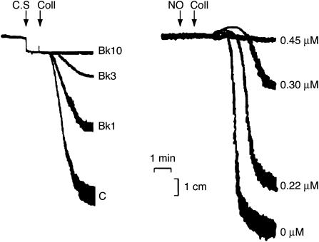
Platelet studies were very important in confirming the nature of EDRF. NO had been known for some years to inhibit platelet aggregation and in 1986 it was shown that EDRF has platelet antiaggregating properties (Azuma et al., 1986). Comparative pharmacological studies between EDRF from vascular tissues and authentic NO demonstrated the resemblance between the two compounds in their actions on platelets (Figure 3; Radomski et al., 1987). Moreover, the antiaggregating and the disaggregating effects of both EDRF and authentic NO were potentiated by subthreshold concentrations of prostacyclin and vice versa, an effect that could be blocked by treatment with indomethacin and partially reversed by treatment with haemoglobin. The antiaggregatory action of NO, but not of prostacyclin, was potentiated by SOD; thus NO is a potent inhibitor of platelet aggregation, whose activity on platelets mimics that of EDRF (Radomski et al., 1987).
Figure 3.
Inhibition of platelet aggregation by EDRF and NO. Control (C) aggregation of human platelet-rich plasma was induced by collagen (Coll). Pretreatment for 1 min with supernatant from endothelial cells stimulated with bradykinin at different concentrations (1–10 nM, C.S, left panel) or with authentic NO (right panel) prevented collagen-induced aggregation in a concentration-dependent manner.
Over the following months, we carried out a variety of experiments to investigate the origin of NO. There were a number of possibilities, including the suggestion that NO2− or NO3− was reduced enzymically to NO or that ammonia or an amino acid was the biological precursor. However, the most interesting possibility was that NO originated from the conversion of L-arginine, since activated macrophages had recently been shown to generate NO2− and NO3− from this amino acid (Hibbs et al., 1987; Iyengar et al., 1987). We surmised that NO might be an unstable intermediate in the synthesis of the stable NO2− and NO3− from L-arginine. Our early experiments in which we infused L-arginine over the tissues were unsuccessful; however, we soon realised that, as the endothelial cells were cultured in a medium rich in amino acids, they were probably already saturated by the time we gave additional amounts. We therefore prepared a culture medium without L-arginine, cultured the cells in it for 24 h before the experiments, put them in our bioassay system, infused L-arginine, and found that we could detect NO not only biologically but chemically by chemiluminescence (Palmer et al., 1988).
We went on to identify the enzyme responsible for the production of NO as NO synthase, which is able to generate NO and L-citrulline from L-arginine, and began to call these biochemical reactions, the L-arginine : NO pathway (Moncada et al., 1989).
Roles of the L-arginine : NO pathway
By 1987 it had become apparent that the generation of NO was not only taking place in the vascular wall, but was probably a widespread mechanism with far-reaching biological significance. The work of Hibbs (Hibbs et al., 1987) and Marletta (Iyengar et al., 1987) pointed to the possibility that NO was formed in macrophages. In addition, we came across some earlier work in which a group in Japan had found that brain homogenates contain a low molecular weight stimulator of the soluble guanylate cyclase which they later identified as L-arginine. This work suggested the existence of the L-arginine :NO pathway in the central nervous system, where we then identified the NO synthase and determined its dependency on calcium. This paper was preceded by a publication from John Garthwaite and his group demonstrating the release of EDRF from cerebellar cells following activation with _N_-methyl-D-aspartate (NMDA; Moncada et al., 1989).
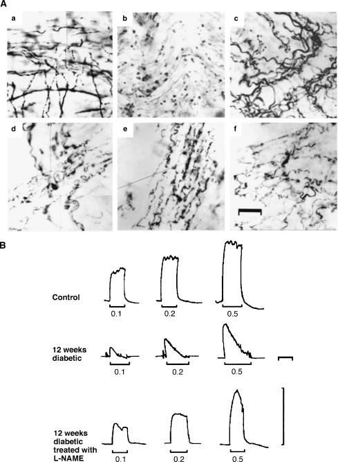
In the late 1980s, NO was identified as the inhibitory mediator of nonadrenergic, noncholinergic (NANC) neurotransmission in peripheral nerves. This ‘nitrergic' neurotransmission has been demonstrated in the gastrointestinal tract where it is responsible for NANC-mediated relaxations of the gastric fundus, in the corpus cavernosum where it brings about relaxation of this tissue, resulting in penile erection, and in the trachea and the bladder where it contributes to NANC-induced relaxation. Thus nitrergic neurotransmission represents a widespread system of peripheral nerves in the body, acting alongside the classical adrenergic and cholinergic systems (Cellek, 1997). Both noradrenergic and cholinergic responses are ‘controlled' by the nitrergic system so that the release of NO (e.g., during electrical field stimulation) counteracts and dominates the response to the noradrenergic or cholinergic stimulus (Cellek & Moncada, 1997). The discovery of nitrergic neurotransmission revealed the mechanism of penile erection in animals and humans and led to pharmacological and therapeutic intervention (Cellek, 1997). In this context, we have shown that degeneration of nitrergic nerves occurs in diabetes while the noradrenergic nerves remain intact (Figure 4; Cellek et al., 1999). Selective damage of nitrergic nerves, which seems to be the direct result of the NO generated interacting with oxygen-derived free radicals or their products, accounts for the erectile dysfunction and gastropathy of diabetes. We therefore proposed that other cells in which NO is generated might be subjected to the same pathophysiological process and therefore to damage, the vascular endothelium being a significant target.
Figure 4.
Effect of diabetes and L-NAME on nitrergic nerves and erectile function in rats. (A) Immunostaining for nNOS in the corpus cavernosum of a control rat (a), in an 8-week-diabetic rat (b, note that the nitrergic nerves became very sparse) and in an 8-week-diabetic rat treated with L-NAME (c, note that the structure and number of the nitrergic nerve fibres were preserved during diabetes). Tyrosine hydroxylase immunostaining in a control rat (d), an 8-week-diabetic rat (e, note that the morphology and density of noradrenergic nerves were unchanged) and an 8-week-diabetic rat treated with L-NAME (f). Scale bar is 33–43 _μ_M. (B) Typical changes in intracavernous pressure (ICP) in response to stimulation in a control rat (upper trace), a 12 week diabetic rat (middle trace: note that the increase in ICP could not be maintained during the stimulation period) and a 12 week diabetic rat treated with L-NAME (lower trace). L-NAME was withdrawn 72 h before the experiments. The vertical scale corresponds to 100 cm H2O ICP. The horizontal scale corresponds to 2 min. From Cellek et al. (1999), with permission.
There are now three main fields of NO research, namely the cardiovascular system, the nervous system, and inflammation/immunology. These are loosely based on three isoforms of the NO synthase (NOS; EC 1.14. 13.39), which were originally named after the tissues in which they were first identified. Thus there are two calcium/calmodulin-dependent, constitutive isoforms, neuronal NOS (nNOS, NOS I) and endothelial NOS (eNOS, NOS-III), and a calcium-independent, inducible NOS (iNOS, NOS-II) which is expressed in macrophages and other tissues following immunological stimulation (Alderton et al., 2005). Since each area of research is now so extensive this review will focus on some of the most significant actions of NO in the cardiovascular system and their potential relevance.
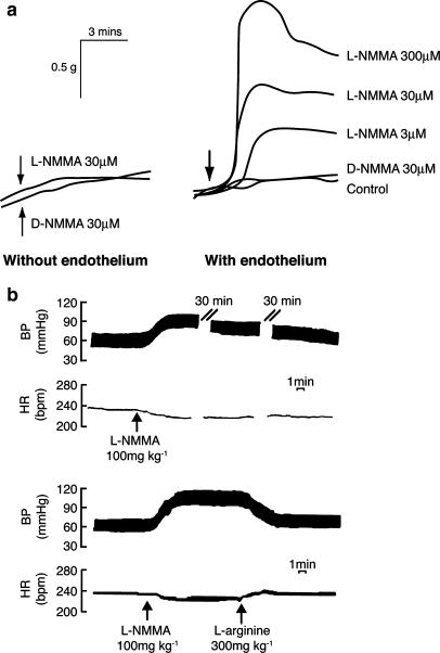
The identification of _N_G-monomethyl-L-arginine (L-NMMA) as an inhibitor of the synthesis of NO provided the most important pharmacological tool for investigating the presence and roles of NO in biological systems. We found that L-NMMA inhibits both endothelium-dependent relaxation and the release of NO. We also observed that L-NMMA by itself produced a dose-dependent and endothelium-dependent contraction of rabbit aorta, suggesting the loss of a continuous NO-dependent relaxing tone (Figure 5a; Rees et al., 1989a). This led us to studies in the whole animal, in which we demonstrated that the intravenous administration of L-NMMA caused an immediate and marked increase in blood pressure that could be reversed by infusion of L-arginine (Figure 5b; Rees et al., 1989b). This simple experiment was arguably one of the most important we ever carried out in relation to the roles of NO since it turned our understanding of the mammalian cardiovascular system the right way round. Prior to our results, it had never been suspected that blood pressure regulation depended so much on a continuously-generated local vasodilator tone, the lack of which would lead to a significant hypertensive response. We went on to suggest that conductance and not resistance was the main determinant of blood pressure regulation and that at least some forms of hypertension could be considered not in terms of an increased resistance, but of a decreased conductance of the system.
Figure 5.
The effect of L-NMMA on vascular tone and blood pressure. (a) The effect of N _G_-monomethyl L-arginine (L-NMMA) and its inactive isomer (D-NMMA) on the basal tone of pre-contracted rabbit aortic rings, with and without endothelium. (b) Long-lasting effect of L-NMMA on blood pressure in the anaesthetised rabbit. The lower part of the trace shows the reversal of this effect by L-arginine. The heart rate is also shown. From (a) Rees et al. (1989a); (b) Rees et al. (1989b), with permission.
We carried out further experiments demonstrating a vasoconstrictor effect of L-NMMA in many vascular beds including the circulation of the human forearm. These observations were confirmed in a variety of studies in animals and humans and later extended to models of genetic manipulation. In these, knocking out the eNOS isoform led to a hypertensive phenotype and over-expression of eNOS in endothelial cells reduced the blood pressure (Huang, 2000). Release of NO is regulated, primarily at a local level, by shear stress produced by blood passing over the endothelium. This activates eNOS through the phosphorylation of a specific tyrosine residue in the enzyme (Fleming & Busse, 2004).
NO, mitochondria and free radicals
The soluble guanylate cyclase has been shown to be the biochemical target through which NO carries out some of its physiological functions. In the vascular system, activation of the enzyme and the subsequent elevation of cyclic GMP concentration accounts for NO-induced vasodilatation and inhibition of platelet aggregation, while it contributes to other effects of NO such as those on vascular smooth muscle and leucocyte-vessel wall interactions. In 1994, however, an additional potential target for NO was identified when it was discovered that NO inhibits the cytochrome c oxidase, the terminal enzyme in the mitochondrial oxidative phosphorylation pathway involved in the reduction of oxygen to water (Moncada & Erusalimsky, 2002). This inhibitory effect was shown to be reversible, in competition with oxygen, and to occur at concentrations of NO likely to be present physiologically. Indeed, the affinity of the cytochrome c oxidase for NO is greater than that for oxygen, such that at, for example, 30 _μ_M oxygen the IC50 of NO is 30 nM. In vascular endothelial cells, endogenous concentrations of NO modulate cell respiration in an oxygen-dependent manner. This led to the suggestion that NO might, on the one hand, modulate cellular bioenergetics by regulating oxygen consumption and on the other, decrease electron flux through the electron transport chain and favour the generation of the superoxide ion, O2−, by inhibiting cytochrome c oxidase. It was further suggested that this mechanism might contribute to cell signalling through the subsequent formation of hydrogen peroxide (H2O2). Increases in NO production were also shown to inhibit cellular respiration irreversibly by selectively inhibiting complex I through a process dependent on the increased generation of O2− and probably peroxynitrite. This might be a way in which cell pathology may be initiated at a mitochondrial level in some situations (Moncada & Erusalimsky, 2002).
We have also shown that NO, by regulating mitochondrial behaviour, plays other intriguing roles related to general homeostatic and host-defence mechanisms, such as the initiation of a Ca2+-related interaction between mitochondria and endoplasmic reticulum leading to the activation of the grp78-dependent stress response (Xu et al., 2005) and the activation of an AMPkinase-dependent glycolytic response (Almeida et al., 2004). Both mechanisms might be relevant in the vasculature. Furthermore, we have recently demonstrated that the inhibitory action of NO on oxygen consumption in the mitochondria has the further consequence of diverting oxygen away from the mitochondria to other areas of the cell and probably to surrounding tissues (Hagen et al., 2003). Whether this is a mechanism utilised to divert oxygen from the vascular endothelium, which is known to be glycolytic, to the vascular smooth muscle, which requires oxygen is also unclear at present. A further finding in this area of research in which we are revealing links between cell bioenergetics and signalling mechanisms is the observation that NO plays a role in mitochondriogenesis (Nisoli et al., 2003), an additional indication that NO might be involved in the regulation of the balance between glycolysis and oxidative phosphorylation in cells. Interestingly, this latter effect is not the result of NO interacting with the cytochrome c oxidase but, unexpectedly, with the soluble guanylate cyclase.
NO and pathophysiology
Lack of NO
In addition to its vasodilator and platelet inhibitory actions, NO was found to inhibit vascular smooth muscle proliferation and to regulate interactions between leucocytes and the blood vessel wall. These findings established NO as a homeostatic regulator in the vasculature, the absence of which plays a role in a number of conditions and pathological states such as hypertension and vasospasm (Moncada & Higgs, 2000; Rees et al., 2000).
The early stages of a number of these conditions share a specific pathophysiological feature, namely endothelial dysfunction. A widely accepted definition of endothelial dysfunction is that of a reduction in endothelial NO, which is detected as a decrease in endothelium-dependent vasodilatation induced either by appropriate agonists or by flow. Such endothelial dysfunction has been observed prior to any other evidence of cardiovascular disease in subjects with a family history of essential hypertension or other risk factors for atherosclerosis. It has also been associated with smoking and in general its presence is predictive of cardiovascular disease. Decreases in NO formation may result either from reduced expression of eNOS or from changes in its substrates or cofactors, such as L-arginine or tetrahydrobiopterin (BH4). However, the most likely mechanism for endothelial dysfunction is that of a reduced bioavailability of NO as a result of its interactions with oxygen-derived species, specifically O2−. Inactivation of NO by O2− contributes to oxidative stress, a term used to describe various deleterious processes resulting from an imbalance between excessive formation of reactive oxygen species (ROS) (and/or the oxidants derived from NO) and limited antioxidant defences (for review and references see Moncada, 2005).
The reaction between NO and O2− leads to the formation of peroxynitrite. This powerful oxidant species has been implicated in established clinical conditions such as hypercholesterolaemia, diabetes and coronary artery disease (Greenacre & Ischiropoulos, 2001). There is also substantial evidence for increased ROS formation in blood vessels in such disorders, and treatment with antioxidants enhances endothelium-dependent vasodilatation in both the forearm and the coronary circulation of individuals with coronary artery disease and diabetes.
The origin of O2− in the vasculature has been the subject of much research. The activation of enzymes such as NADPH oxidase and xanthine oxidase has been implicated, and there is considerable evidence showing that the activity and the expression of these enzymes can be enhanced by pathological stimuli. Vascular cytochrome P450 enzymes that can generate O2− have also been described and their inhibition appears to improve endothelium-dependent, NO-mediated vasodilatation in patients with coronary artery disease. Another potential source of O2− is what has been described as ‘uncoupled NO synthase', a situation in which eNOS can generate O2− when the concentrations of either L-arginine or BH4 are low. The uncoupling of eNOS has been reported to occur in several pathological conditions such as diabetes, hypercholesterolaemia and hypertension (Cai & Harrison, 2000; Moncada, 2005). Uncoupling of eNOS due to depletion of both L-arginine and BH4 is not likely to be an early mechanism of O2− generation, since the lowering of the substrate or the cofactor to critical levels probably requires drastic changes in the vasculature. However, it has recently been suggested that uncoupling of eNOS may result from more subtle changes in the biochemical functioning of the enzyme (Lin et al., 2003).
For many years it has been believed that a small percentage of the oxygen utilised by mitochondria is not completely reduced to water and escapes as O2− and, although it is not clear whether this actually occurs in endothelial cells in vivo at physiological oxygen concentrations, it is possible that the redox status of the mitochondrial respiratory chain is a determinant in the escape of electrons required to generate O2− from oxygen. We have recently shown that NO, by favouring the reduction of the cytochrome c oxidase, facilitates the release of O2− from mitochondria. This is subsequently converted into H2O2 with the resulting signalling consequences (Palacios-Callender et al., 2004). It is likely that such a mechanism, which is an extension of the physiological action of NO on the cytochrome c oxidase, might provide clues to the understanding of the early origins of oxidative stress in the vasculature.
Protection against decreases in the generation of constitutive NO in the vasculature may prevent the development of vascular disease. This may be achieved by the use of antioxidants and the transfection of eNOS. Each of these interventions has shown promise in both animal experiments and in humans. Interestingly, statins have recently been shown to increase the production of endothelial NO in endothelial cell cultures and in animals. Mechanisms proposed for this action include the reduction of oxidative stress by increasing the synthesis of BH4, increasing the coupling of the eNOS or reducing the activation of NADPH oxidase (Laufs, 2003).
Early evidence suggested that administration of oestradiol increased endothelium-dependent relaxation and that aortae from female rabbits generate more NO than those of males. This, together with the fact that NO might be increased during pregnancy, led us to study the effect of oestrogens on NO synthases. Our results indicated that oestrogens not only increased the activity of eNOS but also its expression (Weiner et al., 1994). These results have been confirmed in many studies; indeed it has been demonstrated that ovariectomy of rats dramatically reduces both the amount and activity of eNOS. In addition to an action at the level of eNOS expression or activity, it has been claimed that oestrogens act by reducing the generation of O2− by the vessel wall, leading to a decreased breakdown of available NO (Khalil, 2005).
There appears to be a gender difference in the response to NO, so that agonist-induced NO-dependent dilations are greater in female than in male animals and the degree of hypertension is greater in male than female eNOS−/− mice (Rees et al., 2000). Furthermore, while the vasodilator response to acetylcholine in large conductance vessels is completely abolished in eNOS−/− mice and can be restored by gene transfer of eNOS in vitro, the response in resistance vascular beds is maintained in the knockout animals, in the mesenteric (Rees et al., 2000) and other vascular beds. This ‘remaining' vasodilator response has been extensively investigated and variously attributed to, among others, prostaglandins or the elusive endothelium-derived hyperpolarising factor. Recent studies on a double knockout mouse (eNOS−/− and cyclooxygenase-1−/−), unable to generate either NO or prostacyclin, show that in these animals there is indeed a compensatory vasodilator mechanism, especially in female animals (Scotland et al., 2005), which still requires identification.
Excess of NO
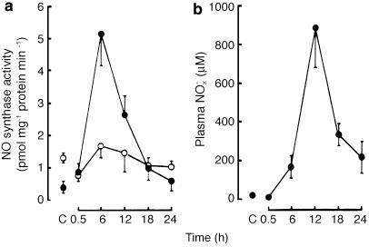
Inflammatory stimuli such as endotoxin lipopolysaccharide and cytokines induce iNOS in many cells and tissues. Induction of this enzyme, which was identified originally in macrophages and contributes to the cytotoxic actions of these cells, is inhibited by anti-inflammatory glucocorticoids. The NO produced by iNOS in the vasculature is involved in the profound vasodilatation of septic shock (Moncada & Higgs, 2000). Endotoxin also induces iNOS in the myocardium (Figure 6; Schulz et al., 1992) where, as in the vasculature, it is responsible for dysfunction and damage. It has become apparent in the last few years that inhibition of mitochondrial respiration is an important component of the NO-induced tissue damage. This inhibition of respiration, which is initially NO-dependent and reversible, becomes persistent with time as a result of oxidative stress (Moncada & Erusalimsky, 2002). This observation has indicated that the inability of the tissues to utilise available oxygen is probably the cornerstone of the pathophysiological events in sepsis. This defect, which we have called metabolic hypoxia, might not be exclusive to septic shock but could also contribute to other inflammatory and degenerative conditions (Moncada & Erusalimsky, 2002).
Figure 6.
Time course of activation of left ventricular NO synthase and changes in plasma NO_x_ − (NO2− plus NO3−) concentration after i.p. injection of endotoxin (LPS) or pyrogen-free saline in rats. (a) Ca2+-dependent (○) and Ca2+-independent (•) NO synthase activity in the left ventricular wall after treatment with LPS. C represents control values 6 h after injection of pyrogen-free saline. (b) Level of NO_x_− in plasma at time rats were killed after treatment with LPS. C Represents control values 6 h after injection of pyrogen-free saline. Reprinted from Schulz et al. (1992), with permission.
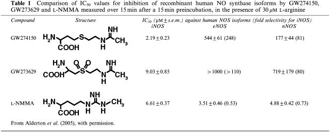
An inhibitor of NOS, L-NMMA, reversed the hypotension and the hyporeactivity to vasoconstriction characteristic of endotoxin shock in animal models. This led to the testing of this compound in a clinical trial in septic shock (Lopez et al., 2004). However, the results of this were disappointing and mortality was actually slightly increased in the treatment group. The reasons for this are not clear, but it is possible that, as L-NMMA is a nonselective inhibitor of NO synthase, inhibition of both eNOS and iNOS might be deleterious. Selective inhibitors of iNOS may prove beneficial for the treatment of the hypotension of shock or cytokine therapy and may also provide a new approach to antiinflammatory therapy. Two such compounds, GW273629 and GW274150, have recently been described as having a high selectivity in vivo in mice, with low potency against nNOS in the rat cerebellum and no effect on blood pressure in the whole animal (Table 1; Alderton et al., 2005). These two compounds are currently in clinical development for inflammatory conditions, such as asthma.
Table 1. Comparison of IC50 values for inhibition of recombinant human NO synthase isoforms by GW274150, GW273629 and L-NMMA measured over 15 min after a 15 min preincubation, in the presence of 30 _μ_M L-arginine.
The inducible isoform, iNOS, is induced in macrophages and smooth muscle cells of atherosclerotic vessels in animals and in humans. In advanced atherosclerotic plaques from human blood vessels, iNOS was found to colocalise with nitrotyrosine, a marker for peroxynitrite-induced damage. The oxidation of low-density lipoprotein (LDL) is thought to be a key factor in the formation of atherosclerotic lesions and it has been shown that peroxynitrite can modify LDL to a potentially atherogenic form (Moncada & Higgs, 2000). Thus, while the low concentrations of NO generated by eNOS protect against atherosclerosis by promoting vasodilatation, inhibiting leucocyte and platelet adhesion and/or aggregation and smooth muscle cell proliferation, higher concentrations of NO generated by iNOS promote atherosclerosis either directly or via the formation of NO adducts, such as peroxynitrite. Such a paradox in the action of NO was apparent from our experiments some years ago in which we found that the acute vascular injury in the ileum and colon following administration of lipopolysaccharide is aggravated by early treatment with a NO synthase inhibitor, whereas delayed administration of such a compound provides protection against the damage to the intestinal vasculature (Laszlo et al., 1994). A prominent example of this comes from experiments in Apo-E mutant mice in which the concomitant knocking out of eNOS leads to an increase in atherosclerosis, while the knocking out of iNOS reduces atherosclerosis (for references see Moncada, 2005).
Conclusion
The discovery of the L-arginine: NO pathway has had a major impact in many areas of research, notably vascular biology. Although much has been learned about the pathway and its interactions, there is still a great deal of work to do before some of this knowledge can be translated into clinical medicine. There are many exciting possibilities and avenues of research and a great deal of benefit to be gained. We look forward to writing about these for the volume to be produced celebrating the 100th anniversary of the British Pharmacological Society!
Glossary
EDRF
endothelium-derived relaxing factor
H2O2
hydrogen peroxide
L-NMMA
_NG_-monomethyl-L-arginine
NANC
nonadrenergic, noncholinergic
NO
nitric oxide
NOS
nitric oxide synthase
NO2−
nitrite
NO3−
nitrate
O2−
superoxide anion
SOD
superoxide dismutase
References
- ALDERTON W.K., ANGELL A.D.R., CRAIG C., DAWSON J., GARVEY E., MONCADA S., MONKHOUSE J., REES D., RUSSELL L.J., RUSSELL R.J., SCHWARTZ S., WASLIDGE N., KNOWLES R.G. GW274150 and GW273629 are potent and highly selective inhibitors of inducible nitric oxide synthase in vitro and in vivo. Br. J. Pharmacol. 2005;145:301–312. doi: 10.1038/sj.bjp.0706168. [DOI] [PMC free article] [PubMed] [Google Scholar]
- ALMEIDA A., MONCADA S., BOLANOS J.P. Nitric oxide switches on glycolysis through the AMP protein kinase and 6-phosphofructo-2-kinase pathway. Nature Cell Biol. 2004;6:45–51. doi: 10.1038/ncb1080. [DOI] [PubMed] [Google Scholar]
- AZUMA H., ISHIKAWA M., SEKIZAKI S. Endothelium-dependent inhibition of platelet aggregation. Br. J. Pharmacol. 1986;88:411–415. doi: 10.1111/j.1476-5381.1986.tb10218.x. [DOI] [PMC free article] [PubMed] [Google Scholar]
- CAI H., HARRISON D.G. Endothelial dysfunction in cardiovascular diseases: the role of oxidant stress. Circ. Res. 2000;87:840–844. doi: 10.1161/01.res.87.10.840. [DOI] [PubMed] [Google Scholar]
- CELLEK S.1997Nitrergic and noradrenergic interaction in dually innervated organs of the genitourinary tractPhD Thesis. University of London [Google Scholar]
- CELLEK S., MONCADA S. Nitrergic modulation of cholinergic responses in the opossum lower oesophageal sphincter. Br. J. Pharmacol. 1997;122:1043–1046. doi: 10.1038/sj.bjp.0701497. [DOI] [PMC free article] [PubMed] [Google Scholar]
- CELLEK S., RODRIGO J., LOBOS E., FERNANDEZ P., SERRANO J., MONCADA S. Selective nitrergic neurodegeneration in diabetes mellitus – a nitric oxide-dependent phenomenon. Br. J. Pharmacol. 1999;128:1804–1812. doi: 10.1038/sj.bjp.0702981. [DOI] [PMC free article] [PubMed] [Google Scholar]
- FLEMING I., BUSSE R. The physiology of nitric oxide: control and consequences. Curr. Med. Chem. 2004;3:189–205. [Google Scholar]
- FURCHGOTT R.F.1988Studies on relaxation of rabbit aorta by sodium nitrite: the basis for the proposal that the acid-activatable inhibitory factor from retractor penis is inorganic nitrite and the endothelium-derived relaxing factor is nitric oxide Vasodilatation: Vascular Smooth Muscle, Peptides, Autonomic Nerves and Endotheliumed. Vanhoutte, P.M., pp. 401–414.New York: Raven Press [Google Scholar]
- FURCHGOTT R.F., ZAWADZKI J.V. The obligatory role of endothelial cells in the relaxation of arterial smooth muscle by acetylcholine. Nature. 1980;288:373–376. doi: 10.1038/288373a0. [DOI] [PubMed] [Google Scholar]
- FURCHGOTT R.F., CHERRY P.D., ZAWADZKI J.V., JOTHIANANDAN D. Endothelial cells as mediators of vasodilation of arteries. J. Cardiovasc. Pharmacol. 1984;6:S336–S343. doi: 10.1097/00005344-198406002-00008. [DOI] [PubMed] [Google Scholar]
- GREENACRE S.A., ISCHIROPOULOS H. Tyrosine nitration: localisation, quantification, consequences for protein function and signal transduction. Free Radic. Res. 2001;34:541–581. doi: 10.1080/10715760100300471. [DOI] [PubMed] [Google Scholar]
- GRYGLEWSKI R.J., MONCADA S., PALMER R.M.J. Bioassay of prostacyclin and endothelium-derived relaxing factor (EDRF) from porcine aortic endothelial cells. Br. J. Pharmacol. 1986a;87:685–694. doi: 10.1111/j.1476-5381.1986.tb14586.x. [DOI] [PMC free article] [PubMed] [Google Scholar]
- GRYGLEWSKI R.J., PALMER R.M.J., MONCADA S. Superoxide anion is involved in the breakdown of endothelium-derived vascular relaxing factor. Nature. 1986b;320:454–456. doi: 10.1038/320454a0. [DOI] [PubMed] [Google Scholar]
- HAGEN T., TAYLOR C.T., LAM F., MONCADA S. Redistribution of intracellular oxygen in hypoxia by nitric oxide: effect on HIF1α. Science. 2003;302:1975–1978. doi: 10.1126/science.1088805. [DOI] [PubMed] [Google Scholar]
- HIBBS J.B., Jr, TAINTOR R.R., VAVRIN Z. Macrophage cytotoxicity: role for L-arginine deiminase and imino nitrogen oxidation to nitrite. Science. 1987;235:473–476. doi: 10.1126/science.2432665. [DOI] [PubMed] [Google Scholar]
- HUANG P.L. Lessons learned from nitric oxide synthase knockout animals. Semin. Perinatol. 2000;24:87–90. doi: 10.1016/s0146-0005(00)80064-6. [DOI] [PubMed] [Google Scholar]
- IGNARRO L.J., BYRNS R.E., WOOD K.S.1988Biochemical and pharmacological properties of endothelium-derived relaxing factor and its similarity to nitric oxide radical Vasodilatation: Vascular Smooth Muscle, Peptide, Autonomic Nerves and Endotheliumed. Vanhoutte, P.M., pp. 427–436.New York: Raven Press [Google Scholar]
- IYENGAR R., STUEHR D.J., MARLETTA M.A. Macrophage synthesis of nitrite, nitrate and N-nitrosamines: precursors and role of the respiratory burst. Proc. Natl. Acad. Sci. U.S.A. 1987;84:6369–6373. doi: 10.1073/pnas.84.18.6369. [DOI] [PMC free article] [PubMed] [Google Scholar]
- KHALIL R.A. Sex hormones as potential modulators of vascular function in hypertension. Hypertension. 2005;46:249–254. doi: 10.1161/01.HYP.0000172945.06681.a4. [DOI] [PMC free article] [PubMed] [Google Scholar]
- LASZLO F., WHITTLE B.J.R., MONCADA S. Time-dependent enhancement or inhibition of endotoxin-induced vascular injury in rat intestine by nitric oxide synthase inhibitors. Br. J. Pharmacol. 1994;111:1309–1315. doi: 10.1111/j.1476-5381.1994.tb14887.x. [DOI] [PMC free article] [PubMed] [Google Scholar]
- LAUFS U. Beyond lipid-lowering: effects of statins on endothelial nitric oxide. Eur. J. Clin. Pharmacol. 2003;58:719–731. doi: 10.1007/s00228-002-0556-0. [DOI] [PubMed] [Google Scholar]
- LIN M.I., FULTON D., BABBITT R., FLEMING I., BUSSE R., PRITCHARD K.A., Jr, SESSA W.C. Phosphorylation of threonine 497 in endothelial nitric-oxide synthase coordinates the coupling of L-arginine metabolism to efficient nitric oxide production. J. Biol. Chem. 2003;278:44719–44726. doi: 10.1074/jbc.M302836200. [DOI] [PubMed] [Google Scholar]
- LOPEZ A., LORENTE J.A., STEINGRUB J., BAKKER J., MCLUCKIE A., WILLATTS S., BROCKWAY M., ANZUETO A., HOLZAPFEL L., BREEN D., SILVERMAN M.S., TAKALA J., DONALDSON J., ARNESON C., GROVE G., GROSSMAN S., GROVER R. Multiple-center, randomized, placebo-controlled, double-blind study of the nitric oxide synthase inhibitor 546C88: effect on survival in patients with septic shock. Crit. Care Med. 2004;32:21–30. doi: 10.1097/01.CCM.0000105581.01815.C6. [DOI] [PubMed] [Google Scholar]
- MONCADA S.2005Adventures in vascular biology: a tale of two mediators Phil. Trans. Roy. Soc. B.in press. [DOI] [PMC free article] [PubMed]
- MONCADA S., ERUSALIMSKY J.D. Does nitric oxide modulate mitochondrial energy generation and apoptosis. Nat. Rev. Mol. Cell Biol. 2002;3:214–220. doi: 10.1038/nrm762. [DOI] [PubMed] [Google Scholar]
- MONCADA S., HIGGS E.A.2000Nitric oxide in cardiovascular function and disease Atherosclerosis XII, Proceedings of the XIIth International Symposium on Atherosclerosiseds. Stemme, S. & Olsson, A.G., pp. 81–89.Stockholm, The Netherlands: Elsevier [Google Scholar]
- MONCADA S., PALMER R.M.J., GRYGLEWSKI R.J. Mechanism of action of some inhibitors of endothelium-derived relaxing factor. Proc. Natl. Acad. Sci. U.S.A. 1986;83:9164–9168. doi: 10.1073/pnas.83.23.9164. [DOI] [PMC free article] [PubMed] [Google Scholar]
- MONCADA S., PALMER R.M.J., HIGGS E.A. Biosynthesis of nitric oxide from L-arginine. A pathway for the regulation of cell function and communication. Biochem. Pharmacol. 1989;38:1709–1715. doi: 10.1016/0006-2952(89)90403-6. [DOI] [PubMed] [Google Scholar]
- NISOLI E., CLEMENTI E., PAOLUCCI C., COZZI V., TONELLO C., SCIORATI C., BRACALE R., VALERIO A., FRANCOLINI M., MONCADA S., CARRUBA M.O. Mitochondrial biogenesis in mammals: the role of endogenous nitric oxide. Science. 2003;299:896–899. doi: 10.1126/science.1079368. [DOI] [PubMed] [Google Scholar]
- PALACIOS-CALLENDER M., QUINTERO M., HOLLIS V.S., SPRINGETT R.J., MONCADA S. Endogenous NO regulates superoxide production at low oxygen concentrations by modifying the redox state of cytochrome c oxidase. Proc. Natl. Acad. Sci. U.S.A. 2004;101:7630–7635. doi: 10.1073/pnas.0401723101. [DOI] [PMC free article] [PubMed] [Google Scholar]
- PALMER R.M.J., FERRIGE A.G., MONCADA S. Nitric oxide release accounts for the biological activity of endothelium-derived relaxing factor. Nature. 1987;327:524–526. doi: 10.1038/327524a0. [DOI] [PubMed] [Google Scholar]
- PALMER R.M.J., ASHTON D.S., MONCADA S. Vascular endothelial cells synthesize nitric oxide from L-arginine. Nature. 1988;333:664–666. doi: 10.1038/333664a0. [DOI] [PubMed] [Google Scholar]
- RADOMSKI M.W., PALMER R.M.J., MONCADA S. The anti-aggregating properties of vascular endothelium: interactions between prostacyclin and nitric oxide. Br. J.Pharmacol. 1987;92:639–646. doi: 10.1111/j.1476-5381.1987.tb11367.x. [DOI] [PMC free article] [PubMed] [Google Scholar]
- REES D.D., PALMER R.M.J., HODSON H.F., MONCADA S. A specific inhibitor of nitric oxide formation from L-arginine attenuates endothelium-dependent relaxation. Br. J. Pharmacol. 1989a;96:418–424. doi: 10.1111/j.1476-5381.1989.tb11833.x. [DOI] [PMC free article] [PubMed] [Google Scholar]
- REES D.D., PALMER R.M.J., MONCADA S. Role of endothelium-derived nitric oxide in the regulation of blood pressure. Proc. Natl. Acad. Sci. U.S.A. 1989b;86:3375–3378. doi: 10.1073/pnas.86.9.3375. [DOI] [PMC free article] [PubMed] [Google Scholar]
- REES D.D., HIGGS E.A., MONCADA S.2000Nitric oxide and the vessel wall Hemostasis and Thrombosiseds. Colman, R.W., Hirsch, J., Marder, V.J., Clowes, A.W. & George, N.J., pp. 673–682.Lippincott Williams & Wilkins: Philadelphia [Google Scholar]
- SCHULZ R., NAVA E., MONCADA S. Induction and potential biological relevance of a Ca2+-independent nitric oxide synthase in the myocardium. Br. J Pharmacol. 1992;105:575–580. doi: 10.1111/j.1476-5381.1992.tb09021.x. [DOI] [PMC free article] [PubMed] [Google Scholar]
- SCOTLAND R.S., MADHANI M., CHAUHAN S., MONCADA S., ANDRESEN J., NILSSON H., HOBBS A.J., AHLUWALIA A. Investigation of vascular responses in endothelial nitric oxide synthase/cyclooxygenase-1 double-knockout mice. Key role for endothelium-derived hyperpolarizing factor in the regulation of blood pressure in vivo. Circulation. 2005;111:796–803. doi: 10.1161/01.CIR.0000155238.70797.4E. [DOI] [PubMed] [Google Scholar]
- WEINER C.P., LIZASOAIN I., BAYLIS S.A., KNOWLES R.G., CHARLES I.G., MONCADA S. Induction of calcium-dependent nitric oxide synthases by sex hormones. Proc. Natl. Acad. Sci. U.S.A. 1994;91:5212–5216. doi: 10.1073/pnas.91.11.5212. [DOI] [PMC free article] [PubMed] [Google Scholar]
- XU W., CHARLES I.G., MONCADA S. Nitric oxide: orchestrating hypoxia regulation through mitochondrial respiration and the endoplasmic reticulum stress response. Cell Res. 2005;15:63–65. doi: 10.1038/sj.cr.7290267. [DOI] [PubMed] [Google Scholar]
