Operational Feasibility of Using Loop-Mediated Isothermal Amplification for Diagnosis of Pulmonary Tuberculosis in Microscopy Centers of Developing Countries (original) (raw)
Abstract
The characteristics of loop-mediated isothermal amplification (LAMP) make it a promising platform for the molecular detection of tuberculosis (TB) in developing countries. Here, we report on the first clinical evaluation of LAMP for the detection of pulmonary TB in microscopy centers in Peru, Bangladesh, and Tanzania to determine its operational applicability in such settings. A prototype LAMP assay with simplified manual DNA extraction was evaluated for accuracy and ease of use. The sensitivity of LAMP in smear- and culture-positive sputum specimens was 97.7% (173/177 specimens; 95% confidence interval [CI], 95.5 to 99.9%), and the sensitivity in smear-negative, culture-positive specimens was 48.8% (21/43 specimens; CI, 33.9 to 63.7%). The specificity in culture-negative samples was 99% (500/505 specimens; CI, 98.1 to 99.9%). The average hands-on time for testing six samples and two controls was 54 min, similar to that of sputum smear microscopy. The optimal amplification time was 40 min. No indeterminate results were reported, and the interreader variability was 0.4%. Despite the use of a single room without biosafety cabinets for all procedures, no DNA contamination was observed. The assay was robust, with high end-point stability and low rates of test failure. Technicians with no prior molecular experience easily performed the assay after 1 week of training, and opportunities for further simplification of the assay were identified.
The lack of early and accurate diagnosis is a critical obstacle to global tuberculosis (TB) control. The minority of the nearly 9 million incident TB cases each year is detected with microscopy, the only confirmatory test widely available in countries where TB is endemic, and delays and misdiagnoses are common. The human immunodeficiency virus epidemic has further diminished the utility of routine microscopy, and smear-negative TB has arisen as a particular problem in sub-Saharan Africa (2). Improved technologies that can abbreviate the diagnostic process and facilitate early diagnosis can save patients time and money, decrease morbidity, improve treatment outcomes, and interrupt the transmission of the disease. Such technologies need to be more sensitive, faster in yielding results, and/or simpler to use than microscopy (3, 15, 16).
Nucleic acid amplification tests (NAATs) for the detection of TB and other mycobacterial diseases have come into common use in industrialized countries because of their great advantage of speed compared to culture. Recent systematic reviews of studies evaluating commercially available technologies confirm very high specificity, with sensitivity approaching, but not reaching, that of culture (4, 8, 11, 17, 19, 20). The complexity and insufficient robustness of existing commercial NAAT formats and their requirement for a precision instrument, a high degree of technical support, and quality assurance make them unsuitable for most developing country settings (8, 12). Furthermore, the technical skill required has resulted in variable performance even in experienced molecular laboratories in both developed and developing countries (9, 20).
A novel molecular amplification method, termed loop-mediated isothermal amplification (LAMP), which has characteristics that may allow its use in less sophisticated settings, has recently been developed (10). LAMP amplifies DNA with high efficiency under isothermal conditions using six sets of primers (7). The large amount of DNA generated and the high specificity of the reaction make it possible to detect amplification by visual inspection of fluorescence or turbidity, without the need for gel electrophoresis or instrument detection of the labeled probe. This allows the use of a closed-tube system, which minimizes the risk of workspace contamination with amplicon. Since its invention, LAMP has been used to detect a number of infectious agents, including severe acute respiratory syndrome (21), West Nile virus (13), dengue virus (14), African trypanosomes (6), and Plasmodium falciparum (18). The speed of the reaction, the lack of a need for a thermocycler, and visual readout make LAMP a promising platform for the development of a simple and sensitive tool for the molecular detection of TB in developing countries.
Proof of principle for a LAMP test for TB was shown with the amplification of a single-copy gene of the Mycobacterium tuberculosis complex, M. avium, or M. intracellulare from DNA extracted from processed sputum specimens or culture isolates with sensitivity and specificity similar to those of commercial PCR (5). Since then, Eiken Chemical Company, in a joint development agreement with the Foundation for Innovative New Diagnostics, has modified the technique and transformed it into a more convenient kit format. The study outlined below is the first clinical evaluation of this test system and focuses on determining the feasibility of using LAMP in one of the settings of intended use, namely, peripheral microscopy centers.
MATERIALS AND METHODS
Study design, setting, and sample size.
This cross-sectional, multicenter study was carried out in three trial sites in Lima, Peru, Dhaka, Bangladesh, and Mbeya, Tanzania. In line with the objective of the study, to assess the applicability of LAMP for peripheral laboratories, LAMP was performed at each study site in one simple room without biosafety cabinets or other special equipment. Two to three benches served as working areas for sample processing, reaction mix preparation, amplification, and detection.
Six technicians without experience in nucleic acid amplification technologies were trained on LAMP for 1 week. The training was divided into two phases. In phase I, technicians practiced the method during a 3-day session with continuous feedback. In phase II, 3 to 4 days of additional training covered theoretical aspects of NAAT and troubleshooting.
Training was followed by an assessment of the clinical performance of LAMP in 725 sputum specimens from 380 patients with suspected TB (1 to 2 samples per patient, with a daily workload of 12 to 24 samples per site). Specimens were collected at outpatient clinics from patients >18 years of age with >2 weeks of cough and clinical symptoms of pulmonary tuberculosis. Operational LAMP criteria, such as “hands-on time” and “repeat rate,” were monitored throughout both training and enrollment phases. Ziehl-Neelsen (ZN) microscopy and Löwenstein-Jensen (LJ) culture were used as comparators. Clinical follow-up of culture-negative patients with suspected TB was not included in the protocol. Technicians performing LAMP were strictly blinded to smear results and vice versa. Blinding to LAMP results was also maintained between readers during the assessment of “interreader variability.”
ZN microscopy and LJ culture.
Following manual stirring for 10 s, untreated sputum samples were split prior to processing: a 250-μl aliquot from each sample was prepared for LAMP testing, and the remainder was kept for culture testing. The portion of the sputum preserved for microscopy and culture was decontaminated using the _N_-acetyl-l-cysteine-sodium hydroxide-sodium citrate method with a final NaOH concentration of 1.5% and an incubation time of 15 min. Centrifugation pellets were resuspended in a final volume of 2 ml and used for the inoculation of LJ cultures (BD, Franklin Lakes, NJ) and preparation of slides for ZN microscopy. Cultures were incubated at 37°C and monitored for growth for 8 weeks.
LAMP.
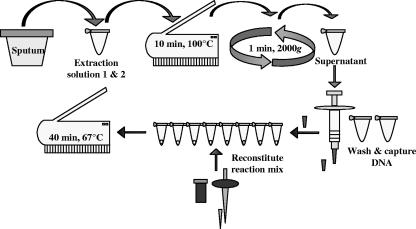
A developmental prototype assay was used in the study that joined manual sputum processing and DNA extraction with isothermal amplification and visual readout with UV fluorescence. As shown in the schematic in Fig. 1, raw sputum was treated with two extraction solutions, heated for 10 min at 100°C, and briefly centrifuged at 2,000 × g. Two hundred fifty microliters of supernatant was aspirated across a detachable filter membrane on the tip of a syringe and washed twice with 1.0 ml of buffer. The filtration tip was placed directly into the reaction tube in which the lyophilized reaction mix, including Bst DNA polymerase, had been reconstituted with 30 μl of buffer. Oligonucleotide primers targeting the gyrB gene were slightly modified from those described previously by Iwamoto et al. (5). Amplification was carried out at 67°C for 40 min in a small heating unit with eight reaction wells and an observation chamber with UV light for visual readout of the results. The reaction was terminated automatically by inactivating the polymerase at 80°C for 2 min. One negative control (buffer) and one positive control (buffer spiked with DNA) were included in each run.
FIG. 1.
Schematic diagram illustrating the LAMP procedural steps followed in the study.
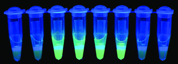
Calcein, a chelating reagent that is fluorescent unless combined with manganese ion, was included in the reaction mix. The binding of manganese by pyrophosphate, produced in abundance as a by-product of the LAMP reaction, released free calcein, resulting in UV fluorescence. The combination of turbidity from manganese pyrophosphate and calcein fluorescence allowed simple naked-eye determinations of reaction results, as shown in Fig. 2.
FIG. 2.
Visual detection of LAMP product under UV light. From left to right, tubes 1, 2, 7, and 8 are negative, and tubes 3, 4, 5, and 6 are positive.
Operational performance.
Primary study end-points were (i) operational performance, as measured by hands-on time, repeat rate, interreader variability, “reading-end-point stability,” ease of use, and satisfaction of laboratory technicians; (ii) priorities for further simplification of the assay; and (iii) optimal amplification time, comparing 30 versus 40 min. The first two end-points were assessed with questionnaires. One was filled by the supervisor after each run, scoring laboratory technicians’ performance and recording problems and such quantitative end-points as hands-on time. The second questionnaire, containing open and multiple-choice questions, was filled by all study team members at three time points during the study period: at the end of phase I training, at the end of phase II training, and after at least 5 weeks of experience performing the assay during the enrollment phase. It recorded qualitative information related to the perceived utility and feasibility of LAMP for routine use in laboratories doing TB microscopy and asked for suggestions on how to further simplify the prototype assay.
Hands-on time was defined as the active working time required to test six samples and two controls, including the sample preparation time, from the beginning of the preparation of reagents to the reading and recording of results. It did not include the 40-min amplification time. Interreader variability was defined as the number of differing results among all samples read by two trained readers blinded to smear results and interpreting LAMP results independently from each other. We classified two levels of error: level I errors (less severe), which were defined as indeterminate readout by one reader versus positive or negative readout by the other reader, and level II errors, defined as positive versus negative readout or vice versa. The repeat rate was defined as the number of runs for which false results in the control tubes forced a repetition of the run. The reading-end-point stability, monitored during the training phase only, was defined as the number of identical results read at 1 h, 3 h, and >12 h after the 40-min amplification period. The temperature of the amplification-and-detection device was maintained at 67°C during this period. At two of the sites, the reaction was terminated at 40 min by a temperature spike to 80°C for 2 min, which was programmed in the amplification device. The optimal reaction time was determined by having two technicians record LAMP results independently at 30 and at 40 min after the start of the reaction.
Clinical performance.
Sensitivity in smear-positive and in smear-negative samples and specificity in culture-negative samples were secondary end-points for the study, which was not powered to determine performance with narrow confidence intervals (CIs). The LAMP results recorded at 40 min by the principal reader were used to determine performance. Patients with smear-positive, culture-negative results were excluded from the analysis.
RESULTS
Operational performance. (i) Hands-on time.
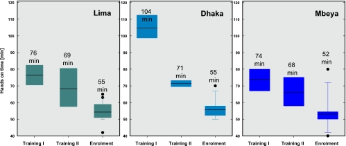
The hands-on time for the assay fell with increasing familiarity of the procedure from a mean of 85 min during the training phase to a mean of 54 min in the enrollment phase, as shown in Fig. 3. Particularly viscous samples increased the hands-on time by slowing the splitting and transfer of sputum into the extraction solution mix and by slowing the aspiration of samples across the DNA capture filter. Outliers in hands-on time of up to 80 min were due primarily to the blocking of the filters with very viscous samples.
FIG. 3.
Hands-on time required per run shown as box plots with the 25th and 75th percentiles (colored boxes) and the means (black lines and text) for the different study phases (basic training phase I, advanced training phase II, and enrollment phase) and different sites. Outliers are shown as black dots.
(ii) Interreader variability.
During the training phase, level I errors occurred in 1.6% of samples tested (3/188), and during the enrollment phase, level I errors occurred in 0.4% of samples tested (1/229). No level II errors were seen. In general, result interpretation was considered to be the easiest and most attractive part of the LAMP procedure by all laboratory personnel involved in the study. Though not specifically studied, visual inspection for turbidity and color was considered to be even more unambiguous than inspection under a black light. Inclusion of a negative control and a positive control in every run was felt to be necessary for result interpretation by all groups.
(iii) Reading-end-point stability.
End-point stability was assessed to exclude nonspecific amplification during any delay in result recording that might occur with routine use. At the two sites where the amplification device was programmed to deliver a temperature spike of 80°C for 2 min at the end of amplification (40 min), the reading-end-point stability was very high (100% correct results at 1, 3, and >12 h). At the site where no temperature spike terminated the reaction, nonspecific amplification at 67°C was observed in all the culture-negative samples between 70 and 240 min after the end of amplification. Positive results were stable, and none of the positive samples turned false negative within 12 h. The stability of positive results was also assessed with continuous exposure to a black light in the observation chamber, as might happen if a technician examined the tubes early and neglected to switch off the detection lamp, to observe potential bleaching of fluorescence. A bleaching effect of the black light on fluorescence was observed only >3 h after the end of amplification.
(iv) Repeat rate.
During the training phase, 5 of 42 runs had to be repeated due to positive results from the negative control, probably due to labeling errors in most cases. This did not occur again during the enrollment phase. At one of the study sites, positive controls gave negative results in 21 reactions during 5 of the 43 runs. A subsequent analysis determined this to be due to errors in the preparation of the DNA spiked control vials in the prototype kit. Other problems were not observed.
Despite repeated power supply and cold-chain interruptions in Mbeya (maximum, 10 h) and Dhaka (maximum, 30 min), reagent stability was not affected during the study duration of 5 to 6 weeks, and no degradation of sensitivity or specificity was seen over time.
(v) Ease of use and implementation.
In questionnaires, the study teams at all sites were asked to comment on the ease of implementation of the current LAMP version in the national TB programs of their countries. Herein, the complexity of the current LAMP version was rated comparable to culture for the sample-processing steps but easier and faster than culture and microscopy for result interpretation. The overall appraisal of the assay in terms of performance and practicability was good. Implementation of the version of the LAMP protocol used in the study in the national health care system was believed to be feasible only at the district level and not at peripheral microscopy laboratories. The main reason given was the perceived lack of skilled and motivated staff at peripheral laboratories to cope with the number and complexity of procedural steps as well as with the risk of DNA contamination. Other reasons were the difficulty of interrupting the LAMP procedure for other tasks, as can be done easily for microscopy, and the need for a continuous power supply. However, all sites identified a significant potential for a further simplification of the method that might lead to an assay suitable for peripheral laboratories.
Clinical performance.
The sensitivity of LAMP for pulmonary TB in smear-positive, culture-positive specimens was 97.7% (173/177 specimens; CI, 95.5 to 99.9). In the small number of smear-negative, culture-positive specimens, the sensitivity of LAMP was 48.8% (21/43 specimens; CI, 33.9% to 63.7%). The overall sensitivity of LAMP in the 220 culture-positive specimens was 88.2% (CI, 83.9 to 92.5). The overall and site-specific performance of LAMP for the detection of M. tuberculosis is shown in Table 1. Of the culture-positive specimens, 15 were reported to have visible heme, none of which were false negative by LAMP, suggesting that blood does not have an important inhibitory effect on the amplification or fluorescence detection. The specificity of LAMP in culture-negative samples was 99.0% (500/505 specimens; 95% CI, 98.1 to 99.9). Clinical follow-up of LAMP-positive, culture-negative patients with suspected TB was not performed, and no discrepant analysis was made. Amplification results were read at 30 min and at 40 min to determine whether the reaction time could be shortened. Of the 194 LAMP assays that were positive at 40 min, 16 (8.2%) were reported to be negative or indeterminate at 30 min.
TABLE 1.
LAMP sensitivity and specificitya
| Area | % Sensitivity (no. of smear-positive samples/no. of LJ-positive samples) | % Sensitivity (no. of smear-negative samples/no. of LJ-positive samples) | % Specificity (no. of smear-negative samples/no. of LJ-negative samples) |
|---|---|---|---|
| Lima | 97.7 (75/78) | 51.8 (14/27) | 99.3 (152/153) |
| Dhaka | 98.4 (61/62) | 50.0 (2/4) | 97.8 (181/185) |
| Mbeya | 100 (37/37) | 41.7 (5/12) | 100 (167/167) |
| Total | 97.7 (173/177) | 48.8 (21/43) | 99.0 (500/505) |
| 95% CI | 95.5-99.9 | 33.9-63.7 | 98.1-99.9 |
DISCUSSION
PCR and other NAAT methods for TB have great advantages of speed (compared to culture) and sensitivity (compared to microscopy). Furthermore, recently developed real-time assays may generate results in less than an hour, a key requirement for point-of-care testing. The utility of current NAAT methods is limited, however, by their cost and complexity, particularly in countries where TB is endemic. The required precision instruments are beyond the capacity of most diagnostic sites to purchase, maintain, or operate, and the complexity of testing obviates the possibility of point-of-care use.
Beyond amplification and detection itself, preparatory steps for NAAT make current formats cumbersome. Existing TB NAAT methods require separate steps for sputum liquefaction, DNA extraction, target amplification, and amplicon detection. This translates into multiple steps, separate workstations, and greater opportunity for cross-contamination with either bacterial DNA or amplicon. Automation of this process may be feasible, but is likely to further drive up the cost of equipment and reagents.
LAMP has inherent properties that make amplification and detection possible in one uninterrupted process, with no need to open the amplification vessel or any need for a luminometer or other detection device. The assay utilizes a single polymerase that is active at relatively high isothermal amplification temperatures, diminishing the likelihood of nonspecific priming. The use of six primers increases specificity at the same time that it enhances speed. In our hands, TB could usually be detected within 30 min, although a 40-min amplification time was optimal.
This paper reports the development and first use of a prototype LAMP assay for TB intended for use in peripheral laboratories. The study demonstrated that technicians without molecular training could perform the test with high reproducibility in a simple laboratory space without specialized equipment. Reader-to-reader variability was negligible, and there were no examples of absolute discordance (positive versus negative). Reaction results were stable for hours following automated enzyme inactivation with a heat spike at the end of amplification. The need for repeat testing for unexpected control tube results did not arise during the enrollment period, except for a limited number of runs in which an inadequately prepared positive control was used.
Quantitatively, the assay significantly outperformed microscopy, detecting M. tuberculosis DNA in almost all smear-positive sputum specimens and half of smear-negative, culture-positive specimens. These results are similar to those reported previously for sophisticated commercialized NAATs for M. tuberculosis (1). The total hands-on time to process and read results on a batch of sputum samples was similar to that of microscopy, with significantly simpler result readout. Notably, the presence of frank blood in the samples did not appear to inhibit the reaction, reinforcing findings reported previously by Poon et al. (18), who were able to detect malaria parasites at low density in boiled whole blood.
Further simplification of the LAMP assay was noted as being a priority by study participants. This is clearly possible, potentially leading to the first molecular test system that can be deployed in developing countries outside of reference laboratories. A significant amount of work has already been done to simplify and optimize the processing and extraction steps. Prior development work on NAAT methods for M. tuberculosis has always been held hostage by the _N_-acetyl-l-cysteine-NaOH processing method that is in standard use prior to culture in industrialized countries. This has been true largely because developers realized that the systems would be used in higher-level laboratories already performing culture and that culture and susceptibility testing would continue. Integration mandated the same initial processing steps. By working toward a method that would replace microscopy, we were able to explore alternative processing methods that could be more easily converted into a few-step process not requiring external equipment.
Eiken and the Foundation for Innovative New Diagnostics are developing an improved version of the test in which the number of steps will be reduced by a third and the number of electrical devices will be reduced to only a single heating block. With these simplifications, the ease of use and the robustness will increase, and the risk of laboratory DNA contamination will drop further. Larger studies are planned for the next version of TB LAMP in which performance, especially in smear-negative TB, will be evaluated and compared to those of other molecular amplification tests in addition to microscopy and culture.
Acknowledgments
We thank the study teams at the trial sites for their efforts in support of this evaluation.
Footnotes
▿
Published ahead of print on 28 March 2007.
REFERENCES
- 1.Bergmann, J. S., and G. L. Woods. 1996. Clinical evaluation of the Roche AMPLICOR PCR Mycobacterium tuberculosis test for detection of M. tuberculosis in respiratory specimens. J. Clin. Microbiol. 34**:**1083-1085. [DOI] [PMC free article] [PubMed] [Google Scholar]
- 2.Dye, C., C. J. Watt, D. M. Bleed, S. M. Hosseini, and M. C. Raviglione. 2005. Evolution of tuberculosis control and prospects for reducing tuberculosis incidence, prevalence, and deaths globally. JAMA 293**:**2767-2775. [DOI] [PubMed] [Google Scholar]
- 3.Foulds, J., and R. O'Brien. 1998. New tools for the diagnosis of tuberculosis: the perspective of developing countries. Int. J. Tuberc. Lung Dis. 2(10)**:**778-783. [PubMed] [Google Scholar]
- 4.Huggett, J. F., T. D. McHugh, and A. Zumla. 2003. Tuberculosis: amplification-based clinical diagnostic techniques. Int. J. Biochem. Cell Biol. 35**:**1407-1412. [DOI] [PubMed] [Google Scholar]
- 5.Iwamoto, T., T. Sonobe, and K. Hayashi. 2003. Loop-mediated isothermal amplification of direct detection of Mycobacterium tuberculosis complex, M. avium, and M. intracellulare in sputum samples. J. Clin. Microbiol. 41**:**2616-2622. [DOI] [PMC free article] [PubMed] [Google Scholar]
- 6.Kuboki, N., N. Inoue, T. Sakuarai, F. Die Cello, D. J. Grab, H. Suzuki, C. Sugimoto, and I. Igarashi. 2003. Loop-mediated isothermal amplification for detection of African trypanosomes. J. Clin. Microbiol. 41**:**5517-5524. [DOI] [PMC free article] [PubMed] [Google Scholar]
- 7.Nagamine, K., T. Hase, T. and Notomi. 2002. Accelerated reaction by loop-mediated isothermal amplification using loop primers. Mol. Cell. Probes 16**:**223-229. [DOI] [PubMed] [Google Scholar]
- 8.Nahid, P., M. Pai, and P. C. Hopewell. 2006. Advances in the diagnosis and treatment of tuberculosis. Proc. Am. Thorac. Soc. 3**:**103-110. [DOI] [PMC free article] [PubMed] [Google Scholar]
- 9.Nordhoek, G. T., J. D. van Embden, and A. H. Kolk. 1996. Reliability of nucleic acid amplification for detection of Mycobacterium tuberculosis: an international collaborative quality control study among 30 laboratories. J. Clin. Microbiol. 34**:**2522-2525. [DOI] [PMC free article] [PubMed] [Google Scholar]
- 10.Notomi, T., H. Okayama, H. Masubuchi, T. Yonekawa, K. Watanabe, N. Amino, and T. Hase. 2000. Loop-mediated isothermal amplification of DNA. Nucleic Acids Res. 28**:**e63. [DOI] [PMC free article] [PubMed] [Google Scholar]
- 11.Pai, M. 2004. The accuracy and reliability of nucleic acid amplification tests in the diagnosis of tuberculosis. Natl. Med. J. India 17(5)**:**233-236. [PubMed] [Google Scholar]
- 12.Pai, M., S. Kalantri, and K. Dheda. 2006. New tools and emerging technologies for the diagnosis of tuberculosis: Part II. Active tuberculosis and drug resistance. Expert Rev. Mol. Diagn. 6**:**423-432. [DOI] [PubMed] [Google Scholar]
- 13.Parida, M., G. Posadas, S. Inoue, F. Hasebe, and K. Morita. 2004. Real-time reverse transcription loop-mediated isothermal amplification for rapid detection of West Nile virus. J. Clin. Microbiol. 42**:**257-263. [DOI] [PMC free article] [PubMed] [Google Scholar]
- 14.Parida, M., K. Horioke, H. Ishida, P. K. Dash, P. Saxena, A. M. Jana, M. A. Islam, S. Inoue, N. Hosaka, and K. Morita. 2005. Rapid detection and differentiation of dengue virus serotypes by a real-time reverse transcription-loop-mediated isothermal amplification assay. J. Clin. Microbiol. 43**:**2895-2903. [DOI] [PMC free article] [PubMed] [Google Scholar]
- 15.Perkins, M. D. 2000. New diagnostic tools for tuberculosis. Int. J. Tuberc. Lung Dis. 4(Suppl. 2)**:**S182-S188. [PubMed] [Google Scholar]
- 16.Perkins, M. D., and P. M. Small. 2006. Admitting defeat. Int. J. Tuberc. Lung Dis. 10(1)**:**1. [PubMed] [Google Scholar]
- 17.Piersimoni, C., and C. Scarparo. 2003. Relevance of commercial amplification methods for direct detection of Mycobacterium tuberculosis complex in clinical samples. J. Clin. Microbiol. 41**:**5355-5365. [DOI] [PMC free article] [PubMed] [Google Scholar]
- 18.Poon, L. L. M., B. W. Y. Wong, E. H. T. Ma, K. H. Chan, L. M. C. Chow, W. Abeyewickreme, N. Tangpukdee, K. Y. Yuen, Y. Guan, S. Looareesuwan, and J. S. M. Peiris. 2006. Sensitive and inexpensive molecular test for falciparum malaria: detecting Plasmodium falciparum DNA directly from heat-treated blood by loop-mediated isothermal amplification. Clin. Chem. 52**:**303-306. [DOI] [PubMed] [Google Scholar]
- 19.Sarmiento, O. L., K. A. Weigle, J. Alexander, D. J. Weber, and W. C. Miller. 2003. Assessment by meta-analysis of PCR for diagnosis of smear-negative pulmonary tuberculosis. J. Clin. Microbiol. 41**:**3233-3240. [DOI] [PMC free article] [PubMed] [Google Scholar]
- 20.Suffys, P., J. C. Palomino, S. Cardaso Leão, C. Espitia, A. Cataldi, A. Alito, M. Velasco, J. Robledo, J. Fernandez, P. S, Rosa, and M. I. Romano. 2000. Evaluation of the polymerase chain reaction for the detection of Mycobacterium tuberculosis. Int. J. Tuberc. Lung Dis. 4(2)**:**179-183. [PubMed] [Google Scholar]
- 21.Thai, H. T. C., Q. L. Mai, C. D. Vuong, M. Parida, H. Minekawa, T. Notomi, F. Hasebe, and K. Morita. 2004. Development and evaluation of a novel loop-mediated isothermal amplification method for rapid detection of severe acute respiratory syndrome coronavirus. J. Clin. Microbiol. 42**:**1956-1961. [DOI] [PMC free article] [PubMed] [Google Scholar]


